Bassoon Antibody (SAP7F407) - BSA Free
Novus Biologicals | Catalog # NB120-13249
![Western Blot: Bassoon Antibody (SAP7F407) [NB120-13249]](https://resources.rndsystems.com/images/products/Bassoon-Antibody-SAP7F407-Western-Blot-NB120-13249-img0001.jpg "Western Blot: Bassoon Antibody (SAP7F407) [NB120-13249]")
Key Product Details
Species Reactivity
Validated:
Cited:
Applications
Validated:
Cited:
Label
Antibody Source
Format
Product Specifications
Immunogen
Reactivity Notes
Specificity
Clonality
Host
Isotype
Theoretical MW
Disclaimer note: The observed molecular weight of the protein may vary from the listed predicted molecular weight due to post translational modifications, post translation cleavages, relative charges, and other experimental factors.
Scientific Data Images for Bassoon Antibody (SAP7F407) - BSA Free
Western Blot: Bassoon Antibody (SAP7F407) [NB120-13249]
Western Blot: Bassoon Antibody (SAP7F407) [NB120-13249] - Rat brain tissue lysate.![Immunocytochemistry/ Immunofluorescence: Bassoon Antibody (SAP7F407) [NB120-13249]](https://resources.rndsystems.com/images/products/Bassoon-Antibody-SAP7F407-Immunocytochemistry-Immunofluorescence-NB120-13249-img0003.jpg "Immunocytochemistry/ Immunofluorescence: Bassoon Antibody (SAP7F407) [NB120-13249]")
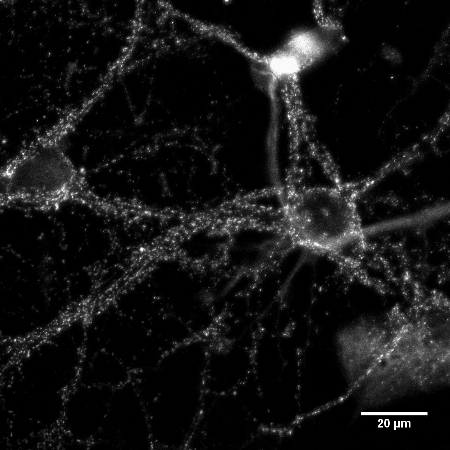
Immunocytochemistry/ Immunofluorescence: Bassoon Antibody (SAP7F407) [NB120-13249]
Immunocytochemistry/Immunofluorescence: Bassoon Antibody (SAP7F407) [NB120-13249] - Rat Hippocampal Neuron, image courtesy of customer.![Immunohistochemistry: Bassoon Antibody (SAP7F407) [NB120-13249]](https://resources.rndsystems.com/images/products/Bassoon-Antibody-SAP7F407-Immunohistochemistry-NB120-13249-img0002.jpg "Immunohistochemistry: Bassoon Antibody (SAP7F407) [NB120-13249]")
Immunohistochemistry: Bassoon Antibody (SAP7F407) [NB120-13249]
Immunohistochemistry: Bassoon Antibody (SAP7F407) [NB120-13249] - Bassoon localization in the rat brain cerebellar molecular layer using Bassoon Monoclonal Antibody.Applications for Bassoon Antibody (SAP7F407) - BSA Free
Immunocytochemistry/ Immunofluorescence
Immunohistochemistry
Immunohistochemistry-Paraffin
Immunoprecipitation
Western Blot
Reviewed Applications
Read 1 review rated 5 using NB120-13249 in the following applications:
Formulation, Preparation, and Storage
Purification
Formulation
Format
Preservative
Concentration
Shipping
Stability & Storage
Background: Bassoon
Alternate Names
Gene Symbol
UniProt
Additional Bassoon Products
Product Documents for Bassoon Antibody (SAP7F407) - BSA Free
Certificate of Analysis
To download a Certificate of Analysis, please enter a lot or batch number in the search box below.
Product Specific Notices for Bassoon Antibody (SAP7F407) - BSA Free
This product is for research use only and is not approved for use in humans or in clinical diagnosis. Primary Antibodies are guaranteed for 1 year from date of receipt.
Citations for Bassoon Antibody (SAP7F407) - BSA Free
 Powered by Bioz
Customer Reviews for Bassoon Antibody (SAP7F407) - BSA Free (1)
Have you used Bassoon Antibody (SAP7F407) - BSA Free?
Submit a review and receive an Amazon gift card!
$25/€18/£15/$25CAN/¥2500 Yen for a review with an image
$10/€7/£6/$10CAN/¥1110 Yen for a review without an image
Submit a review
Customer Images
-
Application: ImmunocytochemistrySample Tested: Cultured rat hippocampal neuronsSpecies: RatVerified Customer | Posted 03/27/2017Fixation: 3% formaldehyde (TAAB) in PBS, 15 minutes RT Blocking: 2% milk powder in PBS, 1 hour RT Permeabilization: 0.2% Triton-X100 Primary Antibody: 1:100 in 1% milk powder in PBS with 0.2% Triton, overnight, 4°C Wash: three times wash with PBS with 0.2% Triton, 10 min each Secondary Antibody: Invitrogen goat anti-mouse Alexa568, 1:500 in PBS with 0.2% Triton, 2 hours, RT Wash: three times wash with PBS, 10 min each Microscope: Confocal (Zeiss LSM710) The antibody labels clusters along the dendrites and somata of the cultured cells as expected.

There are no reviews that match your criteria.
Protocols
Find general support by application which include: protocols, troubleshooting, illustrated assays, videos and webinars.
- Antigen Retrieval Protocol (PIER)
- Antigen Retrieval for Frozen Sections Protocol
- Appropriate Fixation of IHC/ICC Samples
- Cellular Response to Hypoxia Protocols
- Chromogenic IHC Staining of Formalin-Fixed Paraffin-Embedded (FFPE) Tissue Protocol
- Chromogenic Immunohistochemistry Staining of Frozen Tissue
- ClariTSA™ Fluorophore Kits
- Detection & Visualization of Antibody Binding
- Fluorescent IHC Staining of Frozen Tissue Protocol
- Graphic Protocol for Heat-induced Epitope Retrieval
- Graphic Protocol for the Preparation and Fluorescent IHC Staining of Frozen Tissue Sections
- Graphic Protocol for the Preparation and Fluorescent IHC Staining of Paraffin-embedded Tissue Sections
- Graphic Protocol for the Preparation of Gelatin-coated Slides for Histological Tissue Sections
- ICC Cell Smear Protocol for Suspension Cells
- ICC Immunocytochemistry Protocol Videos
- ICC for Adherent Cells
- IHC Sample Preparation (Frozen sections vs Paraffin)
- Immunocytochemistry (ICC) Protocol
- Immunocytochemistry Troubleshooting
- Immunofluorescence of Organoids Embedded in Cultrex Basement Membrane Extract
- Immunofluorescent IHC Staining of Formalin-Fixed Paraffin-Embedded (FFPE) Tissue Protocol
- Immunohistochemistry (IHC) and Immunocytochemistry (ICC) Protocols
- Immunohistochemistry Frozen Troubleshooting
- Immunohistochemistry Paraffin Troubleshooting
- Immunoprecipitation Protocol
- Preparing Samples for IHC/ICC Experiments
- Preventing Non-Specific Staining (Non-Specific Binding)
- Primary Antibody Selection & Optimization
- Protocol for Heat-Induced Epitope Retrieval (HIER)
- Protocol for Making a 4% Formaldehyde Solution in PBS
- Protocol for VisUCyte™ HRP Polymer Detection Reagent
- Protocol for the Fluorescent ICC Staining of Cell Smears - Graphic
- Protocol for the Fluorescent ICC Staining of Cultured Cells on Coverslips - Graphic
- Protocol for the Preparation & Fixation of Cells on Coverslips
- Protocol for the Preparation and Chromogenic IHC Staining of Frozen Tissue Sections
- Protocol for the Preparation and Chromogenic IHC Staining of Frozen Tissue Sections - Graphic
- Protocol for the Preparation and Chromogenic IHC Staining of Paraffin-embedded Tissue Sections
- Protocol for the Preparation and Chromogenic IHC Staining of Paraffin-embedded Tissue Sections - Graphic
- Protocol for the Preparation and Fluorescent ICC Staining of Cells on Coverslips
- Protocol for the Preparation and Fluorescent ICC Staining of Non-adherent Cells
- Protocol for the Preparation and Fluorescent ICC Staining of Stem Cells on Coverslips
- Protocol for the Preparation and Fluorescent IHC Staining of Frozen Tissue Sections
- Protocol for the Preparation and Fluorescent IHC Staining of Paraffin-embedded Tissue Sections
- Protocol for the Preparation of Gelatin-coated Slides for Histological Tissue Sections
- Protocol for the Preparation of a Cell Smear for Non-adherent Cell ICC - Graphic
- R&D Systems Quality Control Western Blot Protocol
- TUNEL and Active Caspase-3 Detection by IHC/ICC Protocol
- The Importance of IHC/ICC Controls
- Troubleshooting Guide: Immunohistochemistry
- Troubleshooting Guide: Western Blot Figures
- Western Blot Conditions
- Western Blot Protocol
- Western Blot Protocol for Cell Lysates
- Western Blot Troubleshooting
- Western Blot Troubleshooting Guide
- View all Protocols, Troubleshooting, Illustrated assays and Webinars
FAQs for Bassoon Antibody (SAP7F407) - BSA Free
-
Q: I am looking for a good antibody to detect Bassoon in mouse brain slices (PFA fixed); can you recommend any one for this?
A:
After searching through our product catalog list, we have found the following antibodies which may be of interest: Bassoo. Using the filters on the left column of the site should allow you to filter by species and desired application. Any antibody that is listed to show validation for use in IHC-P and has also been validated in mouse samples will be backed by our 100% Guarantee. Under the terms and conditions of the guarantee, Novus will provide you with a full refund or replacement should the antibody fail to perform, as specified, in samples expressing the protein.