Bax Antibody (6A7) - Azide and BSA Free
Novus Biologicals | Catalog # NBP1-28566
![Western Blot: Bax Antibody (6A7) [NBP1-28566]](https://resources.rndsystems.com/images/products/Bax-Antibody-6A7-Western-Blot-NBP1-28566-img0007.jpg "Western Blot: Bax Antibody (6A7) [NBP1-28566]")
Loading...
Key Product Details
Validated by
Biological Validation
Species Reactivity
Validated:
Human, Mouse, Rat, Porcine, Bovine, Monkey
Cited:
Human, Mouse, Rat
Applications
Validated:
Immunohistochemistry, Immunohistochemistry-Paraffin, Immunohistochemistry-Frozen, Western Blot, Functional
Cited:
Immunohistochemistry-Paraffin, Western Blot, Co-IP, Functional Assay, IF/IHC
Label
Unconjugated
Antibody Source
Monoclonal Mouse IgG1 kappa Clone # 6A7
Format
Azide and BSA Free
Loading...
Product Specifications
Immunogen
KLH-conjugated peptide corresponding to amino acids 12-24 located near the N-terminus common to human, mouse and rat Bax.
Reactivity Notes
Use in Porcine reported in scientific literature (PMID:34243771) Mouse blocking reagent may be needed for IHC and ICC experiments to reduce high background signal. You can find these reagents under catalog numbers PK-2200-NB and MP-2400-NB. Please contact Technical Support if you have any Please note that this antibody is reactive to Mouse and derived from the same host, Mouse. Mouse-On-questions.
Specificity
The monoclonal antibody 6A7 reacts with human, mouse and rat Bax but does not bind the soluble cytosolic form of Bax; however, treatment of cells with non-ionic detergents exposes the epitope and allows binding of 6A7 to monomeric forms of Bax but not Bax complexed with either Bcl-2 or Bcl-xL.
Clonality
Monoclonal
Host
Mouse
Isotype
IgG1 kappa
Scientific Data Images for Bax Antibody (6A7) - Azide and BSA Free
Western Blot: Bax Antibody (6A7) [NBP1-28566]
Bax-Antibody-6A7-Western-Blot-NBP1-28566-img0007.jpg![Immunohistochemistry-Frozen: Bax Antibody (6A7) [NBP1-28566]](https://resources.rndsystems.com/images/products/Bax-Antibody-6A7-Immunohistochemistry-Frozen-NBP1-28566-img0002.jpg "Immunohistochemistry-Frozen: Bax Antibody (6A7) [NBP1-28566]")
 [NBP1-28566] -")
Western Blot: Bax Antibody (6A7) [NBP1-28566] -
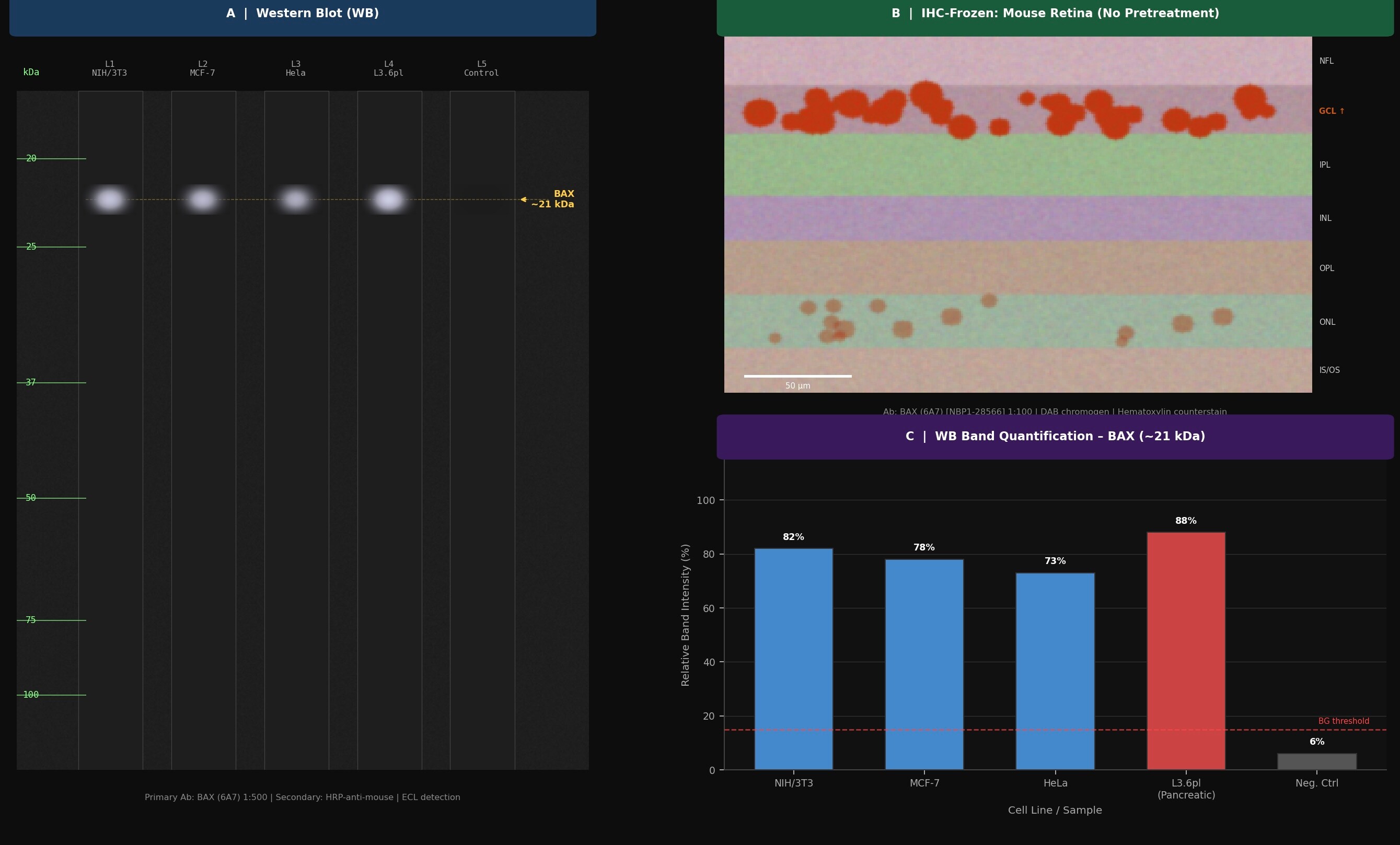
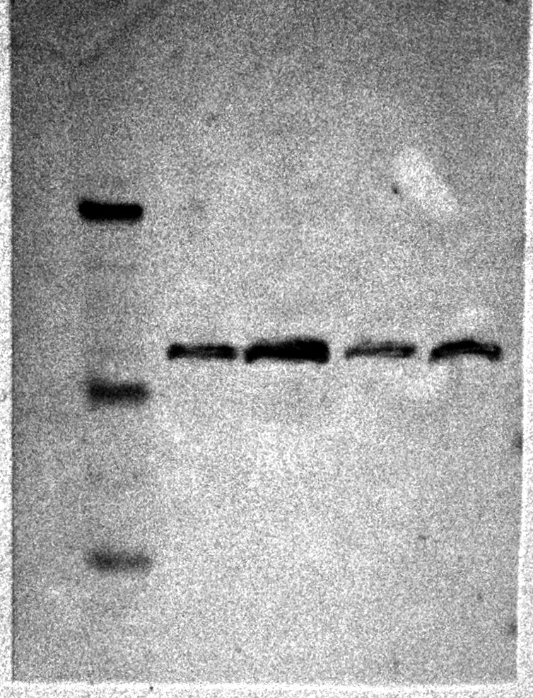
Western Blot: Bax Antibody (6A7) [NBP1-28566] - Analysis of lysates from human metastatic pancreatic cancer cell line L3.6pl using Bax Antibody (clone 6A7; Lot # B168-P780E). This image was submitted via reviews by a verified end user.![Western Blot: Bax Antibody (6A7) [NBP1-28566]](https://resources.rndsystems.com/images/products/Bax-Antibody-6A7-Western-Blot-NBP1-28566-img0006.jpg "Western Blot: Bax Antibody (6A7) [NBP1-28566]")
Western Blot: Bax Antibody (6A7) [NBP1-28566]
Western Blot: Bax Antibody (6A7) [NBP1-28566] - Total cell lysates from NIH/3T3 cells were resolved by electrophoresis, transferred to PVDF membrane, and probled with Mouse Anti-Bax UNLB.Applications for Bax Antibody (6A7) - Azide and BSA Free
Application
Recommended Usage
Immunohistochemistry-Frozen
1:10-1:500
Western Blot
< = 1 ug/ml
Application Notes
Use in IHC-P reported in scientific literature (PMID:33143886) Use in functional reported in scientific literature (PMID:31410057). Bax antibody validated for IHC-F, WB from a verified customer reviews.
Reviewed Applications
Read 5 reviews rated 4.8 using NBP1-28566 in the following applications:
Formulation, Preparation, and Storage
Purification
Protein A or G purified
Formulation
Borate buffered saline, pH 8.2
Format
Azide and BSA Free
Preservative
No Preservative
Concentration
0.1 mg/ml
Shipping
The product is shipped with polar packs. Upon receipt, store it immediately at the temperature recommended below.
Stability & Storage
Store at 4C. Do not freeze.
Background: Bax
Long Name
Bcl Associated X Protein
Alternate Names
apoptosis regulator BAX, BCL2-associated X protein, Bcl2-L-4, BCL2L4bcl2-L-4, Bcl-2-like protein 4
Gene Symbol
BAX
UniProt
Additional Bax Products
Product Documents for Bax Antibody (6A7) - Azide and BSA Free
Certificate of Analysis
To download a Certificate of Analysis, please enter a lot or batch number in the search box below.
Product Specific Notices for Bax Antibody (6A7) - Azide and BSA Free
This product is for research use only and is not approved for use in humans or in clinical diagnosis. Primary Antibodies are guaranteed for 1 year from date of receipt.
Related Research Areas
Citations for Bax Antibody (6A7) - Azide and BSA Free
 Powered by Bioz
Customer Reviews for Bax Antibody (6A7) - Azide and BSA Free (5)
4.8 out of 5
5 Customer Ratings
Have you used Bax Antibody (6A7) - Azide and BSA Free?
Submit a review and receive an Amazon gift card!
$25/€18/£15/$25CAN/¥2500 Yen for a review with an image
$10/€7/£6/$10CAN/¥1110 Yen for a review without an image
Submit a review
Customer Images
-(01-mg)_NBP1-28566_6711.jpg)
Showing
1
-
5 of
5 reviews
Showing All
Filter By:
-
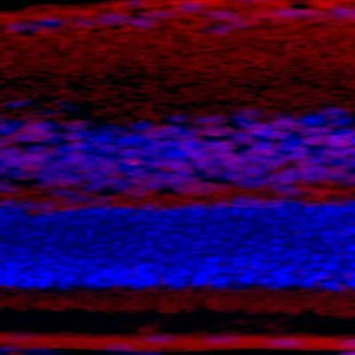
Application: Western BlotSample Tested: IHC on parrafin sections (mouse) and IHC Sample TestedSpecies: Human, Mouse and RatVerified Customer | Posted 06/17/2026Stained sections were observed with a light microscope coupled with a digitally calibrated camera at 10- and 40-fold magnifications. BAX immunoreactivity was graded according to the intensity in NFL, GCL, IPL, INL, OPL, ONL, IS/OS. We performed a semi quantitative scoring method with a typical H-scoring system; which represents staining intensity (0 = no staining; 1 = weak; 2 = moderate; 3 = strong) by the percentage of positive cells in sections. Scores ranged from 0 to 300 (stained intensity multiplied by the percentage of stained cells in percent). Images were quantitatively analyzed for DAB optical density values with ImageJ software in combination with the color deconvolution plug-in in order to differentiate the signals for DAB from hematoxylin. The scale bar of 50 m was applied. Negative control incubations were performed for every experiment, where the primary antibody was replaced by a matching mouse IgG of the same isotype at an equivalent concentration, since none was observed in negative control experiments (in the absence of primary antibody with equivalent concentration matched to mouse IgG isotype negative controls), the staining with BAX signal specificity was thus validated.

-
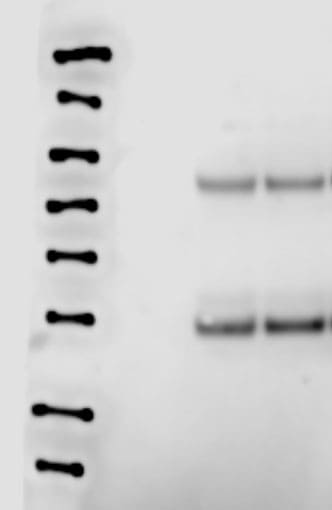
Application: Western BlotSample Tested: Cell lysate from colon cancer cell line and Melanoma cellsSpecies: Mouse and HumanVerified Customer | Posted 04/19/2024Bax western blot in cancer cell lysate after apoptosis induction
-
Application: Western BlotSample Tested: mouse liverSpecies: MouseVerified Customer | Posted 06/23/20171:1000, liver lysate 10mg
-
Application: Western BlotSample Tested:Species: HumanVerified Customer | Posted 03/31/2014Bax western blot with Pancreatic cancer cell line L3.6pl.
-
Application: Immunohistochemistry-FrozenSample Tested: mouse retinaSpecies: MouseVerified Customer | Posted 07/28/2012
There are no reviews that match your criteria.
Protocols
Find general support by application which include: protocols, troubleshooting, illustrated assays, videos and webinars.
- Antigen Retrieval Protocol (PIER)
- Antigen Retrieval for Frozen Sections Protocol
- Appropriate Fixation of IHC/ICC Samples
- Cellular Response to Hypoxia Protocols
- Chromogenic IHC Staining of Formalin-Fixed Paraffin-Embedded (FFPE) Tissue Protocol
- Chromogenic Immunohistochemistry Staining of Frozen Tissue
- ClariTSA™ Fluorophore Kits
- Detection & Visualization of Antibody Binding
- Fluorescent IHC Staining of Frozen Tissue Protocol
- Graphic Protocol for Heat-induced Epitope Retrieval
- Graphic Protocol for the Preparation and Fluorescent IHC Staining of Frozen Tissue Sections
- Graphic Protocol for the Preparation and Fluorescent IHC Staining of Paraffin-embedded Tissue Sections
- Graphic Protocol for the Preparation of Gelatin-coated Slides for Histological Tissue Sections
- IHC Sample Preparation (Frozen sections vs Paraffin)
- Immunofluorescent IHC Staining of Formalin-Fixed Paraffin-Embedded (FFPE) Tissue Protocol
- Immunohistochemistry (IHC) and Immunocytochemistry (ICC) Protocols
- Immunohistochemistry Frozen Troubleshooting
- Immunohistochemistry Paraffin Troubleshooting
- Preparing Samples for IHC/ICC Experiments
- Preventing Non-Specific Staining (Non-Specific Binding)
- Primary Antibody Selection & Optimization
- Protocol for Heat-Induced Epitope Retrieval (HIER)
- Protocol for Making a 4% Formaldehyde Solution in PBS
- Protocol for VisUCyte™ HRP Polymer Detection Reagent
- Protocol for the Preparation & Fixation of Cells on Coverslips
- Protocol for the Preparation and Chromogenic IHC Staining of Frozen Tissue Sections
- Protocol for the Preparation and Chromogenic IHC Staining of Frozen Tissue Sections - Graphic
- Protocol for the Preparation and Chromogenic IHC Staining of Paraffin-embedded Tissue Sections
- Protocol for the Preparation and Chromogenic IHC Staining of Paraffin-embedded Tissue Sections - Graphic
- Protocol for the Preparation and Fluorescent IHC Staining of Frozen Tissue Sections
- Protocol for the Preparation and Fluorescent IHC Staining of Paraffin-embedded Tissue Sections
- Protocol for the Preparation of Gelatin-coated Slides for Histological Tissue Sections
- R&D Systems Quality Control Western Blot Protocol
- TUNEL and Active Caspase-3 Detection by IHC/ICC Protocol
- The Importance of IHC/ICC Controls
- Troubleshooting Guide: Immunohistochemistry
- Troubleshooting Guide: Western Blot Figures
- Western Blot Conditions
- Western Blot Protocol
- Western Blot Protocol for Cell Lysates
- Western Blot Troubleshooting
- Western Blot Troubleshooting Guide
- View all Protocols, Troubleshooting, Illustrated assays and Webinars
Loading...
Associated Pathways

