Bcl-x is a member of the Bcl-2 family of proteins that regulates outer mitochondrial membrane permeability. Alternative splicing results in two distinct Bcl-x isoforms. Bcl-xL (long) is an anti-apoptotic protein that prevents release of Cytochromec from the mitochondrial intermembrane space into the cytosol. Bcl-xs (short) is a pro-apoptotic member that can initiate programmed cell death.
Key Product Details
Species Reactivity
Human, Mouse, Rat, Porcine
Applications
Immunohistochemistry, Immunohistochemistry-Paraffin, Western Blot, Flow Cytometry, Immunocytochemistry/ Immunofluorescence
Label
Unconjugated
Antibody Source
Monoclonal Mouse IgG2a Kappa Clone # 2H12
Loading...
Product Specifications
Immunogen
A synthetic peptide, aa 3-14 (Cys-QSNRELVVDFLS) of human Bcl-xL protein (Uniprot: Q07817)
Localization
Cytoplasmic and cell/nuclear membrane
Marker
Apoptosis Marker
Specificity
Recognizes a protein of 27kDa, identified as the Bcl-X protein. This monoclonal antibody shows no cross-reaction with Bcl-2 or Bax protein. Bcl-X has two isoforms, Bcl-XL (long), a 241 amino acid protein which suppresses cell death. And Bcl-XS (short), a 178 amino acid protein lacking a 63 amino acid domain which functions as a dominant inhibitor of Bcl-2. This monoclonal antibody reacts with both Bcl-XS and Bcl-XL proteins.
Clonality
Monoclonal
Host
Mouse
Isotype
IgG2a Kappa
Theoretical MW
27 kDa.
Disclaimer note: The observed molecular weight of the protein may vary from the listed predicted molecular weight due to post translational modifications, post translation cleavages, relative charges, and other experimental factors.
Disclaimer note: The observed molecular weight of the protein may vary from the listed predicted molecular weight due to post translational modifications, post translation cleavages, relative charges, and other experimental factors.
Description
200ug/ml of antibody purified from Bioreactor Concentrate by Protein A or G. Prepared in 10 mM PBS with 0.05% BSA & 0.05% azide. Also available WITHOUT BSA & azide at 1.0 mg/ml. (NBP2-34531)
Antibody with azide - store at 2 to 8C. Antibody without azide - store at -20 to -80C.
Antibody with azide - store at 2 to 8C. Antibody without azide - store at -20 to -80C.
Scientific Data Images for bcl-x Antibody (2H12)
![Western Blot: bcl-x Antibody (2H12) [NBP2-32917]](https://resources.rndsystems.com/images/products/Bcl-xL-Antibody-2H12-Western-Blot-NBP2-32917-img0004.jpg "Western Blot: bcl-x Antibody (2H12) [NBP2-32917]")
Western Blot: bcl-x Antibody (2H12) [NBP2-32917]
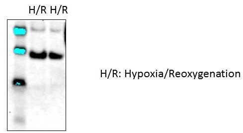
Western Blot: bcl-x Antibody (2H12) [NBP2-32917] - Rat and mouse neonatal cardiomyocytes. Western blot image submitted by a verified customer review.![Immunohistochemistry-Paraffin: bcl-x Antibody (2H12) [NBP2-32917]](https://resources.rndsystems.com/images/products/Bcl-xL-Antibody-2H12-Immunohistochemistry-Paraffin-NBP2-32917-img0001.jpg "Immunohistochemistry-Paraffin: bcl-x Antibody (2H12) [NBP2-32917]")
Immunohistochemistry-Paraffin: bcl-x Antibody (2H12) [NBP2-32917]
Immunohistochemistry-Paraffin: bcl-x Antibody (2H12) [NBP2-32917] - FFPE human Hodgkin's lymphoma stained with bcl-x Ab (2H12). Note cytoplasmic and membrane staining.![Western Blot: bcl-x Antibody (2H12) [NBP2-32917]](https://resources.rndsystems.com/images/products/Bcl-xL-Antibody-2H12-Western-Blot-NBP2-32917-img0003.jpg "Western Blot: bcl-x Antibody (2H12) [NBP2-32917]")
Western Blot: bcl-x Antibody (2H12) [NBP2-32917]
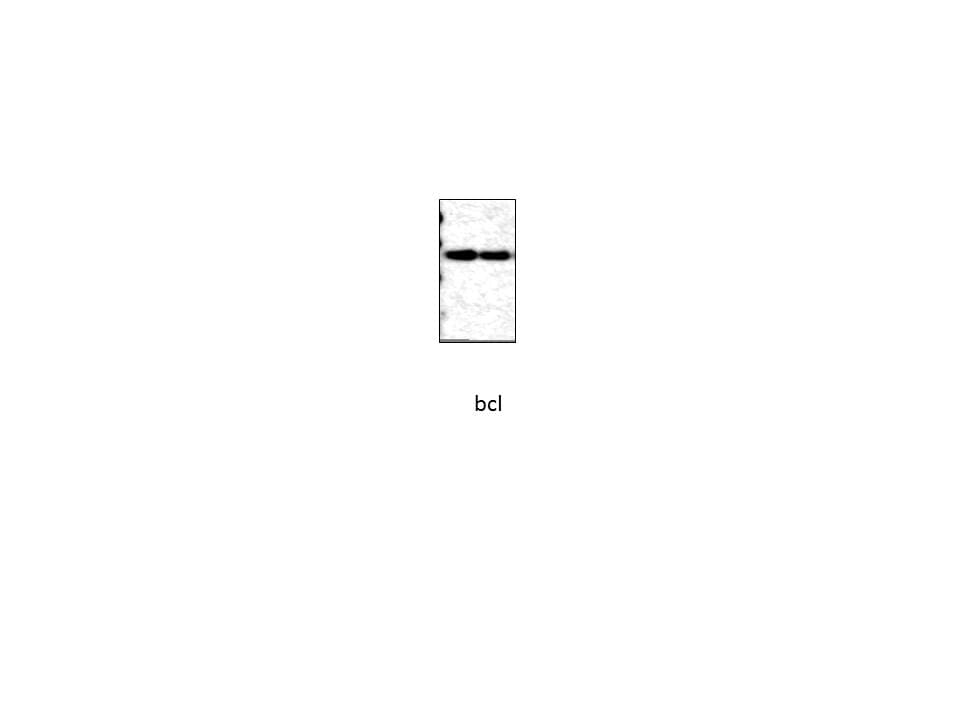
Western Blot: bcl-x Antibody (2H12) [NBP2-32917] - Western blot analysis of Jurkat Cell lysate using bcl-X Ab (2H12).Applications for bcl-x Antibody (2H12)
Application
Recommended Usage
Flow Cytometry
1-2 ug/million cells
Immunocytochemistry/ Immunofluorescence
1-2 ug/ml
Immunohistochemistry-Paraffin
1-2 ug/ml
Western Blot
1-2 ug/ml
Application Notes
Immunohistochemistry (Formalin-fixed): 1-2ug/ml for 30 minutes at RT. Staining of formalin-fixed tissues requires heating tissue sections in 10mM Tris buffer with 1mM EDTA, pH 9.0, for 45 min at 95C followed by cooling at RT for 20 minutes.
Optimal dilution for a specific application should be determined.
Optimal dilution for a specific application should be determined.
Reviewed Applications
Read 2 reviews rated 5 using NBP2-32917 in the following applications:
Flow Cytometry Panel Builder
Bio-Techne Knows Flow Cytometry
Save time and reduce costly mistakes by quickly finding compatible reagents using the Panel Builder Tool.
Advanced Features
- Spectra Viewer - Custom analysis of spectra from multiple fluorochromes
- Spillover Popups - Visualize the spectra of individual fluorochromes
- Antigen Density Selector - Match fluorochrome brightness with antigen density

Formulation, Preparation, and Storage
Purification
Protein A or G purified
Formulation
10 mM PBS with 0.05% BSA
Preservative
0.05% Sodium Azide
Concentration
0.2 mg/ml
Shipping
The product is shipped with polar packs. Upon receipt, store it immediately at the temperature recommended below.
Stability & Storage
Store at 4C.
Background: Bcl-x
Additional Bcl-x Products
Product Documents for bcl-x Antibody (2H12)
Certificate of Analysis
To download a Certificate of Analysis, please enter a lot or batch number in the search box below.
Product Specific Notices for bcl-x Antibody (2H12)
This product is for research use only and is not approved for use in humans or in clinical diagnosis. Primary Antibodies are guaranteed for 1 year from date of receipt.
Related Research Areas
Customer Reviews for bcl-x Antibody (2H12) (2)
5 out of 5
2 Customer Ratings
Have you used bcl-x Antibody (2H12)?
Submit a review and receive an Amazon gift card!
$25/€18/£15/$25CAN/¥2500 Yen for a review with an image
$10/€7/£6/$10CAN/¥1110 Yen for a review without an image
Submit a review
Customer Images
Showing
1
-
2 of
2 reviews
Showing All
Filter By:
-
Application: Western BlotSample Tested: primary neonatal ventricular cardiomyocytesSpecies: Rat neonatal cardiomyocytes and MouseVerified Customer | Posted 10/04/2019Antibody at 1:1000.

-
Application: Western BlotSample Tested: primary neonatal ventricular cardiomyocytesSpecies: MouseVerified Customer | Posted 06/14/2019bcl2
There are no reviews that match your criteria.
Protocols
Find general support by application which include: protocols, troubleshooting, illustrated assays, videos and webinars.
- 7-Amino Actinomycin D (7-AAD) Cell Viability Flow Cytometry Protocol
- Antigen Retrieval Protocol (PIER)
- Antigen Retrieval for Frozen Sections Protocol
- Appropriate Fixation of IHC/ICC Samples
- Cellular Response to Hypoxia Protocols
- Chromogenic IHC Staining of Formalin-Fixed Paraffin-Embedded (FFPE) Tissue Protocol
- Chromogenic Immunohistochemistry Staining of Frozen Tissue
- ClariTSA™ Fluorophore Kits
- Detection & Visualization of Antibody Binding
- Extracellular Membrane Flow Cytometry Protocol
- Flow Cytometry Protocol for Cell Surface Markers
- Flow Cytometry Protocol for Staining Membrane Associated Proteins
- Flow Cytometry Staining Protocols
- Flow Cytometry Troubleshooting Guide
- Fluorescent IHC Staining of Frozen Tissue Protocol
- Graphic Protocol for Heat-induced Epitope Retrieval
- Graphic Protocol for the Preparation and Fluorescent IHC Staining of Frozen Tissue Sections
- Graphic Protocol for the Preparation and Fluorescent IHC Staining of Paraffin-embedded Tissue Sections
- Graphic Protocol for the Preparation of Gelatin-coated Slides for Histological Tissue Sections
- ICC Cell Smear Protocol for Suspension Cells
- ICC Immunocytochemistry Protocol Videos
- ICC for Adherent Cells
- IHC Sample Preparation (Frozen sections vs Paraffin)
- Immunocytochemistry (ICC) Protocol
- Immunocytochemistry Troubleshooting
- Immunofluorescence of Organoids Embedded in Cultrex Basement Membrane Extract
- Immunofluorescent IHC Staining of Formalin-Fixed Paraffin-Embedded (FFPE) Tissue Protocol
- Immunohistochemistry (IHC) and Immunocytochemistry (ICC) Protocols
- Immunohistochemistry Frozen Troubleshooting
- Immunohistochemistry Paraffin Troubleshooting
- Intracellular Flow Cytometry Protocol Using Alcohol (Methanol)
- Intracellular Flow Cytometry Protocol Using Detergents
- Intracellular Nuclear Staining Flow Cytometry Protocol Using Detergents
- Intracellular Staining Flow Cytometry Protocol Using Alcohol Permeabilization
- Intracellular Staining Flow Cytometry Protocol Using Detergents to Permeabilize Cells
- Preparing Samples for IHC/ICC Experiments
- Preventing Non-Specific Staining (Non-Specific Binding)
- Primary Antibody Selection & Optimization
- Propidium Iodide Cell Viability Flow Cytometry Protocol
- Protocol for Heat-Induced Epitope Retrieval (HIER)
- Protocol for Liperfluo
- Protocol for Making a 4% Formaldehyde Solution in PBS
- Protocol for VisUCyte™ HRP Polymer Detection Reagent
- Protocol for the Characterization of Human Th22 Cells
- Protocol for the Characterization of Human Th9 Cells
- Protocol for the Fluorescent ICC Staining of Cell Smears - Graphic
- Protocol for the Fluorescent ICC Staining of Cultured Cells on Coverslips - Graphic
- Protocol for the Preparation & Fixation of Cells on Coverslips
- Protocol for the Preparation and Chromogenic IHC Staining of Frozen Tissue Sections
- Protocol for the Preparation and Chromogenic IHC Staining of Frozen Tissue Sections - Graphic
- Protocol for the Preparation and Chromogenic IHC Staining of Paraffin-embedded Tissue Sections
- Protocol for the Preparation and Chromogenic IHC Staining of Paraffin-embedded Tissue Sections - Graphic
- Protocol for the Preparation and Fluorescent ICC Staining of Cells on Coverslips
- Protocol for the Preparation and Fluorescent ICC Staining of Non-adherent Cells
- Protocol for the Preparation and Fluorescent ICC Staining of Stem Cells on Coverslips
- Protocol for the Preparation and Fluorescent IHC Staining of Frozen Tissue Sections
- Protocol for the Preparation and Fluorescent IHC Staining of Paraffin-embedded Tissue Sections
- Protocol for the Preparation of Gelatin-coated Slides for Histological Tissue Sections
- Protocol for the Preparation of a Cell Smear for Non-adherent Cell ICC - Graphic
- Protocol: Annexin V and PI Staining by Flow Cytometry
- Protocol: Annexin V and PI Staining for Apoptosis by Flow Cytometry
- R&D Systems Quality Control Western Blot Protocol
- TUNEL and Active Caspase-3 Detection by IHC/ICC Protocol
- The Importance of IHC/ICC Controls
- Troubleshooting Guide: Fluorokine Flow Cytometry Kits
- Troubleshooting Guide: Immunohistochemistry
- Troubleshooting Guide: Western Blot Figures
- Western Blot Conditions
- Western Blot Protocol
- Western Blot Protocol for Cell Lysates
- Western Blot Troubleshooting
- Western Blot Troubleshooting Guide
- View all Protocols, Troubleshooting, Illustrated assays and Webinars
Loading...
Associated Pathways

