beta-Actin Antibody (8H10D10) - BSA Free
Novus Biologicals | Catalog # NBP1-47423
Key Product Details
Species Reactivity
Validated:
Human, Mouse, Rat, Chinese Hamster, Primate
Cited:
Human, Mouse, Rat
Applications
Validated:
Western Blot, Immunoblotting, ELISA, Flow Cytometry, Flow (Intracellular), Immunocytochemistry/ Immunofluorescence, Simple Western
Cited:
Western Blot, Immunoblotting, Simple Western
Label
Unconjugated
Antibody Source
Monoclonal Mouse IgG2B Clone # 8H10D10
Format
BSA Free
Loading...
Product Specifications
Immunogen
This beta-Actin Antibody (8H10D10) was made to a synthetic peptide corresponding to amino-terminal residues of human Beta Actin, conjugated to KLH. [UniProt# P60709]
Localization
Cytoplasm, cytoskeleton.
Clonality
Monoclonal
Host
Mouse
Isotype
IgG2B
Theoretical MW
42 kDa.
Disclaimer note: The observed molecular weight of the protein may vary from the listed predicted molecular weight due to post translational modifications, post translation cleavages, relative charges, and other experimental factors.
Disclaimer note: The observed molecular weight of the protein may vary from the listed predicted molecular weight due to post translational modifications, post translation cleavages, relative charges, and other experimental factors.
Scientific Data Images for beta-Actin Antibody (8H10D10) - BSA Free

Detection of Mouse beta-Actin by Simple Western TM.
Left: Simple Western lane view shows lysates of mouse brain, loaded at 1 mg/ml. A specific band was detected for beta ‑Actin at approximately 47 kDa (as indicated) using both 1:500 and 1:100 of Mouse Anti-Human/Mouse/Rat beta ‑Actin Monoclonal Antibody (Catalog # NBP1-47423) followed by HRP-conjugated Goat Anti-Mouse Secondary Antibody (Catalog # 042-205). This experiment was conducted under reducing conditions and using the 12-230kDa separation system. Right: Simple Western electropherogram showing the same Mouse Anti-Human/Mouse/Rat beta ‑Actin Monoclonal Antibody (Catalog # NBP1-47423) tested at 1:500 (blue line) and 1:100 (green line) in the mouse brain.![Western Blot: beta-Actin Antibody (8H10D10)BSA Free [NBP1-47423]](https://resources.rndsystems.com/images/products/beta-Actin-Antibody-8H10D10-Western-Blot-NBP1-47423-img0006.jpg "Western Blot: beta-Actin Antibody (8H10D10)BSA Free [NBP1-47423]")
Western Blot: beta-Actin Antibody (8H10D10)BSA Free [NBP1-47423]
Western Blot: beta-Actin Antibody (8H10D10) [NBP1-47423] - Analysis using anti-Beta Actin mAb against human, mouse, hamster, rat, and primate cell lystate(s): (1) NIH/3T3, (2) Jurkat, (3) HeLa, (4) CHO, (5) PC12, (6) HEK293, (7) COS, (8) A549, and (9) MCF-7. A specific band can be observed at a molecular weight of approximately 43 kDa in all lanes.![Immunocytochemistry/ Immunofluorescence: beta-Actin Antibody (8H10D10) - BSA Free [NBP1-47423]](https://resources.rndsystems.com/images/products/beta-Actin-Antibody-8H10D10-Immunocytochemistry-Immunofluorescence-NBP1-47423-img0011.jpg "Immunocytochemistry/ Immunofluorescence: beta-Actin Antibody (8H10D10) - BSA Free [NBP1-47423]")
Immunocytochemistry/ Immunofluorescence: beta-Actin Antibody (8H10D10) - BSA Free [NBP1-47423]
Immunocytochemistry/Immunofluorescence: beta-Actin Antibody (8H10D10) [NBP1-47423] - Beta actin was detected in NIH-3T3 cells fixed with methanol using mouse anti-mouse beta-Actin monoclonal antibody (NBP1-47423) at 1:5400 dilution. Cells were stained using NorthernLights 557 conjugated anti-mouse secondary antibody (NL007) and counterstained with DAPI.![Simple Western: beta-Actin Antibody (8H10D10)BSA Free [NBP1-47423]](https://resources.rndsystems.com/images/products/beta-Actin-Antibody-8H10D10-Simple-Western-NBP1-47423-img0009.jpg "Simple Western: beta-Actin Antibody (8H10D10)BSA Free [NBP1-47423]")
Simple Western: beta-Actin Antibody (8H10D10)BSA Free [NBP1-47423]
Simple Western: beta-Actin Antibody (8H10D10) [NBP1-47423] - Image shows a specific band for Beta Actin in 1.0 mg/mL of HeLa lysate. This experiment was performed under reducing conditions using the 12-230 kDa separation system.![Flow Cytometry: beta-Actin Antibody (8H10D10) - BSA Free [NBP1-47423]](https://resources.rndsystems.com/images/products/beta-Actin-Antibody-8H10D10-Flow-Cytometry-NBP1-47423-img0010.jpg "Flow Cytometry: beta-Actin Antibody (8H10D10) - BSA Free [NBP1-47423]")
Flow Cytometry: beta-Actin Antibody (8H10D10) - BSA Free [NBP1-47423]
Flow Cytometry: beta-Actin Antibody (8H10D10) [NBP1-47423] - Analysis of HeLa cells using mouse Monoclonal beta-Actin antibody (Orange) and Isotype control Antibody (Blue).![Western Blot: beta-Actin Antibody (8H10D10)BSA Free [NBP1-47423]](https://resources.rndsystems.com/images/products/beta-Actin-Antibody-8H10D10-Western-Blot-NBP1-47423-img0014.jpg "Western Blot: beta-Actin Antibody (8H10D10)BSA Free [NBP1-47423]")
Western Blot: beta-Actin Antibody (8H10D10)BSA Free [NBP1-47423]
beta-Actin-Antibody-8H10D10-Western-Blot-NBP1-47423-img0014.jpg![Immunocytochemistry/ Immunofluorescence: beta-Actin Antibody (8H10D10) - BSA Free [NBP1-47423]](https://resources.rndsystems.com/images/products/beta-Actin-Antibody-8H10D10-Immunocytochemistry-Immunofluorescence-NBP1-47423-img0015.jpg "Immunocytochemistry/ Immunofluorescence: beta-Actin Antibody (8H10D10) - BSA Free [NBP1-47423]")
Immunocytochemistry/ Immunofluorescence: beta-Actin Antibody (8H10D10) - BSA Free [NBP1-47423]
Immunocytochemistry/Immunofluorescence: beta-Actin Antibody (8H10D10) [NBP1-47423] - HeLa cells were fixed and permeabilized for 10 minutes with -20C MeOH. The cells were incubated with beta-Actin Antibody [8H10D10] conjugated to Biotin (NBP1-47423B) at 5ug/ml overnight at 4C and detected with Streptavidin conjugated to DyLight 550. Nuclei were counterstained with DAPI (Blue). Cells were imaged using a 100X objective and digitally deconvolved.![Flow Cytometry: beta-Actin Antibody (8H10D10) - BSA Free [NBP1-47423]](https://resources.rndsystems.com/images/products/beta-Actin-Antibody-8H10D10-BSA-Free-Flow-Cytometry-NBP1-47423-img0017.jpg "Flow Cytometry: beta-Actin Antibody (8H10D10) - BSA Free [NBP1-47423]")
Flow Cytometry: beta-Actin Antibody (8H10D10) - BSA Free [NBP1-47423]
Flow Cytometry: beta-Actin Antibody (8H10D10) - BSA Free [NBP1-47423] - An intracellular stain was performed on NIH3T3 cells with beta-Actin Antibody (8H10D10) NBP1-47423 (blue) and a matched isotype control MAB004 (orange). Cells were fixed with 4% PFA and then permeabilized with 0.1% saponin. Cells were incubated in an antibody dilution of 1 ug/mL for 30 minutes at room temperature, followed by Mouse IgG (H+L) Cross-Adsorbed Secondary Antibody, Dylight 550 (84540, Thermo Fisher).![Western Blot: beta-Actin Antibody (8H10D10)BSA Free [NBP1-47423]](https://resources.rndsystems.com/images/products/beta-Actin-Antibody-8H10D10-Western-Blot-NBP1-47423-img0007.jpg "Western Blot: beta-Actin Antibody (8H10D10)BSA Free [NBP1-47423]")
Western Blot: beta-Actin Antibody (8H10D10)BSA Free [NBP1-47423]
Western Blot: beta-Actin Antibody (8H10D10) [NBP1-47423] - Western blot of pancreatic cell lines (CAPAN2, PATU8988T, DANG, and PANC1005). Primary antibody: beta-Actin (8H10D10), NBP1-47423, 1:1000 dilution. Secondary antibody: HRP conjugated Mouse IgG (H+L), NB7539. A specific band can be observed at a molecular weight of approximately 42 kDa. Image from verified customer review.![Western Blot: beta-Actin Antibody (8H10D10)BSA Free [NBP1-47423]](https://resources.rndsystems.com/images/products/beta-Actin-Antibody-8H10D10-Western-Blot-NBP1-47423-img0008.jpg "Western Blot: beta-Actin Antibody (8H10D10)BSA Free [NBP1-47423]")
Western Blot: beta-Actin Antibody (8H10D10)BSA Free [NBP1-47423]
Western Blot: beta-Actin Antibody (8H10D10) [NBP1-47423] - Analysis of Beta Actin expression in 1) HeLa 2) HepG2 and 3) Cos7 whole cell lysates.![Western Blot: beta-Actin Antibody (8H10D10)BSA Free [NBP1-47423]](https://resources.rndsystems.com/images/products/beta-Actin-Antibody-8H10D10-Western-Blot-NBP1-47423-img0012.jpg "Western Blot: beta-Actin Antibody (8H10D10)BSA Free [NBP1-47423]")
Western Blot: beta-Actin Antibody (8H10D10)BSA Free [NBP1-47423]
Western Blot: beta-Actin Antibody (8H10D10) [NBP1-47423] - Lysates of HeLa human cervical epithelial carcinoma cell line, A431 human epithelial carcinoma cell line, K562 human chronic myelogenous leukemia cell line, NIH-3T3 mouse embryonic fibroblast cell line, and C6 rat glioma cell line were probed with 1:2500 mouse anti-beta-Actin monoclonal (NBP1-47423) followed by 1:2000 dilution of donkey anti-mouse IgG-HRP secondary antibody (HAF018).![Western Blot: beta-Actin Antibody (8H10D10)BSA Free [NBP1-47423]](https://resources.rndsystems.com/images/products/beta-Actin-Antibody-8H10D10-Western-Blot-NBP1-47423-img0013.jpg "Western Blot: beta-Actin Antibody (8H10D10)BSA Free [NBP1-47423]")
Western Blot: beta-Actin Antibody (8H10D10)BSA Free [NBP1-47423]
Western Blot: beta-Actin Antibody (8H10D10) [NBP1-47423] - Three different human cancer cell lines (SW780, BT-20, and CLS439) were probed with the antibody. Image from verified customer review.![Flow Cytometry: beta-Actin Antibody (8H10D10) - BSA Free [NBP1-47423]](https://resources.rndsystems.com/images/products/beta-Actin-Antibody-8H10D10-Flow-Cytometry-NBP1-47423-img0004.jpg "Flow Cytometry: beta-Actin Antibody (8H10D10) - BSA Free [NBP1-47423]")
Flow Cytometry: beta-Actin Antibody (8H10D10) - BSA Free [NBP1-47423]
Flow Cytometry: beta-Actin Antibody (8H10D10) [NBP1-47423] - Analysis of MCF-7 cells using anti-Beta Actin mAb (right) and negative control (left).![Flow Cytometry: beta-Actin Antibody (8H10D10) - BSA Free [NBP1-47423]](https://resources.rndsystems.com/images/products/beta-Actin-Antibody-8H10D10-BSA-Free-Flow-Cytometry-NBP1-47423-img0016.jpg "Flow Cytometry: beta-Actin Antibody (8H10D10) - BSA Free [NBP1-47423]")
Flow Cytometry: beta-Actin Antibody (8H10D10) - BSA Free [NBP1-47423]
Flow Cytometry: beta-Actin Antibody (8H10D10) - BSA Free [NBP1-47423] - An intracellular stain was performed on U-251 MG cells with beta-Actin Antibody (8H10D10) NBP1-47423 (blue) and a matched isotype control MAB004 (orange). Cells were fixed with 4% PFA and then permeabilized with 0.1% saponin. Cells were incubated in an antibody dilution of 1 ug/mL for 30 minutes at room temperature, followed by Mouse IgG (H+L) Cross-Adsorbed Secondary Antibody, Dylight 550 (84540, Thermo Fisher). in MCF7 Human Cell Line.")
Beta-Actin (8H10D10) in MCF7 Human Cell Line.
Beta-Actin (8H10D10) was detected in immersion fixed MCF7 human breast cancer cell line using Mouse anti-beta-Actin (8H10D10) Protein G Purified Monoclonal Antibody conjugated to Biotin (Catalog # NBP1-47423B) at 5 µg/mL overnight at 4C. Cells were stained using Streptavidin conjugated to DyLight 550 (red) and counterstained with DAPI (blue). Cells were imaged using a 100X objective and digitally deconvolved. [NBP1-47423]")
Western Blot: Mouse Monoclonal beta-Actin Antibody (8H10D10) [NBP1-47423]
Four mouse melanoma cell lines were analyzed by Western blot using the HRP-conjugated anti– beta -actin antibody for a loading control. No secondary antibody was required, reducing assay time. Image from a verified customer review. The customer used the HRP-conjugated version of NBP1-47423, which is listed under NBP1-47423H.Applications for beta-Actin Antibody (8H10D10) - BSA Free
Application
Recommended Usage
ELISA
1:10000
Flow (Intracellular)
1:200 - 1:400
Flow Cytometry
1:200-1:400
Immunoblotting
reported in scientific literature (PMID 27903531)
Immunocytochemistry/ Immunofluorescence
1:200-1:1000
Simple Western
12.5
Western Blot
1:1000-1:5000
Application Notes
In Simple Western only 10 - 15 uL of the recommended dilution is used per data point.
See Simple Western Antibody Database for Simple Western validation: Tested in HeLa lysate 1.0 mg/mL, separated by Size, antibody dilution of 12.5, apparent MW was 49 kDa. Separated by Size-Wes, Sally Sue/Peggy Sue.
See Simple Western Antibody Database for Simple Western validation: Tested in HeLa lysate 1.0 mg/mL, separated by Size, antibody dilution of 12.5, apparent MW was 49 kDa. Separated by Size-Wes, Sally Sue/Peggy Sue.
Reviewed Applications
Read 4 reviews rated 4.5 using NBP1-47423 in the following applications:
Flow Cytometry Panel Builder
Bio-Techne Knows Flow Cytometry
Save time and reduce costly mistakes by quickly finding compatible reagents using the Panel Builder Tool.
Advanced Features
- Spectra Viewer - Custom analysis of spectra from multiple fluorochromes
- Spillover Popups - Visualize the spectra of individual fluorochromes
- Antigen Density Selector - Match fluorochrome brightness with antigen density

Formulation, Preparation, and Storage
Purification
Protein G purified
Formulation
PBS
Format
BSA Free
Preservative
0.02% Sodium Azide
Concentration
1.0 mg/ml
Shipping
The product is shipped with polar packs. Upon receipt, store it immediately at the temperature recommended below.
Stability & Storage
Store at 4C short term. Aliquot and store at -20C long term. Avoid freeze-thaw cycles.
Background: beta-Actin
References
1. Vandekerckhove J, Weber K. 1978. At least six different actins are expressed in a higher mammal: an analysis based on the amino acid sequence of the amino-terminal tryptic peptide. J Mol Biol. 126(4):783-802. PMID: 745245
2. Gimona M, Vandekerckhove J, Goethals M, Herzog M, Lando Z, Small JV. (1994) Beta-actin specific monoclonal antibody. Cell Motil Cytoskeleton. 27(2):108-16. PMID: 8162619
3. Holden VI, Lenio S, Kuick R, Ramakrishnan SK, Shah YM, Bachman MA. (2014) Bacterial siderophores that evade or overwhelm Lipocalin 2 induce HIF-1a and pro-inflammatory cytokine secretion in cultured respiratory epithelial cells Infect Immun. 82(9):3826-36 PMID: 24980968
4. Tsai WL, Yeh PH, Tsai CY, Ting CT, Chiu YH, Tao MH, Li WC, Hung SC. (2016) Efficient Programming of Human Mesenchymal Stem Cell Derived Hepatocytes by Epigenetic Regulations. J. Gastroenterol. Hepatol. 32(1):261-269. PMID: 27218433
Additional beta-Actin Products
Product Documents for beta-Actin Antibody (8H10D10) - BSA Free
Certificate of Analysis
To download a Certificate of Analysis, please enter a lot or batch number in the search box below.
Product Specific Notices for beta-Actin Antibody (8H10D10) - BSA Free
This product is for research use only and is not approved for use in humans or in clinical diagnosis. Primary Antibodies are guaranteed for 1 year from date of receipt.
Related Research Areas
Citations for beta-Actin Antibody (8H10D10) - BSA Free
 Powered by Bioz
Customer Reviews for beta-Actin Antibody (8H10D10) - BSA Free (4)
4.5 out of 5
4 Customer Ratings
Have you used beta-Actin Antibody (8H10D10) - BSA Free?
Submit a review and receive an Amazon gift card!
$25/€18/£15/$25CAN/¥2500 Yen for a review with an image
$10/€7/£6/$10CAN/¥1110 Yen for a review without an image
Submit a review
Customer Images

-(01-ml)_NBP1-47423_9306.jpg)
Showing
1
-
4 of
4 reviews
Showing All
Filter By:
-
Application: Western BlotSample Tested: Human trabecular meshwork cellsSpecies: HumanVerified Customer | Posted 11/06/2018

-
Application: Western BlotSample Tested: lysates of neutrophilsSpecies: MouseVerified Customer | Posted 06/28/2018This antibody worked well for immunoblotting.
-
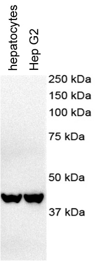
Application: Western BlotSample Tested: HepG2Species: Human and MouseVerified Customer | Posted 11/25/2017
-
Application: Western BlotSample Tested: Human Pancreatic Cancer Cell lineSpecies: HumanVerified Customer | Posted 08/14/2014beta-actin NBP1-47423
There are no reviews that match your criteria.
Protocols
View specific protocols for beta-Actin Antibody (8H10D10) - BSA Free (NBP1-47423):
Protocol for Flow Cytometry Intracellular Staining
Sample Preparation.
1. Grow cells to 60-85% confluency. Flow cytometry requires between 2 x 10⁵ and 1 x 10⁶ cells for optimal performance.
2. If cells are adherent, harvest gently by washing once with staining buffer and then scraping. Avoid using trypsin as this can disrupt certain epitopes of interest. If enzymatic harvest is required, use Accutase, Collagenase, or TrypLE Express for a less damaging option.
3. Reserve 100 uL for counting, then transfer cell volume into a 50 mL conical tube and centrifuge for 8 minutes at 400 RCF.
a. Count cells using a hemocytometer and a 1:1 trypan blue exclusion stain to determine cell viability before starting the flow protocol. If cells appear blue, do not proceed.
4. Re-suspend cells to a concentration of 1 x 10⁶ cells/mL in staining buffer (NBP2-26247).
5. Aliquot out 100 uL samples in accordance with your experimental samples.
Tip: When cell surface and intracellular staining are required in the same sample, it is advisable that the cell surface staining be performed first since the fixation and permeablization steps might reduce the availability of surface antigens.
Intracellular Staining.
Tip: When performing intracellular staining, it is important to use appropriate fixation and permeabilization reagents based upon the target and its subcellular location. Generally, our Intracellular Flow Assay Kit (NBP2-29450) is a good place to start as it contains an optimized combination of reagents for intracellular staining as well as an inhibitor of intracellular protein transport (necessary if staining secreted proteins). Certain targets may require more gentle or transient permeabilization protocols such as the commonly employed methanol or saponin-based methods.
Protocol for Cytoplasmic Targets:
1. Fix the cells by adding 100 uL fixation solution (such as 4% PFA) to each sample for 10-15 minutes.
2. Permeabilize cells by adding 100 uL of a permeabization buffer to every 1 x 10⁶ cells present in the sample. Mix well and incubate at room temperature for 15 minutes.
a. For cytoplasmic targets, use a gentle permeabilization solution such as 1X PBS + 0.5% Saponin or 1X PBS + 0.5% Tween-20.
b. To maintain the permeabilized state throughout your experiment, use staining buffer + 0.1% of the permeabilization reagent (i.e. 0.1% Tween-20 or 0.1% Saponin).
3. Following the 15 minute incubation, add 2 mL of the staining buffer + 0.1% permeabilizer to each sample.
4. Centrifuge for 1 minute at 400 RCF.
5. Discard supernatant and re-suspend in 100 uL of staining buffer + 0.1% permeabilizer.
6. Add appropriate amount of each antibody (eg. 1 test or 1 ug per sample, as experimentally determined).
7. Mix well and incubate at room temperature for 30 minutes- 1 hour. Gently mix samples every 10-15 minutes.
8. Following the primary/conjugate incubation, add 1-2 mL/sample of staining buffer +0.1% permeabilizer and centrifuge for 1 minute at 400 RCF.
9. Wash twice by re-suspending cells in staining buffer (2 mL for tubes or 200 uL for wells) and centrifuging at 400 RCF for 5 minutes. Discard supernatant.
10. Add appropriate amount of secondary antibody (as experimentally determined) to each sample.
11. Incubate at room temperature in dark for 20 minutes.
12. Add 1-2 mL of staining buffer and centrifuge at 400 RCF for 1 minute and discard supernatant.
13. Wash twice by re-suspending cells in staining buffer (2 mL for tubes or 200 uL for wells) and centrifuging at 400 RCF for 5 minutes. Discard supernatant.
14. Resuspend in an appropriate volume of staining buffer (usually 500 uL per sample) and proceed with analysis on your flow cytometer.
Immunocytochemistry Protocol
Culture cells to appropriate density in 35 mm culture dishes or 6-well plates.
1. Remove culture medium and wash the cells briefly in PBS. Add 10% formalin to the dish and fix at room temperature for 10 minutes.
2. Remove the formalin and wash the cells in PBS.
3. Permeablize the cells with 0.1% Triton X100 or other suitable detergent for 10 min.
4. Remove the permeablization buffer and wash three times for 10 minutes each in PBS. Be sure to not let the specimen dry out.
5. To block nonspecific antibody binding, incubate in 10% normal goat serum from 1 hour to overnight at room temperature.
6. Add primary antibody at appropriate dilution and incubate overnight at 4C.
7. Remove primary antibody and replace with PBS. Wash three times for 10 minutes each.
8. Add secondary antibody at appropriate dilution. Incubate for 1 hour at room temperature.
9. Remove secondary antibody and replace with PBS. Wash three times for 10 minutes each.
10. Counter stain DNA with DAPi if required.
Immunohistochemistry-Paraffin Embedded Sections
Antigen Unmasking:
Bring slides to a boil in 10 mM sodium citrate buffer (pH 6.0) then maintain at a sub-boiling temperature for 10 minutes. Cool slides on bench-top for 30 minutes (keep slides in the sodium citrate buffer all the time).
Staining:
1. Wash sections in deionized water three times for 5 minutes each.
2. Wash sections in PBS for 5 minutes.
3. Block each section with 100-400 ul blocking solution (1% BSA in PBS) for 1 hour at room temperature.
4. Remove blocking solution and add 100-400 ul diluted primary antibody. Incubate overnight at 4 C.
5. Remove antibody solution and wash sections in wash buffer three times for 5 minutes each.
6. Add 100-400 ul HRP polymer conjugated secondary antibody. Incubate 30 minutes at room temperature.
7. Wash sections three times in wash buffer for 5 minutes each.
8. Add 100-400 ul DAB substrate to each section and monitor staining closely.
9. As soon as the sections develop, immerse slides in deionized water.
10. Counterstain sections in hematoxylin.
11. Wash sections in deionized water two times for 5 minutes each.
12. Dehydrate sections.
13. Mount coverslips.
Western Blot Protocol
1. Perform SDS-PAGE on samples to be analyzed, loading 10-25 ug of total protein per lane.
2. Transfer proteins to PVDF membrane according to the instructions provided by the manufacturer of the membrane and transfer apparatus.
3. Stain the membrane with Ponceau S (or similar product) to assess transfer success, and mark molecular weight standards where appropriate.
4. Rinse the blot TBS -0.05% Tween 20 (TBST).
5. Block the membrane in 5% Non-fat milk in TBST (blocking buffer) for at least 1 hour.
6. Wash the membrane in TBST three times for 10 minutes each.
7. Dilute primary antibody in blocking buffer and incubate overnight at 4C with gentle rocking.
8. Wash the membrane in TBST three times for 10 minutes each.
9. Incubate the membrane in diluted HRP conjugated secondary antibody in blocking buffer (as per manufacturer's instructions) for 1 hour at room temperature.
10. Wash the blot in TBST three times for 10 minutes each (this step can be repeated as required to reduce background).
11. Apply the detection reagent of choice in accordance with the manufacturers instructions.
Find general support by application which include: protocols, troubleshooting, illustrated assays, videos and webinars.
- 7-Amino Actinomycin D (7-AAD) Cell Viability Flow Cytometry Protocol
- Appropriate Fixation of IHC/ICC Samples
- Cellular Response to Hypoxia Protocols
- ClariTSA™ Fluorophore Kits
- Detection & Visualization of Antibody Binding
- ELISA Sample Preparation & Collection Guide
- ELISA Troubleshooting Guide
- Extracellular Membrane Flow Cytometry Protocol
- Flow Cytometry Protocol for Cell Surface Markers
- Flow Cytometry Protocol for Staining Membrane Associated Proteins
- Flow Cytometry Staining Protocols
- Flow Cytometry Troubleshooting Guide
- How to Run an R&D Systems DuoSet ELISA
- How to Run an R&D Systems Quantikine ELISA
- How to Run an R&D Systems Quantikine™ QuicKit™ ELISA
- ICC Cell Smear Protocol for Suspension Cells
- ICC Immunocytochemistry Protocol Videos
- ICC for Adherent Cells
- Immunocytochemistry (ICC) Protocol
- Immunocytochemistry Troubleshooting
- Immunofluorescence of Organoids Embedded in Cultrex Basement Membrane Extract
- Immunohistochemistry (IHC) and Immunocytochemistry (ICC) Protocols
- Intracellular Flow Cytometry Protocol Using Alcohol (Methanol)
- Intracellular Flow Cytometry Protocol Using Detergents
- Intracellular Nuclear Staining Flow Cytometry Protocol Using Detergents
- Intracellular Staining Flow Cytometry Protocol Using Alcohol Permeabilization
- Intracellular Staining Flow Cytometry Protocol Using Detergents to Permeabilize Cells
- Preparing Samples for IHC/ICC Experiments
- Preventing Non-Specific Staining (Non-Specific Binding)
- Primary Antibody Selection & Optimization
- Propidium Iodide Cell Viability Flow Cytometry Protocol
- Protocol for Liperfluo
- Protocol for VisUCyte™ HRP Polymer Detection Reagent
- Protocol for the Characterization of Human Th22 Cells
- Protocol for the Characterization of Human Th9 Cells
- Protocol for the Fluorescent ICC Staining of Cell Smears - Graphic
- Protocol for the Fluorescent ICC Staining of Cultured Cells on Coverslips - Graphic
- Protocol for the Preparation and Fluorescent ICC Staining of Cells on Coverslips
- Protocol for the Preparation and Fluorescent ICC Staining of Non-adherent Cells
- Protocol for the Preparation and Fluorescent ICC Staining of Stem Cells on Coverslips
- Protocol for the Preparation of a Cell Smear for Non-adherent Cell ICC - Graphic
- Protocol: Annexin V and PI Staining by Flow Cytometry
- Protocol: Annexin V and PI Staining for Apoptosis by Flow Cytometry
- Quantikine HS ELISA Kit Assay Principle, Alkaline Phosphatase
- Quantikine HS ELISA Kit Principle, Streptavidin-HRP Polymer
- R&D Systems Quality Control Western Blot Protocol
- Sandwich ELISA (Colorimetric) – Biotin/Streptavidin Detection Protocol
- Sandwich ELISA (Colorimetric) – Direct Detection Protocol
- TUNEL and Active Caspase-3 Detection by IHC/ICC Protocol
- The Importance of IHC/ICC Controls
- Troubleshooting Guide: ELISA
- Troubleshooting Guide: Fluorokine Flow Cytometry Kits
- Troubleshooting Guide: Western Blot Figures
- Western Blot Conditions
- Western Blot Protocol
- Western Blot Protocol for Cell Lysates
- Western Blot Troubleshooting
- Western Blot Troubleshooting Guide
- View all Protocols, Troubleshooting, Illustrated assays and Webinars
FAQs for beta-Actin Antibody (8H10D10) - BSA Free
Showing
1
-
5 of
6 FAQs
Showing All
-
Q: Are the beta-Actin antibodies validated in Simple Western?
A: Yes, we offer a few beta actin antibodies that have been tested in Simple Western: NBP1-47423, NB600-501, NB600-532, NB600-503, and NB100-56874.
-
Q: Do Beta-Actin antibodies come in lyophilized format?
A: ACTB antibodies like MAB8969 AND MAB8929 come in lyophilized form.
-
Q: I wanted to know which of the two housekeeping genes B-actin or GAPDH can be used for best results. I intend to do an experiment to determine expression of IL-6 and TNF-alpha in liver and kidney tissues.
A: For homogenized tissue, beta-actin and GAPDH are both fine.
-
Q: What is the immunogen sequence of this Beta-Actin antibody?
A: A synthetic sequence corresponding to N-terminal region of Human beta Actin, conjugate to KLH. The exact sequence is proprietary.
-
Q: What is the theoretical molecular weight for Beta-Actin antibodies?
A: The TMW for beta Actin antibodies is approximately 42 kDa.
-
Q: Why is beta actin used as a loading control in blotting ?
A: Beta actin is a highly-expressed protein found in all cells, and is considered a house keeping protein whose expression is necessary for any cell's proper functioning. For this reason it is used as a loading control to confirm that the same amount of total protein is loaded in each lane of an SDS-PAGE gel so that appropriate comparisons can be made between the actual proteins of interest in different samples.
-
Q: Are the beta-Actin antibodies validated in Simple Western?
A: Yes, we offer a few beta actin antibodies that have been tested in Simple Western: NBP1-47423, NB600-501, NB600-532, NB600-503, and NB100-56874.
-
Q: Do Beta-Actin antibodies come in lyophilized format?
A: ACTB antibodies like MAB8969 AND MAB8929 come in lyophilized form.
-
Q: I wanted to know which of the two housekeeping genes B-actin or GAPDH can be used for best results. I intend to do an experiment to determine expression of IL-6 and TNF-alpha in liver and kidney tissues.
A: For homogenized tissue, beta-actin and GAPDH are both fine.
-
Q: What is the immunogen sequence of this Beta-Actin antibody?
A: A synthetic sequence corresponding to N-terminal region of Human beta Actin, conjugate to KLH. The exact sequence is proprietary.
-
Q: What is the theoretical molecular weight for Beta-Actin antibodies?
A: The TMW for beta Actin antibodies is approximately 42 kDa.
-
Q: Why is beta actin used as a loading control in blotting ?
A: Beta actin is a highly-expressed protein found in all cells, and is considered a house keeping protein whose expression is necessary for any cell's proper functioning. For this reason it is used as a loading control to confirm that the same amount of total protein is loaded in each lane of an SDS-PAGE gel so that appropriate comparisons can be made between the actual proteins of interest in different samples.
Loading...
Associated Pathways


