beta-Actin Antibody - BSA Free
Novus Biologicals | Catalog # NB600-532
![Simple Western: beta-Actin Antibody [NB600-532]](https://resources.rndsystems.com/images/products/beta-Actin-Antibody-Simple-Western-NB600-532-img0016.jpg "Simple Western: beta-Actin Antibody [NB600-532]")
Key Product Details
Validated by
Biological Validation
Species Reactivity
Validated:
Human, Mouse
Cited:
Human, Mouse, Avian
Predicted:
Bovine (100%), Canine (100%), Chicken (100%), Chimpanzee (100%), Golden Syrian Hamster (100%), Guinea Pig (100%), Orangutan (100%), Porcine (100%), Rabbit (100%), Rat (100%), Sheep (100%), Xenopus (100%). Backed by our 100% Guarantee.
Applications
Validated:
Immunohistochemistry, Western Blot, Simple Western, ICC/IF (Negative), Immunoprecipitation (Negative)
Cited:
Western Blot, Simple Western, IF/IHC
Label
Unconjugated
Antibody Source
Polyclonal Rabbit IgG
Format
BSA Free
Loading...
Product Specifications
Immunogen
This beta-Actin Antibody maps to a region corresponding to the N-terminus of human Beta Actin. [UniProt# P60709]
Reactivity Notes
Based on 100% sequence identity, this antibody is predicted to react with Rat, X. tropicalis, Chicken, Sheep, Bovine, Dog, Horse, Rabbit, Guinea pig, Pig, Golden hamster, Orangutan, and Chimpanzee.
Clonality
Polyclonal
Host
Rabbit
Isotype
IgG
Theoretical MW
42 kDa.
Disclaimer note: The observed molecular weight of the protein may vary from the listed predicted molecular weight due to post translational modifications, post translation cleavages, relative charges, and other experimental factors.
Disclaimer note: The observed molecular weight of the protein may vary from the listed predicted molecular weight due to post translational modifications, post translation cleavages, relative charges, and other experimental factors.
Scientific Data Images for beta-Actin Antibody - BSA Free
Simple Western: beta-Actin Antibody [NB600-532]
Simple Western: beta-Actin Antibody [NB600-532] - Simple Western lane view shows a specific band for Beta Actin using NB600-532 at 1:200 in A431, C2C12 and C6 cell lysates. This experiment was performed under reducing conditions using the 12-230 kDa separation system.![Western Blot: beta-Actin Antibody [NB600-532]](https://resources.rndsystems.com/images/products/beta-Actin-Antibody-Western-Blot-NB600-532-img0009.jpg "Western Blot: beta-Actin Antibody [NB600-532]")
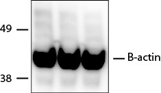
Western Blot: beta-Actin Antibody [NB600-532]
Western Blot: beta-Actin Antibody [NB600-532] - Detection of actin in 3T3 lysates (20ug). ECL detection 30 seconds. A specific band is seen using different dilutions: Lane 1 (1:15,000), Lane 2 (1:10,000), and Lane 3 (1:15,000).![Western Blot: beta-Actin Antibody [NB600-532]](https://resources.rndsystems.com/images/products/beta-Actin-Antibody-Western-Blot-NB600-532-img0014.jpg "Western Blot: beta-Actin Antibody [NB600-532]")
Western Blot: beta-Actin Antibody [NB600-532]
Western Blot: beta-Actin Antibody [NB600-532] - Detection of human and mouse Cytoskeletal Actin by western blot. Samples: Whole cell lysate (50 ug) from HeLa, HEK293T, Jurkat, mouse TCMK-1, and mouse NIH 3T3 cells prepared using NETN lysis buffer. Antibody: Affinity purified rabbit anti-Cytoskeletal Actin antibody NB600-532 used for WB at 0.1 ug/ml. Detection: Chemiluminescence with an exposure time of 1 second.![Western Blot: beta-Actin Antibody [NB600-532]](https://resources.rndsystems.com/images/products/beta-Actin-Antibody-Western-Blot-NB600-532-img0019.jpg "Western Blot: beta-Actin Antibody [NB600-532]")
Western Blot: beta-Actin Antibody [NB600-532]
Western Blot: beta-Actin Antibody [NB600-532] - Mouse colon whole cell lysate. PVDF membrane was probed with Rabbit Anti-B actin Antibody (Catalog # NB600-532) followed by HRP-conjugated Anti-Rabbit IgG Secondary Antibody. WB image submitted by a verified customer review.![Immunohistochemistry: beta-Actin Antibody [NB600-532]](https://resources.rndsystems.com/images/products/beta-Actin-Antibody-Immunohistochemistry-NB600-532-img0015.jpg "Immunohistochemistry: beta-Actin Antibody [NB600-532]")
Immunohistochemistry: beta-Actin Antibody [NB600-532]
Immunohistochemistry: beta-Actin Antibody [NB600-532] - Detection of human Cytoskeletal Actin by immunohistochemistry. Sample: FFPE section of human ovarian cancer. Antibody: Affinity purified rabbit anti-Cytoskeletal Actin (NB600-532). Detection: DAB![Western Blot: beta-Actin Antibody [NB600-532]](https://resources.rndsystems.com/images/products/beta-Actin-Antibody-Western-Blot-NB600-532-img0006.jpg "Western Blot: beta-Actin Antibody [NB600-532]")
Western Blot: beta-Actin Antibody [NB600-532]
Western Blot: beta-Actin Antibody [NB600-532] - Western blot analysis of Actin (NB600-532) using RCC4 whole cell lysate [NBP1-30412].![Western Blot: beta-Actin Antibody [NB600-532]](https://resources.rndsystems.com/images/products/beta-Actin-Antibody-Western-Blot-NB600-532-img0007.jpg "Western Blot: beta-Actin Antibody [NB600-532]")
Western Blot: beta-Actin Antibody [NB600-532]
Western Blot: beta-Actin Antibody [NB600-532] - Western blot analysis of Actin (NB600-532) using HepG2 whole cell lysate [NBP1-42569].![Western Blot: beta-Actin Antibody [NB600-532]](https://resources.rndsystems.com/images/products/beta-Actin-Antibody-Western-Blot-NB600-532-img0011.jpg "Western Blot: beta-Actin Antibody [NB600-532]")
Western Blot: beta-Actin Antibody [NB600-532]
Western Blot: beta-Actin Antibody [NB600-532] - Analysis using the HRP conjugate of NB600-532. Detection of Beta Actin in MCF-7 cell lysate (20ug) using anti-Beta Actin antibody. WB image submitted by a verified customer review.![Western Blot: beta-Actin Antibody [NB600-532]](https://resources.rndsystems.com/images/products/beta-Actin-Antibody-Western-Blot-NB600-532-img0018.jpg "Western Blot: beta-Actin Antibody [NB600-532]")
![Western Blot: beta-Actin Antibody [NB600-532]](https://resources.rndsystems.com/images/products/beta-Actin-Antibody-Western-Blot-NB600-532-img0017.jpg "Western Blot: beta-Actin Antibody [NB600-532]")
Western Blot: beta-Actin Antibody [NB600-532]
beta-Actin-Antibody-Western-Blot-NB600-532-img0017.jpg![Immunohistochemistry: beta-Actin Antibody [NB600-532]](https://resources.rndsystems.com/images/products/beta-Actin-Antibody-Immunohistochemistry-NB600-532-img0012.jpg "Immunohistochemistry: beta-Actin Antibody [NB600-532]")
Immunohistochemistry: beta-Actin Antibody [NB600-532]
Immunohistochemistry: beta-Actin Antibody [NB600-532] - Sample: FFPE section of human lung carcinoma. Antibody: Affinity purified rabbit anti-Cytoskeletal Actin used at a dilution of 1:1,000 (1ug/ml). Detection: DAB
Western Blot: beta-Actin Antibody [NB600-532] -
Western Blot: beta-Actin Antibody [NB600-532] - Cotreatment of periplocin & TRAIL activated IAP. (a) The expression levels of Bax, Bad, Mcl-1, apaf-1, & caspase 9 in HA22T/VGH in response to 1 μM periplocin and/or 100 ng/mL TRAIL treatment were examined by western blot. (b) The expression levels of Bcl-2, cIAP-1, cIAP-2, XIAP, & survivin in HA22T/VGH in response to 1 μM periplocin and/or 100 ng/mL TRAIL treatment were examined by western blot. The original blots are shown in supplemental Figure 2. Image collected & cropped by CiteAb from the following publication (https://pubmed.ncbi.nlm.nih.gov/23365613), licensed under a CC-BY license. Not internally tested by Novus Biologicals.
Western Blot: beta-Actin Antibody [NB600-532] -
Western Blot: beta-Actin Antibody [NB600-532] - Treatments of periplocin and/or TRAIL activate DR4, FADD, & proapoptotic proteins in HCC cells. (a) The effect of periplocin treatment on the expression of DR4, DR5, & FADD was analyzed by western blot. (b) HA22T/VGH cells were treated with different doses of the indicated compounds for 24 h. Expressions of both proforms & cleaved forms of caspase-8, caspase 3, PARP, & BID were analyzed by western blotting. (c) Huh-7 cells were treated with different concentrations of the indicated compounds for 4 h or 24 h. The expression of caspase-8, caspase-9 was analyzed after indicated compounds treatment for 4 h, & the expression of caspase 3, PARP, BID was analyzed after the treatment of indicated compounds for 24 h. (d) HA22T/VGH cells were pretreated with 20 μM or 50 μM inhibitors against caspase-3 (Z-DEVD-FMK), caspasae-8 (Z-IETD-FMK), caspase-9 (Z-LEHD-FMK), & general caspase inhibitor (Z-VAD-FMK) for 3 hours prior to periplocin and/or TRAIL treatment. Cell viability was examined by MTT assay. Image collected & cropped by CiteAb from the following publication (https://pubmed.ncbi.nlm.nih.gov/23365613), licensed under a CC-BY license. Not internally tested by Novus Biologicals.
Western Blot: beta-Actin Antibody [NB600-532] -
Western Blot: beta-Actin Antibody [NB600-532] - GRN PTC readthrough rescues FTLD/NCL lysosomal pathological CSTD maturation phenotype in hiPSC-derived R493X−/− KI cortical neurons. a DIV 80 WT & R493X−/− KI hiPSC-derived cortical neurons were treated with vehicle solution, G418 alone, G418 in combination with CDX5–288, & rec. Human PGRN at the indicated concentrations for 72 h. Expression of mature CSTD in treated WT & R493X−/− KI cortical neuron lysates analyzed by western blotting, using actin as the loading control. b Densitometric quantification of CSTD expression in the aforementioned cortical neuron lysates normalized to vehicle-treated WT levels. n = 3–6 independent cultures; values are shown as mean ± SEM; * p < 0.05, ** p < 0.01, was determined by one-way ANOVA with Tukey’s multiple comparison test Image collected & cropped by CiteAb from the following publication (https://pubmed.ncbi.nlm.nih.gov/32178712), licensed under a CC-BY license. Not internally tested by Novus Biologicals.
Western Blot: beta-Actin Antibody [NB600-532] -
Western Blot: beta-Actin Antibody [NB600-532] - Induction of PTC readthrough by G418 & enhancers in hiPSC-derived R493X−/− KI astrocytes. a R493X−/− KI hiPSC-derived astrocytes were treated with vehicle solution, G418 alone, G418 in combination with CDX5–288, & rec. Human PGRN at the indicated concentrations for 72 h. Expression of PGRN & GRN-2,3 peptides in treated WT & R493X−/− KI astrocyte samples were analyzed by western blotting, using actin as the loading control. b Densitometric quantification of ~ 70 kDa PGRN (i) & GRN-2,3 peptide (ii) in astrocyte lysates (a) normalized to vehicle-treated (VT) WT levels. VT WT was excluded from ii due to oversaturation of GRN-2,3 signal in long exposure blot. For clarity, rec. Human PGRN treated R493X−/− KI astrocytes expressed 20.9% ± 0.027 of VT WT GRN-2,3 levels based on quantification of the short exposure blot (data not shown). c R493X−/− KI hiPSC-derived astrocytes were treated with vehicle solution, G418 in combination with CDX5–288, & G418 CDX5–288 combination with either 10 or 30 μM of Z-Phe-Phe-FMK for 72 h. Again, expression of PGRN in WT & R493X−/− KI astrocyte lysates was also analyzed by western blotting, using actin as the loading control. d Densitometric quantification of full-length PGRN in astrocyte lysates (c) normalized to VT WT levels. n = 3 independent cultures (except in dn = 2); values are shown as mean ± SEM; p < 0.05, ** p < 0.01, *** p < 0.0001 was determined by one-way ANOVA with Tukey’s multiple comparison test Image collected & cropped by CiteAb from the following publication (https://pubmed.ncbi.nlm.nih.gov/32178712), licensed under a CC-BY license. Not internally tested by Novus Biologicals.
Simple Western: beta-Actin Antibody [NB600-532] -
Simple Western: beta-Actin Antibody [NB600-532] - Induction of PTC readthrough by G418 & CDX5 enhancers in cells expressing GRN-V5. a Schematic of full-length PGRN highlighting the position of the S116X (UAA), R418X (UGA), & R493X (UGA) nonsense mutations in relation to the position of individual granulin peptides & the C-terminal V5 tag. b HEK293 cell lines stably expressing GRN-V5 with the indicated nonsense mutations were treated with G418 & the indicated concentrations of CDX5–1, CDX5–196, & CDX5–288 for 72 h. Cell culture supernatants (extracellular) & cell lysates (intracellular) were subjected to automated capillary electrophoresis western analysis. Full-length PGRN was detected with a V5 antibody. Actin was measured in cell lysates as a loading control. The readthrough enhancement ratios are indicated under the lanes. The proportion loaded was 15–20 fold lower for the extracellular samples than for the intracellular samples Image collected & cropped by CiteAb from the following publication (https://pubmed.ncbi.nlm.nih.gov/32178712), licensed under a CC-BY license. Not internally tested by Novus Biologicals.Applications for beta-Actin Antibody - BSA Free
Application
Recommended Usage
Immunohistochemistry
1:2000-1:10000
Simple Western
1:2000
Western Blot
1:2000-1:10000
Application Notes
This antibody is useful for Western Blot. A 40 kDa band is detected in HeLa whole cell lysate and mouse NIH3T3 cells. For IHC, epitope retrieval with citrate buffer pH6.0 is recommended for FFPE tissue sections.
In Simple Western only 10 - 15 uL of the recommended dilution is used per data point.
See Simple Western Antibody Database for Simple Western validation: Tested in Skin, separated by Size, antibody dilution of 1:2000, apparent MW was 49 kDa. Separated by Size-Wes, Sally Sue/Peggy Sue.
In Simple Western only 10 - 15 uL of the recommended dilution is used per data point.
See Simple Western Antibody Database for Simple Western validation: Tested in Skin, separated by Size, antibody dilution of 1:2000, apparent MW was 49 kDa. Separated by Size-Wes, Sally Sue/Peggy Sue.
Reviewed Applications
Read 6 reviews rated 4.5 using NB600-532 in the following applications:
Formulation, Preparation, and Storage
Purification
Immunogen affinity purified
Formulation
Tris-Citrate/Phosphate (pH 7.0 - 8.0)
Format
BSA Free
Preservative
0.09% Sodium Azide
Concentration
1 mg/ml
Shipping
The product is shipped with polar packs. Upon receipt, store it immediately at the temperature recommended below.
Stability & Storage
Store at 4C. Do not freeze.
Background: beta-Actin
References
1. Vandekerckhove J, Weber K. 1978. At least six different actins are expressed in a higher mammal: an analysis based on the amino acid sequence of the amino-terminal tryptic peptide. J Mol Biol. 126(4):783-802. PMID: 745245
2. Gimona M, Vandekerckhove J, Goethals M, Herzog M, Lando Z, Small JV. (1994) Beta-actin specific monoclonal antibody. Cell Motil Cytoskeleton. 27(2):108-16. PMID: 8162619
3. Holden VI, Lenio S, Kuick R, Ramakrishnan SK, Shah YM, Bachman MA. (2014) Bacterial siderophores that evade or overwhelm Lipocalin 2 induce HIF-1a and pro-inflammatory cytokine secretion in cultured respiratory epithelial cells Infect Immun. 82(9):3826-36 PMID: 24980968
4. Tsai WL, Yeh PH, Tsai CY, Ting CT, Chiu YH, Tao MH, Li WC, Hung SC. (2016) Efficient Programming of Human Mesenchymal Stem Cell Derived Hepatocytes by Epigenetic Regulations. J. Gastroenterol. Hepatol. 32(1):261-269. PMID: 27218433
Alternate Names
ACTB, betaActin
Gene Symbol
ACTB
Additional beta-Actin Products
Product Documents for beta-Actin Antibody - BSA Free
Certificate of Analysis
To download a Certificate of Analysis, please enter a lot or batch number in the search box below.
Product Specific Notices for beta-Actin Antibody - BSA Free
This product is for research use only and is not approved for use in humans or in clinical diagnosis. Primary Antibodies are guaranteed for 1 year from date of receipt.
Related Research Areas
Citations for beta-Actin Antibody - BSA Free
 Powered by Bioz
Customer Reviews for beta-Actin Antibody - BSA Free (6)
4.5 out of 5
6 Customer Ratings
Have you used beta-Actin Antibody - BSA Free?
Submit a review and receive an Amazon gift card!
$25/€18/£15/$25CAN/¥2500 Yen for a review with an image
$10/€7/£6/$10CAN/¥1110 Yen for a review without an image
Submit a review
Customer Images

Showing
1
-
5 of
6 reviews
Showing All
Filter By:
-
Application: Western BlotSample Tested: Mouse Colon and Mouse whole cell lysateSpecies: MouseVerified Customer | Posted 08/16/2021PVDF membrane was probed with Rabbit Anti-B actin Antibody (Catalog # NB600-532) followed by HRP-conjugated Anti-Rabbit IgG Secondary Antibody (CST, Catalog # 7074)

-
Application: Western BlotSample Tested: Human ovarian teratocarcinomaSpecies: HumanVerified Customer | Posted 03/28/2016NB600-532SS primary 1:5000 secondary 1:2000
-
Application: Simple WesternSample Tested: whole brain homogenates from miceSpecies: MouseVerified Customer | Posted 01/21/2016Simple Western: beta-Actin in mouse brain lysate
-
Application: Immunohistochemistry-ParaffinSample Tested: Human TonsilsSpecies: HumanVerified Customer | Posted 07/23/2014
-
Application: Western BlotSample Tested: Mouse total lysate, macrophagesSpecies: MouseVerified Customer | Posted 07/09/2013
-
Application: Western BlotVerified Customer | Posted 07/11/2012
There are no reviews that match your criteria.
Protocols
View specific protocols for beta-Actin Antibody - BSA Free (NB600-532):
Western Blot Protocol
1. Perform SDS-PAGE (4-12%) on samples to be analyzed, loading 20 ug of total protein per lane.
2. Transfer proteins to Nitrocellulose according to the instructions provided by the manufacturer of the transfer apparatus.
3. Stain the blot using ponceau S for 1-2 minutes to access the transfer of proteins onto the nitrocellulose membrane. Rinse the blot in water to remove excess stain and mark the lane locations and locations of molecular weight markers using a pencil.
4. Rinse the blot in TBS for approximately 5 minutes.
5. Block the membrane using 5% non-fat dry milk + 1% BSA in TBS for 1 hour.
6. Dilute the rabbit anti-actin primary antibody (NB 600-532) in blocking buffer and incubate 2 hours at room temperature.
7. Wash the membrane in water for 5 minutes and apply the diluted rabbit-IgG HRP-conjugated secondary antibody in blocking buffer (as per manufacturer's instructions) and incubate 1 hour at room temperature.
8. Wash the blot in TBS containing 0.05-0.1% Tween-20 for 10-20 minutes.
9. Wash the blot in type I water for an additional 10-20 minutes (this step can be repeated as required to reduce background).
10. Apply the detection reagent of choice in accordance with the manufacturer's instructions (Amersham's ECL is the standard reagent used at Novus Biologicals).
Note: Tween-20 can be added to the blocking buffer at a final concentration of 0.05-0.2%, provided it does not interfere
with antibody-antigen binding.
Find general support by application which include: protocols, troubleshooting, illustrated assays, videos and webinars.
- Antigen Retrieval Protocol (PIER)
- Antigen Retrieval for Frozen Sections Protocol
- Appropriate Fixation of IHC/ICC Samples
- Cellular Response to Hypoxia Protocols
- Chromogenic IHC Staining of Formalin-Fixed Paraffin-Embedded (FFPE) Tissue Protocol
- Chromogenic Immunohistochemistry Staining of Frozen Tissue
- ClariTSA™ Fluorophore Kits
- Detection & Visualization of Antibody Binding
- Fluorescent IHC Staining of Frozen Tissue Protocol
- Graphic Protocol for Heat-induced Epitope Retrieval
- Graphic Protocol for the Preparation and Fluorescent IHC Staining of Frozen Tissue Sections
- Graphic Protocol for the Preparation and Fluorescent IHC Staining of Paraffin-embedded Tissue Sections
- Graphic Protocol for the Preparation of Gelatin-coated Slides for Histological Tissue Sections
- IHC Sample Preparation (Frozen sections vs Paraffin)
- Immunofluorescent IHC Staining of Formalin-Fixed Paraffin-Embedded (FFPE) Tissue Protocol
- Immunohistochemistry (IHC) and Immunocytochemistry (ICC) Protocols
- Immunohistochemistry Frozen Troubleshooting
- Immunohistochemistry Paraffin Troubleshooting
- Preparing Samples for IHC/ICC Experiments
- Preventing Non-Specific Staining (Non-Specific Binding)
- Primary Antibody Selection & Optimization
- Protocol for Heat-Induced Epitope Retrieval (HIER)
- Protocol for Making a 4% Formaldehyde Solution in PBS
- Protocol for VisUCyte™ HRP Polymer Detection Reagent
- Protocol for the Preparation & Fixation of Cells on Coverslips
- Protocol for the Preparation and Chromogenic IHC Staining of Frozen Tissue Sections
- Protocol for the Preparation and Chromogenic IHC Staining of Frozen Tissue Sections - Graphic
- Protocol for the Preparation and Chromogenic IHC Staining of Paraffin-embedded Tissue Sections
- Protocol for the Preparation and Chromogenic IHC Staining of Paraffin-embedded Tissue Sections - Graphic
- Protocol for the Preparation and Fluorescent IHC Staining of Frozen Tissue Sections
- Protocol for the Preparation and Fluorescent IHC Staining of Paraffin-embedded Tissue Sections
- Protocol for the Preparation of Gelatin-coated Slides for Histological Tissue Sections
- R&D Systems Quality Control Western Blot Protocol
- TUNEL and Active Caspase-3 Detection by IHC/ICC Protocol
- The Importance of IHC/ICC Controls
- Troubleshooting Guide: Immunohistochemistry
- Troubleshooting Guide: Western Blot Figures
- Western Blot Conditions
- Western Blot Protocol
- Western Blot Protocol for Cell Lysates
- Western Blot Troubleshooting
- Western Blot Troubleshooting Guide
- View all Protocols, Troubleshooting, Illustrated assays and Webinars
FAQs for beta-Actin Antibody - BSA Free
Showing
1
-
5 of
7 FAQs
Showing All
-
Q: Are the beta-Actin antibodies validated in Simple Western?
A: Yes, we offer a few beta actin antibodies that have been tested in Simple Western: NBP1-47423, NB600-501, NB600-532, NB600-503, and NB100-56874.
-
Q: Do Beta-Actin antibodies come in lyophilized format?
A: ACTB antibodies like MAB8969 AND MAB8929 come in lyophilized form.
-
Q: I wanted to know which of the two housekeeping genes B-actin or GAPDH can be used for best results. I intend to do an experiment to determine expression of IL-6 and TNF-alpha in liver and kidney tissues.
A: For homogenized tissue, beta-actin and GAPDH are both fine.
-
Q: I would like to know whether the antibody anti-actin (NB600-532) from rabbit can be also used for immunofluorescence and which dilution you recommend?
A: This antibody is listed as being negative for immunocytochemistry/immunofluorescence, so is not recommended for your assay. As it has been validated in ICC/IF, I would highly recommend NB600-501 instead of NB600-532. Full information about this product and recommended dilutions can be found on the datasheet. For ICC/IF, we recommend a dilution of 1:1000-1:2000.
-
Q: What is the immunogen sequence of this Beta-Actin antibody?
A: A sequence within the N-terminal region of Human beta Actin. The exact sequence is proprietary.
-
Q: What is the theoretical molecular weight for Beta-Actin antibodies?
A: The TMW for beta Actin antibodies is approximately 42 kDa.
-
Q: Why is beta actin used as a loading control in blotting ?
A: Beta actin is a highly-expressed protein found in all cells, and is considered a house keeping protein whose expression is necessary for any cell's proper functioning. For this reason it is used as a loading control to confirm that the same amount of total protein is loaded in each lane of an SDS-PAGE gel so that appropriate comparisons can be made between the actual proteins of interest in different samples.
-
Q: Are the beta-Actin antibodies validated in Simple Western?
A: Yes, we offer a few beta actin antibodies that have been tested in Simple Western: NBP1-47423, NB600-501, NB600-532, NB600-503, and NB100-56874.
-
Q: Do Beta-Actin antibodies come in lyophilized format?
A: ACTB antibodies like MAB8969 AND MAB8929 come in lyophilized form.
-
Q: I wanted to know which of the two housekeeping genes B-actin or GAPDH can be used for best results. I intend to do an experiment to determine expression of IL-6 and TNF-alpha in liver and kidney tissues.
A: For homogenized tissue, beta-actin and GAPDH are both fine.
-
Q: I would like to know whether the antibody anti-actin (NB600-532) from rabbit can be also used for immunofluorescence and which dilution you recommend?
A: This antibody is listed as being negative for immunocytochemistry/immunofluorescence, so is not recommended for your assay. As it has been validated in ICC/IF, I would highly recommend NB600-501 instead of NB600-532. Full information about this product and recommended dilutions can be found on the datasheet. For ICC/IF, we recommend a dilution of 1:1000-1:2000.
-
Q: What is the immunogen sequence of this Beta-Actin antibody?
A: A sequence within the N-terminal region of Human beta Actin. The exact sequence is proprietary.
-
Q: What is the theoretical molecular weight for Beta-Actin antibodies?
A: The TMW for beta Actin antibodies is approximately 42 kDa.
-
Q: Why is beta actin used as a loading control in blotting ?
A: Beta actin is a highly-expressed protein found in all cells, and is considered a house keeping protein whose expression is necessary for any cell's proper functioning. For this reason it is used as a loading control to confirm that the same amount of total protein is loaded in each lane of an SDS-PAGE gel so that appropriate comparisons can be made between the actual proteins of interest in different samples.
Loading...
Associated Pathways


