beta-Arrestin 2 Antibody - BSA Free
Novus Biologicals | Catalog # NBP2-24569
![Western Blot: beta-Arrestin 2 AntibodyBSA Free [NBP2-24569]](https://resources.rndsystems.com/images/products/beta-Arrestin-2-Antibody-Western-Blot-NBP2-24569-img0004.jpg "Western Blot: beta-Arrestin 2 AntibodyBSA Free [NBP2-24569]")
Key Product Details
Species Reactivity
Human, Mouse, Rat, Bovine, Canine, Chicken, Equine, Primate, Xenopus, Zebrafish
Applications
Immunohistochemistry, Immunohistochemistry-Paraffin, Western Blot, Simple Western
Label
Unconjugated
Antibody Source
Polyclonal Rabbit IgG
Format
BSA Free
Loading...
Product Specifications
Immunogen
A peptide sequence corresponding to an area between amino acids 1 and 50 of human ARRB2 was used as the immunogen for the antibody, Gen Bank no. gb:ABG47460.1.
Reactivity Notes
Chimpanzee, platypus, opssoum, boar (100%), Tetradon (94%).
Clonality
Polyclonal
Host
Rabbit
Isotype
IgG
Scientific Data Images for beta-Arrestin 2 Antibody - BSA Free
Western Blot: beta-Arrestin 2 AntibodyBSA Free [NBP2-24569]
Western Blot: beta-Arrestin 2 Antibody [NBP2-24569] - Analysis of ARRB2 in HUVEC cell lysate in the 1) absence and 2) presence of immunizing peptide using ARRB2 antibody at 0.25 ug/mL. Goat anti-rabbit Ig HRP secondary antibody and PicoTect ECL substrate solution were used.![Immunohistochemistry-Paraffin: beta-Arrestin 2 Antibody - BSA Free [NBP2-24569]](https://resources.rndsystems.com/images/products/beta-Arrestin-2-Antibody-Immunohistochemistry-Paraffin-NBP2-24569-img0005.jpg "Immunohistochemistry-Paraffin: beta-Arrestin 2 Antibody - BSA Free [NBP2-24569]")
Immunohistochemistry-Paraffin: beta-Arrestin 2 Antibody - BSA Free [NBP2-24569]
Immunohistochemistry-Paraffin: beta-Arrestin 2 Antibody [NBP2-24569] - Human thyroid tissue stained with ARRB2 antibody at 5 ug/mL. Staining of formalin-fixed tissues is enhanced by boiling tissue sections in 10 mM sodium citrate buffer, pH 6.0 for 10-20 min followed by cooling at RT for 20 min.![Simple Western: beta-Arrestin 2 AntibodyBSA Free [NBP2-24569]](https://resources.rndsystems.com/images/products/beta-Arrestin-2-Antibody-Simple-Western-NBP2-24569-img0003.jpg "Simple Western: beta-Arrestin 2 AntibodyBSA Free [NBP2-24569]")
Simple Western: beta-Arrestin 2 AntibodyBSA Free [NBP2-24569]
Simple Western: beta-Arrestin 2 Antibody [NBP2-24569] - Simple Western lane view shows a specific band for beta Arrestin in 0.5 mg/mL of Jurkat lysate. This experiment was performed under reducing conditions using the 12-230 kDa separation system.![Simple Western: beta-Arrestin 2 AntibodyBSA Free [NBP2-24569]](https://resources.rndsystems.com/images/products/beta-Arrestin-2-Antibody-Simple-Western-NBP2-24569-img0006.jpg "Simple Western: beta-Arrestin 2 AntibodyBSA Free [NBP2-24569]")
Simple Western: beta-Arrestin 2 AntibodyBSA Free [NBP2-24569]
Simple Western: beta-Arrestin 2 Antibody [NBP2-24569] - Beta-arrestin antibody detects 2 peaks at 32 and 45 kDa using Simple Western. Traditional size of beta-arrestin 2 is ~55 kDa. Image from verified customer review.Applications for beta-Arrestin 2 Antibody - BSA Free
Application
Recommended Usage
Immunohistochemistry-Paraffin
5 ug/mL
Simple Western
1:100
Western Blot
0.25 - 0.5 ug/mL
Application Notes
In Simple Western only 10 - 15 uL of the recommended dilution is used per data point.
See Simple Western Antibody Database for Simple Western validation: Tested in Jurkat lysate 0.5 mg/mL, separated by Size, antibody dilution of 1:100, apparent MW was 54 kDa
See Simple Western Antibody Database for Simple Western validation: Tested in Jurkat lysate 0.5 mg/mL, separated by Size, antibody dilution of 1:100, apparent MW was 54 kDa
Reviewed Applications
Read 1 review rated 3 using NBP2-24569 in the following applications:
Formulation, Preparation, and Storage
Purification
Immunogen affinity purified
Formulation
PBS
Format
BSA Free
Preservative
0.05% Sodium Azide
Concentration
1.0 mg/ml
Shipping
The product is shipped with polar packs. Upon receipt, store it immediately at the temperature recommended below.
Stability & Storage
Store at -20C. Avoid freeze-thaw cycles.
Background: beta-Arrestin 2
Alternate Names
ARB2, ARR2, ARRB2, betaArrestin 2
Gene Symbol
ARRB2
Additional beta-Arrestin 2 Products
Product Documents for beta-Arrestin 2 Antibody - BSA Free
Certificate of Analysis
To download a Certificate of Analysis, please enter a lot or batch number in the search box below.
Product Specific Notices for beta-Arrestin 2 Antibody - BSA Free
This product is for research use only and is not approved for use in humans or in clinical diagnosis. Primary Antibodies are guaranteed for 1 year from date of receipt.
Citations for beta-Arrestin 2 Antibody - BSA Free
 Powered by Bioz
Customer Reviews for beta-Arrestin 2 Antibody - BSA Free (1)
3 out of 5
1 Customer Rating
Have you used beta-Arrestin 2 Antibody - BSA Free?
Submit a review and receive an Amazon gift card!
$25/€18/£15/$25CAN/¥2500 Yen for a review with an image
$10/€7/£6/$10CAN/¥1110 Yen for a review without an image
Submit a review
Customer Images
Showing
1
-
1 of
1 review
Showing All
Filter By:
-
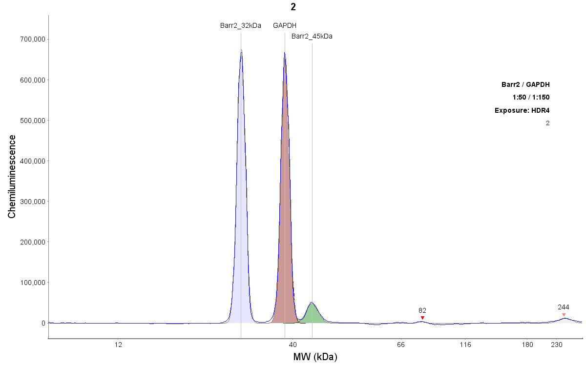
Application: Simple WesternSample Tested: Mouse skeletal muscleSpecies: MouseVerified Customer | Posted 11/16/2018Beta-arrestin antibody detects 2 peaks at 32 and 45 kDa using Simple Western. Traditional size of beta-arrestin 2 is ~55 kDa.antibody used at 1:50 dilution 0.5 mg/ml protein concentration of the sample GAPDH, 1:150 dilution, (14C10, HRP Conjugate, #3683 CST) used as loading control (~39 kDa)

There are no reviews that match your criteria.
Protocols
Find general support by application which include: protocols, troubleshooting, illustrated assays, videos and webinars.
- Antigen Retrieval Protocol (PIER)
- Antigen Retrieval for Frozen Sections Protocol
- Appropriate Fixation of IHC/ICC Samples
- Cellular Response to Hypoxia Protocols
- Chromogenic IHC Staining of Formalin-Fixed Paraffin-Embedded (FFPE) Tissue Protocol
- Chromogenic Immunohistochemistry Staining of Frozen Tissue
- ClariTSA™ Fluorophore Kits
- Detection & Visualization of Antibody Binding
- Fluorescent IHC Staining of Frozen Tissue Protocol
- Graphic Protocol for Heat-induced Epitope Retrieval
- Graphic Protocol for the Preparation and Fluorescent IHC Staining of Frozen Tissue Sections
- Graphic Protocol for the Preparation and Fluorescent IHC Staining of Paraffin-embedded Tissue Sections
- Graphic Protocol for the Preparation of Gelatin-coated Slides for Histological Tissue Sections
- IHC Sample Preparation (Frozen sections vs Paraffin)
- Immunofluorescent IHC Staining of Formalin-Fixed Paraffin-Embedded (FFPE) Tissue Protocol
- Immunohistochemistry (IHC) and Immunocytochemistry (ICC) Protocols
- Immunohistochemistry Frozen Troubleshooting
- Immunohistochemistry Paraffin Troubleshooting
- Preparing Samples for IHC/ICC Experiments
- Preventing Non-Specific Staining (Non-Specific Binding)
- Primary Antibody Selection & Optimization
- Protocol for Heat-Induced Epitope Retrieval (HIER)
- Protocol for Making a 4% Formaldehyde Solution in PBS
- Protocol for VisUCyte™ HRP Polymer Detection Reagent
- Protocol for the Preparation & Fixation of Cells on Coverslips
- Protocol for the Preparation and Chromogenic IHC Staining of Frozen Tissue Sections
- Protocol for the Preparation and Chromogenic IHC Staining of Frozen Tissue Sections - Graphic
- Protocol for the Preparation and Chromogenic IHC Staining of Paraffin-embedded Tissue Sections
- Protocol for the Preparation and Chromogenic IHC Staining of Paraffin-embedded Tissue Sections - Graphic
- Protocol for the Preparation and Fluorescent IHC Staining of Frozen Tissue Sections
- Protocol for the Preparation and Fluorescent IHC Staining of Paraffin-embedded Tissue Sections
- Protocol for the Preparation of Gelatin-coated Slides for Histological Tissue Sections
- R&D Systems Quality Control Western Blot Protocol
- TUNEL and Active Caspase-3 Detection by IHC/ICC Protocol
- The Importance of IHC/ICC Controls
- Troubleshooting Guide: Immunohistochemistry
- Troubleshooting Guide: Western Blot Figures
- Western Blot Conditions
- Western Blot Protocol
- Western Blot Protocol for Cell Lysates
- Western Blot Troubleshooting
- Western Blot Troubleshooting Guide
- View all Protocols, Troubleshooting, Illustrated assays and Webinars
Loading...
Associated Pathways
