c-Maf Antibody - BSA Free
Novus Biologicals | Catalog # NBP2-24551
![Western Blot: c-Maf AntibodyBSA Free [NBP2-24551]](https://resources.rndsystems.com/images/products/c-Maf-Antibody-Western-Blot-NBP2-24551-img0005.jpg "Western Blot: c-Maf AntibodyBSA Free [NBP2-24551]")
Key Product Details
Species Reactivity
Validated:
Human, Mouse, Rat, Bovine
Cited:
Human, Mouse
Predicted:
Monkey (93%). Backed by our 100% Guarantee.
Applications
Validated:
Immunohistochemistry, Immunohistochemistry-Paraffin, Western Blot
Cited:
Western Blot, IF/IHC
Label
Unconjugated
Antibody Source
Polyclonal Rabbit IgG
Format
BSA Free
Loading...
Product Specifications
Immunogen
A synthetic peptide corresponding to amino acids 75-110 of human c-maf was used as the immunogen, GenBank no. NP_001026974.1|.
Reactivity Notes
Chimpanzee (87%), Bovine, Equine (81%).
Specificity
100% homologous in human isoforms CRA_a, CRA_b, and CRA_c).
Clonality
Polyclonal
Host
Rabbit
Isotype
IgG
Scientific Data Images for c-Maf Antibody - BSA Free
Western Blot: c-Maf AntibodyBSA Free [NBP2-24551]
Western Blot: c-Maf Antibody [NBP2-24551] - Total protein from Human Pancreas and Human Small Intestine was separated on a 7.5% gel by SDS-PAGE, transferred to PVDF membrane and blocked in 5% non-fat milk in TBST. The membrane was probed with 2.0 ug/ml anti-cMaf in 1% block buffer and detected with an anti-rabbit HRP secondary antibody using chemiluminescence.![Western Blot: c-Maf AntibodyBSA Free [NBP2-24551]](https://resources.rndsystems.com/images/products/c-Maf-Antibody-Western-Blot-NBP2-24551-img0006.jpg "Western Blot: c-Maf AntibodyBSA Free [NBP2-24551]")
Western Blot: c-Maf AntibodyBSA Free [NBP2-24551]
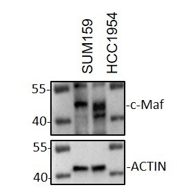
Western Blot: c-Maf Antibody [NBP2-24551] - SUM159 and HCC1954 whole cell lysates were loaded with 50 ug/lane. 10% SDS-PAGE. c-Maf Antibody (NBP2-24551) was used for primary antibody: 1:500, 4C, overnight. WB image submitted by a verified customer review.![Immunohistochemistry-Paraffin: c-Maf Antibody - BSA Free [NBP2-24551]](https://resources.rndsystems.com/images/products/c-Maf-Antibody-Immunohistochemistry-Paraffin-NBP2-24551-img0004.jpg "Immunohistochemistry-Paraffin: c-Maf Antibody - BSA Free [NBP2-24551]")
Immunohistochemistry-Paraffin: c-Maf Antibody - BSA Free [NBP2-24551]
Immunohistochemistry-Paraffin: c-Maf Antibody [NBP2-24551] - IHC analysis of a formalin fixed paraffin embedded (FFPE) tissue section of human liver cancer using c-MAF antibody at 2ug/ml concentration (1:500 dilution). The primary antibody binding to c-MAF protein was detected using HRP conjugated anti-rabbit secondary antibody with DAB reagent, and the sections were further counterstained with hematoxylin for labeling cellular nuclei. This c-MAF antibody showed a very strong nuclear and moderate to strong staining in the endothelial cells (blood vessels) and in a subset of apparently infiltrating inflammatory cells. Weak to moderate cytoplasmic and nuclear staining was observed in liver cancer cells and the cells of tumor stroma including fibroblasts.![Western Blot: c-Maf AntibodyBSA Free [NBP2-24551]](https://resources.rndsystems.com/images/products/c-Maf-Antibody-Western-Blot-NBP2-24551-img0001.jpg "Western Blot: c-Maf AntibodyBSA Free [NBP2-24551]")
Western Blot: c-Maf AntibodyBSA Free [NBP2-24551]
Western Blot: c-Maf Antibody [NBP2-24551] - Analysis of Lysate from human brain in the 1) absence, 2) presence of immunizing peptide, 3) mouse brain and 4) rat brain probed at 3 ug/ml.![Immunohistochemistry-Paraffin: c-Maf Antibody - BSA Free [NBP2-24551]](https://resources.rndsystems.com/images/products/c-Maf-Antibody-Immunohistochemistry-Paraffin-NBP2-24551-img0002.jpg "Immunohistochemistry-Paraffin: c-Maf Antibody - BSA Free [NBP2-24551]")
Immunohistochemistry-Paraffin: c-Maf Antibody - BSA Free [NBP2-24551]
Immunohistochemistry-Paraffin: c-Maf Antibody [NBP2-24551] - Staining of Human kidney probed with c-maf antibody at 10 ug/ml.![Immunohistochemistry-Paraffin: c-Maf Antibody - BSA Free [NBP2-24551]](https://resources.rndsystems.com/images/products/c-Maf-Antibody-Immunohistochemistry-Paraffin-NBP2-24551-img0003.jpg "Immunohistochemistry-Paraffin: c-Maf Antibody - BSA Free [NBP2-24551]")
Immunohistochemistry-Paraffin: c-Maf Antibody - BSA Free [NBP2-24551]
Immunohistochemistry-Paraffin: c-Maf Antibody [NBP2-24551] - Staining of Human placenta probed with c-maf antibody at 10 ug/ml.Applications for c-Maf Antibody - BSA Free
Application
Recommended Usage
Immunohistochemistry-Paraffin
1-2 ug/ml
Western Blot
2-4 ug/ml
Reviewed Applications
Read 3 reviews rated 3.3 using NBP2-24551 in the following applications:
Formulation, Preparation, and Storage
Purification
Immunogen affinity purified
Formulation
PBS
Format
BSA Free
Preservative
0.02% Sodium Azide
Concentration
1.0 mg/ml
Shipping
The product is shipped with polar packs. Upon receipt, store it immediately at the temperature recommended below.
Stability & Storage
Store at 4C short term. Aliquot and store at -20C long term. Avoid freeze-thaw cycles.
Background: c-Maf
Long Name
v-maf Musculoaponeurotic Fibrosarcoma Oncogene Homolog (Avian)
Alternate Names
CCA4, cMaf, MAF2
Gene Symbol
MAF
UniProt
Additional c-Maf Products
Product Documents for c-Maf Antibody - BSA Free
Certificate of Analysis
To download a Certificate of Analysis, please enter a lot or batch number in the search box below.
Product Specific Notices for c-Maf Antibody - BSA Free
This product is for research use only and is not approved for use in humans or in clinical diagnosis. Primary Antibodies are guaranteed for 1 year from date of receipt.
Citations for c-Maf Antibody - BSA Free
 Powered by Bioz
Customer Reviews for c-Maf Antibody - BSA Free (3)
3.3 out of 5
3 Customer Ratings
Have you used c-Maf Antibody - BSA Free?
Submit a review and receive an Amazon gift card!
$25/€18/£15/$25CAN/¥2500 Yen for a review with an image
$10/€7/£6/$10CAN/¥1110 Yen for a review without an image
Submit a review
Customer Images
Showing
1
-
3 of
3 reviews
Showing All
Filter By:
-
Application: Western BlotSample Tested: Breast cancer cellsSpecies: HumanVerified Customer | Posted 09/13/2021Western Blot: SUM159 and HCC1954 whole cell lysates were loaded with 50 ug/lane. 10% SDS-PAGE. c-Maf Antibody (NBP2-24551) was used for primary antibody: 1:500, 4℃, overnight.

-
Application: Immunohistochemistry-ParaffinSample Tested: M1/M2 cell linesSpecies: HumanVerified Customer | Posted 11/11/2015
-
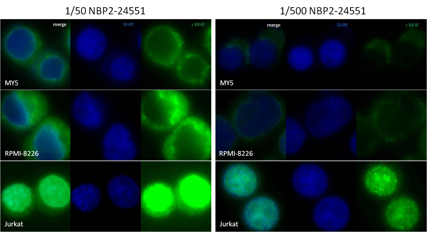
Application: ImmunocytochemistrySample Tested: Multiple myeloma cells lines MY5 & RPMI-8226 and T cell leukemia cell line Jurkat cytospinsSpecies: HumanVerified Customer | Posted 07/28/2015IFF/ICC staining of MY5, RPMI-8226 and Jurkat cells with NBP2-24551
There are no reviews that match your criteria.
Protocols
View specific protocols for c-Maf Antibody - BSA Free (NBP2-24551):
Western Blot Protocol
1. Perform SDS-PAGE on samples to be analyzed, loading 10-25 ug of total protein per lane.
2. Transfer proteins to PVDF membrane according to the instructions provided by the manufacturer of the membrane and transfer apparatus.
3. Stain the membrane with Ponceau S (or similar product) to assess transfer success, and mark molecular weight standards where appropriate.
4. Rinse the blot TBS -0.05% Tween 20 (TBST).
5. Block the membrane in 5% Non-fat milk in TBST (blocking buffer) for at least 1 hour.
6. Wash the membrane in TBST three times for 10 minutes each.
7. Dilute primary antibody in blocking buffer and incubate overnight at 4C with gentle rocking.
8. Wash the membrane in TBST three times for 10 minutes each.
9. Incubate the membrane in diluted HRP conjugated secondary antibody in blocking buffer (as per manufacturer's instructions) for 1 hour at room temperature.
10. Wash the blot in TBST three times for 10 minutes each (this step can be repeated as required to reduce background).
11. Apply the detection reagent of choice in accordance with the manufacturer's instructions.
Find general support by application which include: protocols, troubleshooting, illustrated assays, videos and webinars.
- Antigen Retrieval Protocol (PIER)
- Antigen Retrieval for Frozen Sections Protocol
- Appropriate Fixation of IHC/ICC Samples
- Cellular Response to Hypoxia Protocols
- Chromogenic IHC Staining of Formalin-Fixed Paraffin-Embedded (FFPE) Tissue Protocol
- Chromogenic Immunohistochemistry Staining of Frozen Tissue
- ClariTSA™ Fluorophore Kits
- Detection & Visualization of Antibody Binding
- Fluorescent IHC Staining of Frozen Tissue Protocol
- Graphic Protocol for Heat-induced Epitope Retrieval
- Graphic Protocol for the Preparation and Fluorescent IHC Staining of Frozen Tissue Sections
- Graphic Protocol for the Preparation and Fluorescent IHC Staining of Paraffin-embedded Tissue Sections
- Graphic Protocol for the Preparation of Gelatin-coated Slides for Histological Tissue Sections
- IHC Sample Preparation (Frozen sections vs Paraffin)
- Immunofluorescent IHC Staining of Formalin-Fixed Paraffin-Embedded (FFPE) Tissue Protocol
- Immunohistochemistry (IHC) and Immunocytochemistry (ICC) Protocols
- Immunohistochemistry Frozen Troubleshooting
- Immunohistochemistry Paraffin Troubleshooting
- Preparing Samples for IHC/ICC Experiments
- Preventing Non-Specific Staining (Non-Specific Binding)
- Primary Antibody Selection & Optimization
- Protocol for Heat-Induced Epitope Retrieval (HIER)
- Protocol for Making a 4% Formaldehyde Solution in PBS
- Protocol for VisUCyte™ HRP Polymer Detection Reagent
- Protocol for the Preparation & Fixation of Cells on Coverslips
- Protocol for the Preparation and Chromogenic IHC Staining of Frozen Tissue Sections
- Protocol for the Preparation and Chromogenic IHC Staining of Frozen Tissue Sections - Graphic
- Protocol for the Preparation and Chromogenic IHC Staining of Paraffin-embedded Tissue Sections
- Protocol for the Preparation and Chromogenic IHC Staining of Paraffin-embedded Tissue Sections - Graphic
- Protocol for the Preparation and Fluorescent IHC Staining of Frozen Tissue Sections
- Protocol for the Preparation and Fluorescent IHC Staining of Paraffin-embedded Tissue Sections
- Protocol for the Preparation of Gelatin-coated Slides for Histological Tissue Sections
- R&D Systems Quality Control Western Blot Protocol
- TUNEL and Active Caspase-3 Detection by IHC/ICC Protocol
- The Importance of IHC/ICC Controls
- Troubleshooting Guide: Immunohistochemistry
- Troubleshooting Guide: Western Blot Figures
- Western Blot Conditions
- Western Blot Protocol
- Western Blot Protocol for Cell Lysates
- Western Blot Troubleshooting
- Western Blot Troubleshooting Guide
- View all Protocols, Troubleshooting, Illustrated assays and Webinars
Loading...
Associated Pathways
