Key Product Details
Species Reactivity
Human, Chicken (Negative), Mouse (Negative), Rat (Negative)
Applications
Immunohistochemistry, Immunohistochemistry-Paraffin, Flow Cytometry, Immunofluorescence, Immunocytochemistry/ Immunofluorescence, Simple Western
Label
Unconjugated
Antibody Source
Monoclonal Mouse IgG1 kappa Clone # 9E10.3
Loading...
Product Specifications
Immunogen
A synthetic peptide, corresponding to aa 408-439 (AEEQKLISEEDLLRKRREQLKHKLEQLRNSCA) from C-terminus of human c-Myc, coupled to KLH. (Uniprot: P01106)
Epitope
aa 410-419 (EQKLISEEDL)
Reactivity Notes
Does not react with Mouse, Rat or Chicken.
Localization
Nuclear
Specificity
It recognizes a transcription factor of 64-67kDa, identified as c-myc. Its epitope spans between aa 410-419 (EQKLISEEDL) which is a specific portion of an alpha helical region of human c-myc protein. This monoclonal antibody shows no cross-reaction with v-myc. c-myc is involved in the control of cell proliferation and differentiation and is amplified and/or overexpressed in a variety of tumors. Over-expression of c-myc protein occurs frequently in luminal cells of prostate intraepithelial neoplasia as well as in most primary carcinomas and metastatic disease.
Clonality
Monoclonal
Host
Mouse
Isotype
IgG1 kappa
Theoretical MW
63 kDa.
Disclaimer note: The observed molecular weight of the protein may vary from the listed predicted molecular weight due to post translational modifications, post translation cleavages, relative charges, and other experimental factors.
Disclaimer note: The observed molecular weight of the protein may vary from the listed predicted molecular weight due to post translational modifications, post translation cleavages, relative charges, and other experimental factors.
Description
200ug/ml of antibody purified from Bioreactor Concentrate by Protein A or G. Prepared in 10 mM PBS with 0.05% BSA & 0.05% azide. Also available WITHOUT BSA & azide at 1.0 mg/ml. (NBP2-47738)
Antibody with azide - store at 2 to 8C. Antibody without azide - store at -20 to -80C.
Antibody with azide - store at 2 to 8C. Antibody without azide - store at -20 to -80C.
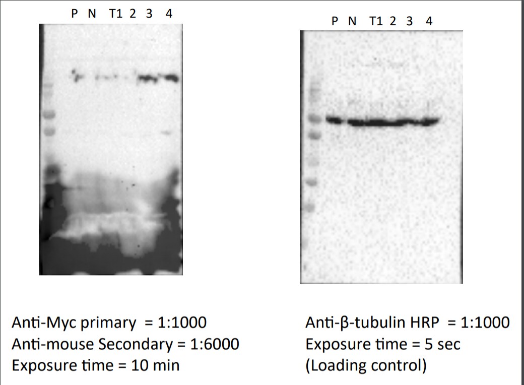
Scientific Data Images for c-Myc Antibody (9E10.3)
![Immunohistochemistry-Paraffin: c-Myc Antibody (9E10.3) [NBP2-45144]](https://resources.rndsystems.com/images/products/c-Myc-Antibody-9E10-3-Immunohistochemistry-Paraffin-NBP2-45144-img0001.jpg "Immunohistochemistry-Paraffin: c-Myc Antibody (9E10.3) [NBP2-45144]")
Immunohistochemistry-Paraffin: c-Myc Antibody (9E10.3) [NBP2-45144]
Immunohistochemistry-Paraffin: c-Myc Antibody (9E10.3) [NBP2-45144] - Human Cervical Carcinoma stained with c-myc Monoclonal Antibody (9E10.3).![Simple Western: c-Myc Antibody (9E10.3) [NBP2-45144]](https://resources.rndsystems.com/images/products/c-Myc-Antibody-9E10-3-Simple-Western-NBP2-45144-img0002.jpg "Simple Western: c-Myc Antibody (9E10.3) [NBP2-45144]")
Simple Western: c-Myc Antibody (9E10.3) [NBP2-45144]
Simple Western: c-Myc Antibody (9E10.3) [NBP2-45144] - Simple Western lane view shows a specific band for c-Myc in 0.2 mg/ml of HeLa lysate(s). This experiment was performed under reducing conditions using the 12-230 kDa separation system.![Simple Western: c-Myc Antibody (9E10.3) [NBP2-45144]](https://resources.rndsystems.com/images/products/c-Myc-Antibody-9E10-3-Simple-Western-NBP2-45144-img0005.jpg "Simple Western: c-Myc Antibody (9E10.3) [NBP2-45144]")
Simple Western: c-Myc Antibody (9E10.3) [NBP2-45144]
Simple Western: c-Myc Antibody (9E10.3) [NBP2-45144] - Electropherogram image of the corresponding Simple Western lane. c-Myc antibody was used at 50 ug/ml dilution of HeLa lysates(s) respectively. [NBP2-45144] -")
Simple Western: c-Myc Antibody (9E10.3) [NBP2-45144] -
Simple Western: c-Myc Antibody (9E10.3) [NBP2-45144] - Lane view shows a specific band for c-Myc in 0.2 mg/mL of HeLa lysate and antibody at 20 ug/mL. Electropherogram image of corresponding Simple Western lane view at WES molecular weight of 74 kDa. Image reported by internal validation.Applications for c-Myc Antibody (9E10.3)
Application
Recommended Usage
Flow Cytometry
1-2 ug/million cells
Immunocytochemistry/ Immunofluorescence
1-2 ug/ml
Immunofluorescence
1 - 2 ug/ml
Immunohistochemistry-Paraffin
1-2 ug/ml
Simple Western
50 ug/ml
Application Notes
Immunohistochemistry (Formalin-fixed): 1-2ug/ml for 30 minutes at RT. Staining of formalin-fixed tissues requires heating tissue sections in 10mM Tris with 1mM EDTA, pH 9.0, for 45 min at 95C followed by cooling at RT for 20 minutes.
Optimal dilution for a specific application should be determined.
In Simple Western only 10 - 15 ul of the recommended dilution is used per data point.
See Simple Western Antibody Database for Simple Western validation: Tested in HeLa lysate, separated by Size, antibody dilution of 50 ug/mL, apparent MW was 78 kDa.
Reviewed Applications
Read 1 review rated 1 using NBP2-45144 in the following applications:
Flow Cytometry Panel Builder
Bio-Techne Knows Flow Cytometry
Save time and reduce costly mistakes by quickly finding compatible reagents using the Panel Builder Tool.
Advanced Features
- Spectra Viewer - Custom analysis of spectra from multiple fluorochromes
- Spillover Popups - Visualize the spectra of individual fluorochromes
- Antigen Density Selector - Match fluorochrome brightness with antigen density

Formulation, Preparation, and Storage
Purification
Protein A or G purified
Formulation
10 mM PBS with 0.05% BSA
Preservative
0.05% Sodium Azide
Concentration
0.2 mg/ml
Shipping
The product is shipped with polar packs. Upon receipt, store it immediately at the temperature recommended below.
Stability & Storage
Store at 4C.
Background: c-Myc
A basic Helix-Loop-Helix, Leucine Zipper domain (bHLH/LZ), designated Max, specifically associates with c-Myc, N-Myc and L-Myc proteins. The Myc-Max complex binds to DNA in a sequence-specific manner under conditions where neither Max nor Myc exhibit appreciable binding. Max can also form heterodimers with other bHLH-Zip proteins, Mad and Mxi1. c-Myc plays a role in cell cycle progression, apoptosis, cellular transformation and angiogenesis (2). Mutations, overexpression, rearrangement and translocation of this gene have been associated with a variety of cancers including B-cell Lymphomas, acute myeloid leukemia, glioblastoma, stomach adenocarcinoma, and prostate adenocarcinoma (3).
References
1. Wilkinson, D. S., Tsai, W. W., Schumacher, M. A., & Barton, M. C. (2008). Chromatin-bound p53 anchors activated Smads and the mSin3A corepressor to confer transforming-growth-factor-beta-mediated transcription repression. Mol Cell Biol, 28(6), 1988-1998. doi:10.1128/mcb.01442-07
2. Pedrosa, A. R., Bodrug, N., Gomez-Escudero, J., Carter, E. P., Reynolds, L. E., Georgiou, P. N.,... Hodivala-Dilke, K. M. (2019). Tumor Angiogenesis Is Differentially Regulated by Phosphorylation of Endothelial Cell Focal Adhesion Kinase Tyrosines-397 and -861. Cancer Res, 79(17), 4371-4386. doi:10.1158/0008-5472.Can-18-3934
3. Nagasaka, M., Tsuzuki, K., Ozeki, Y., Tokugawa, M., Ohoka, N., Inoue, Y., & Hayashi, H. (2019). Lysine-Specific Demethylase 1 (LSD1/KDM1A) Is a Novel Target Gene of c-Myc. Biol Pharm Bull, 42(3), 481-488. doi:10.1248/bpb.b18-00892
Long Name
v-Myc Avian Myelocytomatosis Viral Oncogene Homolog (Avian)
Alternate Names
cMyc, Myc, Myc2, Niard, Nird
Gene Symbol
MYC
UniProt
Additional c-Myc Products
Product Documents for c-Myc Antibody (9E10.3)
Certificate of Analysis
To download a Certificate of Analysis, please enter a lot or batch number in the search box below.
Product Specific Notices for c-Myc Antibody (9E10.3)
This product is for research use only and is not approved for use in humans or in clinical diagnosis. Primary Antibodies are guaranteed for 1 year from date of receipt.
Customer Reviews for c-Myc Antibody (9E10.3) (1)
1 out of 5
1 Customer Rating
Have you used c-Myc Antibody (9E10.3)?
Submit a review and receive an Amazon gift card!
$25/€18/£15/$25CAN/¥2500 Yen for a review with an image
$10/€7/£6/$10CAN/¥1110 Yen for a review without an image
Submit a review
Customer Images
Showing
1
-
1 of
1 review
Showing All
Filter By:
-
Application: Western BlotSample Tested: 293T cell lysateSpecies: HumanVerified Customer | Posted 12/16/2020low signal for western blot

There are no reviews that match your criteria.
Protocols
Find general support by application which include: protocols, troubleshooting, illustrated assays, videos and webinars.
- 7-Amino Actinomycin D (7-AAD) Cell Viability Flow Cytometry Protocol
- Antigen Retrieval Protocol (PIER)
- Antigen Retrieval for Frozen Sections Protocol
- Appropriate Fixation of IHC/ICC Samples
- Cellular Response to Hypoxia Protocols
- Chromogenic IHC Staining of Formalin-Fixed Paraffin-Embedded (FFPE) Tissue Protocol
- Chromogenic Immunohistochemistry Staining of Frozen Tissue
- ClariTSA™ Fluorophore Kits
- Detection & Visualization of Antibody Binding
- Extracellular Membrane Flow Cytometry Protocol
- Flow Cytometry Protocol for Cell Surface Markers
- Flow Cytometry Protocol for Staining Membrane Associated Proteins
- Flow Cytometry Staining Protocols
- Flow Cytometry Troubleshooting Guide
- Fluorescent IHC Staining of Frozen Tissue Protocol
- Graphic Protocol for Heat-induced Epitope Retrieval
- Graphic Protocol for the Preparation and Fluorescent IHC Staining of Frozen Tissue Sections
- Graphic Protocol for the Preparation and Fluorescent IHC Staining of Paraffin-embedded Tissue Sections
- Graphic Protocol for the Preparation of Gelatin-coated Slides for Histological Tissue Sections
- ICC Cell Smear Protocol for Suspension Cells
- ICC Immunocytochemistry Protocol Videos
- ICC for Adherent Cells
- IHC Sample Preparation (Frozen sections vs Paraffin)
- Immunocytochemistry (ICC) Protocol
- Immunocytochemistry Troubleshooting
- Immunofluorescence of Organoids Embedded in Cultrex Basement Membrane Extract
- Immunofluorescent IHC Staining of Formalin-Fixed Paraffin-Embedded (FFPE) Tissue Protocol
- Immunohistochemistry (IHC) and Immunocytochemistry (ICC) Protocols
- Immunohistochemistry Frozen Troubleshooting
- Immunohistochemistry Paraffin Troubleshooting
- Intracellular Flow Cytometry Protocol Using Alcohol (Methanol)
- Intracellular Flow Cytometry Protocol Using Detergents
- Intracellular Nuclear Staining Flow Cytometry Protocol Using Detergents
- Intracellular Staining Flow Cytometry Protocol Using Alcohol Permeabilization
- Intracellular Staining Flow Cytometry Protocol Using Detergents to Permeabilize Cells
- Preparing Samples for IHC/ICC Experiments
- Preventing Non-Specific Staining (Non-Specific Binding)
- Primary Antibody Selection & Optimization
- Propidium Iodide Cell Viability Flow Cytometry Protocol
- Protocol for Heat-Induced Epitope Retrieval (HIER)
- Protocol for Liperfluo
- Protocol for Making a 4% Formaldehyde Solution in PBS
- Protocol for VisUCyte™ HRP Polymer Detection Reagent
- Protocol for the Characterization of Human Th22 Cells
- Protocol for the Characterization of Human Th9 Cells
- Protocol for the Fluorescent ICC Staining of Cell Smears - Graphic
- Protocol for the Fluorescent ICC Staining of Cultured Cells on Coverslips - Graphic
- Protocol for the Preparation & Fixation of Cells on Coverslips
- Protocol for the Preparation and Chromogenic IHC Staining of Frozen Tissue Sections
- Protocol for the Preparation and Chromogenic IHC Staining of Frozen Tissue Sections - Graphic
- Protocol for the Preparation and Chromogenic IHC Staining of Paraffin-embedded Tissue Sections
- Protocol for the Preparation and Chromogenic IHC Staining of Paraffin-embedded Tissue Sections - Graphic
- Protocol for the Preparation and Fluorescent ICC Staining of Cells on Coverslips
- Protocol for the Preparation and Fluorescent ICC Staining of Non-adherent Cells
- Protocol for the Preparation and Fluorescent ICC Staining of Stem Cells on Coverslips
- Protocol for the Preparation and Fluorescent IHC Staining of Frozen Tissue Sections
- Protocol for the Preparation and Fluorescent IHC Staining of Paraffin-embedded Tissue Sections
- Protocol for the Preparation of Gelatin-coated Slides for Histological Tissue Sections
- Protocol for the Preparation of a Cell Smear for Non-adherent Cell ICC - Graphic
- Protocol: Annexin V and PI Staining by Flow Cytometry
- Protocol: Annexin V and PI Staining for Apoptosis by Flow Cytometry
- TUNEL and Active Caspase-3 Detection by IHC/ICC Protocol
- The Importance of IHC/ICC Controls
- Troubleshooting Guide: Fluorokine Flow Cytometry Kits
- Troubleshooting Guide: Immunohistochemistry
- View all Protocols, Troubleshooting, Illustrated assays and Webinars


