CACNA2D1 Antibody (20A)
Novus Biologicals | Catalog # NB120-2864
![Western Blot: CACNA2D1 Antibody (20A) [NB120-2864]](https://resources.rndsystems.com/images/products/CACNA2D1-Antibody-20A-Western-Blot-NB120-2864-img0014.jpg "Western Blot: CACNA2D1 Antibody (20A) [NB120-2864]")
Key Product Details
Species Reactivity
Validated:
Cited:
Applications
Validated:
Cited:
Label
Antibody Source
Product Specifications
Immunogen
Reactivity Notes
Specificity
Clonality
Host
Isotype
Scientific Data Images for CACNA2D1 Antibody (20A)
Western Blot: CACNA2D1 Antibody (20A) [NB120-2864]
Western Blot: CACNA2D1 Antibody (20A) [NB120-2864] - Primary antibody dilution- 1:1000 for 2 Hours at RT, in blocking buffer, 5% Milk in TBS-0.05% Tween : Blocking Overnight at +4 degree C. Secondary antibody : 1:2000 in blocking buffer, 1 hour at RT. Image from verified customer review.![Immunocytochemistry/ Immunofluorescence: CACNA2D1 Antibody (20A) [NB120-2864]](https://resources.rndsystems.com/images/products/CACNA2D1-Antibody-20A-Immunocytochemistry-Immunofluorescence-NB120-2864-img0005.jpg "Immunocytochemistry/ Immunofluorescence: CACNA2D1 Antibody (20A) [NB120-2864]")
Immunocytochemistry/ Immunofluorescence: CACNA2D1 Antibody (20A) [NB120-2864]
Immunocytochemistry/Immunofluorescence: CACNA2D1 Antibody (20A) [NB120-2864] - Cells were grown on chamber slides and fixed with formaldehyde prior to staining. Cells were probed without (control) or with a Dihydropyridine Receptor alpha-2 monoclonal antibody at a dilution of 1:100 overnight at 4 C, washed with PBS and incubated with a DyLight-488 conjugated secondary antibody and nuclei with DAPI (blue) is shown.![Immunohistochemistry-Paraffin: CACNA2D1 Antibody (20A) [NB120-2864]](https://resources.rndsystems.com/images/products/CACNA2D1-Antibody-20A-Immunohistochemistry-Paraffin-NB120-2864-img0002.jpg "Immunohistochemistry-Paraffin: CACNA2D1 Antibody (20A) [NB120-2864]")
Immunohistochemistry-Paraffin: CACNA2D1 Antibody (20A) [NB120-2864]
Immunohistochemistry-Paraffin: CACNA2D1 Antibody (20A) [NB120-2864] - Normal biopsies of deparaffinized mouse skeletal muscle tissue.![Flow Cytometry: CACNA2D1 Antibody (20A) [NB120-2864]](https://resources.rndsystems.com/images/products/CACNA2D1-Antibody-20A-Flow-Cytometry-NB120-2864-img0012.jpg "Flow Cytometry: CACNA2D1 Antibody (20A) [NB120-2864]")
Flow Cytometry: CACNA2D1 Antibody (20A) [NB120-2864]
Flow Cytometry: CACNA2D1 Antibody (20A) [NB120-2864] - Analysis of C6 cells compared to an isotype control (blue).![Western Blot: CACNA2D1 Antibody (20A) [NB120-2864]](https://resources.rndsystems.com/images/products/CACNA2D1-Antibody-20A-Western-Blot-NB120-2864-img0013.jpg "Western Blot: CACNA2D1 Antibody (20A) [NB120-2864]")
Western Blot: CACNA2D1 Antibody (20A) [NB120-2864]
Western Blot: CACNA2D1 Antibody (20A) [NB120-2864] - Analysis of 25 ug of U87-MG (lane 1) and mouse brain (lane 2) cell lysates.![Immunocytochemistry/ Immunofluorescence: CACNA2D1 Antibody (20A) [NB120-2864]](https://resources.rndsystems.com/images/products/CACNA2D1-Antibody-20A-Immunocytochemistry-Immunofluorescence-NB120-2864-img0003.jpg "Immunocytochemistry/ Immunofluorescence: CACNA2D1 Antibody (20A) [NB120-2864]")
Immunocytochemistry/ Immunofluorescence: CACNA2D1 Antibody (20A) [NB120-2864]
Immunocytochemistry/Immunofluorescence: CACNA2D1 Antibody (20A) [NB120-2864] - Cells were grown on chamber slides and fixed with formaldehyde prior to staining. Cells were probed without (control) or with a Dihydropyridine Receptor alpha-2 monoclonal antibody at a dilution of 1:100 overnight at 4 C, washed with PBS and incubated with a DyLight-488 conjugated secondary antibody and nuclei with DAPI (blue) is shown.![Immunocytochemistry/ Immunofluorescence: CACNA2D1 Antibody (20A) [NB120-2864]](https://resources.rndsystems.com/images/products/CACNA2D1-Antibody-20A-Immunocytochemistry-Immunofluorescence-NB120-2864-img0004.jpg "Immunocytochemistry/ Immunofluorescence: CACNA2D1 Antibody (20A) [NB120-2864]")
Immunocytochemistry/ Immunofluorescence: CACNA2D1 Antibody (20A) [NB120-2864]
Immunocytochemistry/Immunofluorescence: CACNA2D1 Antibody (20A) [NB120-2864] - Cells were grown on chamber slides and fixed with formaldehyde prior to staining. Cells were probed without (control) or with a Dihydropyridine Receptor alpha-2 monoclonal antibody at a dilution of 1:20 overnight at 4 C, washed with PBS and incubated with a DyLight-488 conjugated secondary antibody and nuclei with DAPI (blue) is shown.![Immunohistochemistry-Paraffin: CACNA2D1 Antibody (20A) [NB120-2864]](https://resources.rndsystems.com/images/products/CACNA2D1-Antibody-20A-Immunohistochemistry-Paraffin-NB120-2864-img0001.jpg "Immunohistochemistry-Paraffin: CACNA2D1 Antibody (20A) [NB120-2864]")
Immunohistochemistry-Paraffin: CACNA2D1 Antibody (20A) [NB120-2864]
Immunohistochemistry-Paraffin: CACNA2D1 Antibody (20A) [NB120-2864] - Normal biopsies of deparaffinized human skeletal muscle tissue.![Flow Cytometry: CACNA2D1 Antibody (20A) [NB120-2864]](https://resources.rndsystems.com/images/products/CACNA2D1-Antibody-20A-Flow-Cytometry-NB120-2864-img0010.jpg "Flow Cytometry: CACNA2D1 Antibody (20A) [NB120-2864]")
Flow Cytometry: CACNA2D1 Antibody (20A) [NB120-2864]
Flow Cytometry: CACNA2D1 Antibody (20A) [NB120-2864] - Analysis of SH-SY5Y cells compared to an isotype control (blue).![Flow Cytometry: CACNA2D1 Antibody (20A) [NB120-2864]](https://resources.rndsystems.com/images/products/CACNA2D1-Antibody-20A-Flow-Cytometry-NB120-2864-img0011.jpg "Flow Cytometry: CACNA2D1 Antibody (20A) [NB120-2864]")
Flow Cytometry: CACNA2D1 Antibody (20A) [NB120-2864]
Flow Cytometry: CACNA2D1 Antibody (20A) [NB120-2864] - Analysis of Neuro-2a cells compared to an isotype control (blue).Applications for CACNA2D1 Antibody (20A)
Flow Cytometry
Immunocytochemistry/ Immunofluorescence
Immunohistochemistry
Immunohistochemistry-Frozen
Immunohistochemistry-Paraffin
Western Blot
Reviewed Applications
Read 1 review rated 5 using NB120-2864 in the following applications:
Flow Cytometry Panel Builder
Bio-Techne Knows Flow Cytometry
Save time and reduce costly mistakes by quickly finding compatible reagents using the Panel Builder Tool.
Advanced Features
- Spectra Viewer - Custom analysis of spectra from multiple fluorochromes
- Spillover Popups - Visualize the spectra of individual fluorochromes
- Antigen Density Selector - Match fluorochrome brightness with antigen density

Formulation, Preparation, and Storage
Purification
Formulation
Preservative
Concentration
Shipping
Stability & Storage
Background: CACNA2D1
Long Name
Alternate Names
Gene Symbol
UniProt
Additional CACNA2D1 Products
Product Documents for CACNA2D1 Antibody (20A)
Certificate of Analysis
To download a Certificate of Analysis, please enter a lot or batch number in the search box below.
Product Specific Notices for CACNA2D1 Antibody (20A)
This product is for research use only and is not approved for use in humans or in clinical diagnosis. Primary Antibodies are guaranteed for 1 year from date of receipt.
Citations for CACNA2D1 Antibody (20A)
 Powered by Bioz
Customer Reviews for CACNA2D1 Antibody (20A) (1)
Have you used CACNA2D1 Antibody (20A)?
Submit a review and receive an Amazon gift card!
$25/€18/£15/$25CAN/¥2500 Yen for a review with an image
$10/€7/£6/$10CAN/¥1110 Yen for a review without an image
Submit a review
Customer Images
-
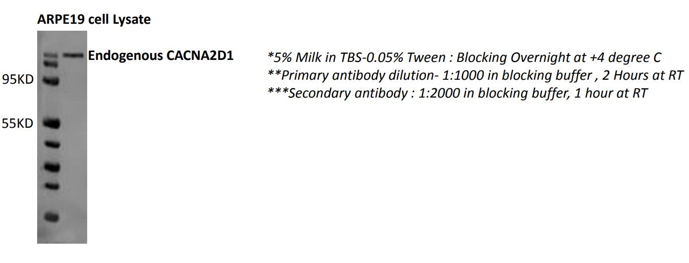
Application: Western BlotSample Tested: ARPE19 cell lysateSpecies: HumanVerified Customer | Posted 06/06/2018ARPE19 cell lysate

There are no reviews that match your criteria.
Protocols
Find general support by application which include: protocols, troubleshooting, illustrated assays, videos and webinars.
- 7-Amino Actinomycin D (7-AAD) Cell Viability Flow Cytometry Protocol
- Antigen Retrieval Protocol (PIER)
- Antigen Retrieval for Frozen Sections Protocol
- Appropriate Fixation of IHC/ICC Samples
- Cellular Response to Hypoxia Protocols
- Chromogenic IHC Staining of Formalin-Fixed Paraffin-Embedded (FFPE) Tissue Protocol
- Chromogenic Immunohistochemistry Staining of Frozen Tissue
- ClariTSA™ Fluorophore Kits
- Detection & Visualization of Antibody Binding
- Extracellular Membrane Flow Cytometry Protocol
- Flow Cytometry Protocol for Cell Surface Markers
- Flow Cytometry Protocol for Staining Membrane Associated Proteins
- Flow Cytometry Staining Protocols
- Flow Cytometry Troubleshooting Guide
- Fluorescent IHC Staining of Frozen Tissue Protocol
- Graphic Protocol for Heat-induced Epitope Retrieval
- Graphic Protocol for the Preparation and Fluorescent IHC Staining of Frozen Tissue Sections
- Graphic Protocol for the Preparation and Fluorescent IHC Staining of Paraffin-embedded Tissue Sections
- Graphic Protocol for the Preparation of Gelatin-coated Slides for Histological Tissue Sections
- ICC Cell Smear Protocol for Suspension Cells
- ICC Immunocytochemistry Protocol Videos
- ICC for Adherent Cells
- IHC Sample Preparation (Frozen sections vs Paraffin)
- Immunocytochemistry (ICC) Protocol
- Immunocytochemistry Troubleshooting
- Immunofluorescence of Organoids Embedded in Cultrex Basement Membrane Extract
- Immunofluorescent IHC Staining of Formalin-Fixed Paraffin-Embedded (FFPE) Tissue Protocol
- Immunohistochemistry (IHC) and Immunocytochemistry (ICC) Protocols
- Immunohistochemistry Frozen Troubleshooting
- Immunohistochemistry Paraffin Troubleshooting
- Immunoprecipitation Protocol
- Intracellular Flow Cytometry Protocol Using Alcohol (Methanol)
- Intracellular Flow Cytometry Protocol Using Detergents
- Intracellular Nuclear Staining Flow Cytometry Protocol Using Detergents
- Intracellular Staining Flow Cytometry Protocol Using Alcohol Permeabilization
- Intracellular Staining Flow Cytometry Protocol Using Detergents to Permeabilize Cells
- Preparing Samples for IHC/ICC Experiments
- Preventing Non-Specific Staining (Non-Specific Binding)
- Primary Antibody Selection & Optimization
- Propidium Iodide Cell Viability Flow Cytometry Protocol
- Protocol for Heat-Induced Epitope Retrieval (HIER)
- Protocol for Liperfluo
- Protocol for Making a 4% Formaldehyde Solution in PBS
- Protocol for VisUCyte™ HRP Polymer Detection Reagent
- Protocol for the Characterization of Human Th22 Cells
- Protocol for the Characterization of Human Th9 Cells
- Protocol for the Fluorescent ICC Staining of Cell Smears - Graphic
- Protocol for the Fluorescent ICC Staining of Cultured Cells on Coverslips - Graphic
- Protocol for the Preparation & Fixation of Cells on Coverslips
- Protocol for the Preparation and Chromogenic IHC Staining of Frozen Tissue Sections
- Protocol for the Preparation and Chromogenic IHC Staining of Frozen Tissue Sections - Graphic
- Protocol for the Preparation and Chromogenic IHC Staining of Paraffin-embedded Tissue Sections
- Protocol for the Preparation and Chromogenic IHC Staining of Paraffin-embedded Tissue Sections - Graphic
- Protocol for the Preparation and Fluorescent ICC Staining of Cells on Coverslips
- Protocol for the Preparation and Fluorescent ICC Staining of Non-adherent Cells
- Protocol for the Preparation and Fluorescent ICC Staining of Stem Cells on Coverslips
- Protocol for the Preparation and Fluorescent IHC Staining of Frozen Tissue Sections
- Protocol for the Preparation and Fluorescent IHC Staining of Paraffin-embedded Tissue Sections
- Protocol for the Preparation of Gelatin-coated Slides for Histological Tissue Sections
- Protocol for the Preparation of a Cell Smear for Non-adherent Cell ICC - Graphic
- Protocol: Annexin V and PI Staining by Flow Cytometry
- Protocol: Annexin V and PI Staining for Apoptosis by Flow Cytometry
- R&D Systems Quality Control Western Blot Protocol
- TUNEL and Active Caspase-3 Detection by IHC/ICC Protocol
- The Importance of IHC/ICC Controls
- Troubleshooting Guide: Fluorokine Flow Cytometry Kits
- Troubleshooting Guide: Immunohistochemistry
- Troubleshooting Guide: Western Blot Figures
- Western Blot Conditions
- Western Blot Protocol
- Western Blot Protocol for Cell Lysates
- Western Blot Troubleshooting
- Western Blot Troubleshooting Guide
- View all Protocols, Troubleshooting, Illustrated assays and Webinars
FAQs for CACNA2D1 Antibody (20A)
-
Q: We bought a vial of CACNA2D1 Antibody (20A) (NB120-2864) from you. But the datasheet doesn't tell us the antibody's concentration and positive control tissues. Can you supply the information for me? I wonder if the antibody has a total concentration or concentration range. You know if we don't have the concentration information, we can't ascertain the IgG's concentration for negative control.
A: This product is unpurified (diluted ascites) and therefore, concentration is not relevant. I talked to the lab team regarding your concerns and they told me that the total protein concentration in the ascites was around 2.2 mg/mL. Generally, 90% of the ascites is IgG, therefore, I assume that the IgG concentration in your vial should be around 1.98 mg/ml. You can use our CACNA2D1 overexpression lysate (# NBP2-08998) or skeletal/cardiac muscle tissue membrane preparations as positive control for Western Blot assay. As far as IHC application is concerned, you may use skeletal/cardiac muscle tissue sections as positive control wherein this product will show selective cytoplasmic expression of CACNA2D1.