Calbindin D-28K Antibody - BSA Free
Novus Biologicals | Catalog # NBP2-50028
![Western Blot: Calbindin D-28K Antibody [NBP2-50028]](https://resources.rndsystems.com/images/products/Calbindin-D-28K-Antibody-Western-Blot-NBP2-50028-img0004.jpg "Western Blot: Calbindin D-28K Antibody [NBP2-50028]")
Key Product Details
Species Reactivity
Validated:
Cited:
Applications
Validated:
Cited:
Label
Antibody Source
Format
Product Specifications
Immunogen
Reactivity Notes
Clonality
Host
Isotype
Theoretical MW
Disclaimer note: The observed molecular weight of the protein may vary from the listed predicted molecular weight due to post translational modifications, post translation cleavages, relative charges, and other experimental factors.
Scientific Data Images for Calbindin D-28K Antibody - BSA Free
Western Blot: Calbindin D-28K Antibody [NBP2-50028]
Western Blot: Calbindin D-28K Antibody [NBP2-50028] - Analysis of different tissue lysates and recombinant protein solutions using chicken pAb to calbindin, NBP2-50028, dilution 1:5,000 in green: [1] protein standard (red), [2] rat cerebellum, [3] pig hippocampus, [4] cow cerebellum, [5] protein standard (red). Next lanes are full length recombinant human proteins, [6] Parvalbumin, [7] Calretinin, [8] Calbindin. Bands at 25kDa in tissue lysates and ~30 kDa in protein solutions correspond to calbindin, the recombinant form being slightly larger due to the presence of a His tag and other vector derived sequence. NBP2-50028 antibody specifically recognizes calbindin protein and does not react with the closely related proteins parvalbumin and calretinin.![Immunocytochemistry/ Immunofluorescence: Calbindin D-28K Antibody [NBP2-50028]](https://resources.rndsystems.com/images/products/Calbindin-D-28K-Antibody-Immunocytochemistry-Immunofluorescence-NBP2-50028-img0005.jpg "Immunocytochemistry/ Immunofluorescence: Calbindin D-28K Antibody [NBP2-50028]")
Immunocytochemistry/ Immunofluorescence: Calbindin D-28K Antibody [NBP2-50028]
Immunocytochemistry/Immunofluorescence: Calbindin D-28K Antibody [NBP2-50028] - Cultured mouse embryonic kidney stained with Calbindin antibody (1:200) and DAPI. Image from verified customer review.![Immunohistochemistry: Calbindin D-28K Antibody [NBP2-50028]](https://resources.rndsystems.com/images/products/Calbindin-D-28K-Antibody-Immunohistochemistry-NBP2-50028-img0007.jpg "Immunohistochemistry: Calbindin D-28K Antibody [NBP2-50028]")
Immunohistochemistry: Calbindin D-28K Antibody [NBP2-50028]
Calbindin-D-28K-Antibody-Immunohistochemistry-NBP2-50028-img0007.jpg![Western Blot: Calbindin D-28K Antibody [NBP2-50028]](https://resources.rndsystems.com/images/products/Calbindin-D-28K-Antibody-Western-Blot-NBP2-50028-img0002.jpg "Western Blot: Calbindin D-28K Antibody [NBP2-50028]")
Western Blot: Calbindin D-28K Antibody [NBP2-50028]
Western Blot: Calbindin D-28K Antibody [NBP2-50028] - Analysis of rat brain lysate (left), 0.5 ug of His-tagged recombinant proteins (right) were probed with NBP2-50028 at 1:5,000. Lane 1: Parvalbumin, Lane 2: Calretinin, Lane 3: Calbindin.![Immunocytochemistry/ Immunofluorescence: Calbindin D-28K Antibody [NBP2-50028]](https://resources.rndsystems.com/images/products/Calbindin-D-28K-Antibody-Immunocytochemistry-Immunofluorescence-NBP2-50028-img0003.jpg "Immunocytochemistry/ Immunofluorescence: Calbindin D-28K Antibody [NBP2-50028]")
Immunocytochemistry/ Immunofluorescence: Calbindin D-28K Antibody [NBP2-50028]
Immunocytochemistry/Immunofluorescence: Calbindin D-28K Antibody [NBP2-50028] - Adult rat brain cortex (Left) and striatum (Right) sections (45 uM; fixed by transcardial perfusion with 4% paraformaldehyde) were stained with NBP2-50028 (1:1,000, red), and monoclonal mouse anti-RBFOX3/NeuN (NBP1-92693; green). Calbindin labels a subset of sparsely-distributed interneurons (calbindin-positive interneurons) in the cortex (Left), and more densely-distributed neurons in the striatum (Right). Since neurons also express RBFOX3/NeuN, calbindin-positive cells appear to be gold to yellow. Insets are high magnification images of boxed area of each image. Blue is Dapi nucleus staining.![Immunohistochemistry Free-Floating: Calbindin D-28K Antibody [NBP2-50028]](https://resources.rndsystems.com/images/products/Calbindin-D-28K-Antibody-Immunohistochemistry-Free-Floating-NBP2-50028-img0006.jpg "Immunohistochemistry Free-Floating: Calbindin D-28K Antibody [NBP2-50028]")
Immunohistochemistry Free-Floating: Calbindin D-28K Antibody [NBP2-50028]
Immunohistochemistry Free-Floating: Calbindin D-28K Antibody [NBP2-50028] - Analysis of rat cerebellum section stained with chicken Calbindin D-28K pAb, dilution 1:2,000 (Green), and costained with rabbit MeCP2 pAb, dilution 1:5,000 (Red). Following transcardial perfusion with 4% paraformaldehyde, brain was post fixed for 24 hours, cut to 45uM, and free-floating sections were stained with above antibodies. Calbindin D-28K, often used as a Purkinje cell marker, is prominently detected in dendrites and perikarya of these cells in the cerebellar molecular layer. The MeCP2 antibody selectively stains nuclei of neuronal cells.
Immunohistochemistry: Calbindin D-28K Antibody [NBP2-50028] -
Immunohistochemistry: Calbindin D-28K Antibody [NBP2-50028] - C1q is expressed in retina microglia during development. (A) qRT-PCR for C1q at P9, P14, & 14 weeks. Values represent the fold mRNA expression level relative to the levels detected in adult animals following normalization to GAPDH. (B) Representative fluorescent in situ hybridization images at P13 of C1q (green) co-stained with either Iba1 (magenta) or calbindin (red) to label microglia & horizontal cells, respectively. C1q colocalizes with microglia but not with horizontal cells in the outer retina at this time (arrowheads). Inset shows C1q expression in Iba1-positive microglia that is absent in calbindin-positive horizontal cells. Scale bars = 25 & 10 μm (inset). Image collected & cropped by CiteAb from the following publication (https://pubmed.ncbi.nlm.nih.gov/33177995), licensed under a CC-BY license. Not internally tested by Novus Biologicals.
Immunocytochemistry/ Immunofluorescence: Calbindin D-28K Antibody [NBP2-50028] -
Immunocytochemistry/ Immunofluorescence: Calbindin D-28K Antibody [NBP2-50028] - Myh14 localizes to apical membranes of Atp6v1b1‐positive intercalated cells in the distal tubule. Adult kidney sections were stained for calbindin‐D28K, Atp6v1b1, Aqp2, & Myh14 to identify intercalated cells within the distal & connecting tubule segments as well as to confirm Myh14 localization to the intercalated cell type. Cells with positive staining for Atp6v1b1 (red) can be found in tubule segments positive for calbindin‐D28K only (A–D) as well as in tubules positive for both calbindin‐D28K & Aqp2 (E–H); in both cases Atp6v1b1 staining is observed along the basolateral membrane indicating beta ‐ intercalated cell type (arrowhead, D&H). We also observed Atp6v1b1 staining in majority of the tubules along the apical membrane indicating the alpha ‐ intercalated cell type (arrowhead, I–L). Myh14 localized to the apical membrane of the intercalated cell type in the distal tubule (arrowhead, M–P). Sections were mounted with vectashield containing DAPI to stain the nucleus (blue; D, H, L & P). Scale bar = 10 μm. Image collected & cropped by CiteAb from the following publication (https://pubmed.ncbi.nlm.nih.gov/29208685), licensed under a CC-BY license. Not internally tested by Novus Biologicals.
Immunohistochemistry: Calbindin D-28K Antibody [NBP2-50028] -
Immunohistochemistry: Calbindin D-28K Antibody [NBP2-50028] - Horizontal cells develop normally with the loss of either C3aR or CR3. (A,B) qRT-PCR for complement proteins C3aR & CR3 at P6, P13, & adult in wild type mice. Values represent the fold mRNA expression level relative to the levels detected in control animals following normalization to GAPDH. N = 3 animals. Representative images of horizontal cells (C, anti-calbindin, cyan) & quantifications (D,E) of ectopic horizontal cell neurites in control & C3aR–/– or CR3–/– mice at P13. Horizontal cell neurites in C3aR–/– & CR3–/– mice remain confined to the OPL, showing no increase in the number (D) or length (E) of ectopic neurites (n = 52 sprouts from four control mice; n = 84 sprouts from four C3aR–/– mice; n = 67 sprouts from four CR3–/– mice). Scale bars = 25 μm. Data are represented as the mean ± SEM (D) or as beeswarm SuperPlots (E) in which individual horizontal cell sprout values are presented together with the mean from each animal ± the SEM. Image collected & cropped by CiteAb from the following publication (https://pubmed.ncbi.nlm.nih.gov/33177995), licensed under a CC-BY license. Not internally tested by Novus Biologicals.
Immunocytochemistry/ Immunofluorescence: Calbindin D-28K Antibody [NBP2-50028] -
Immunocytochemistry/ Immunofluorescence: Calbindin D-28K Antibody [NBP2-50028] - Myh10 & Myh14 are the NM2 isoforms expressed in the distal & connecting renal tubules. Adult kidney sections were stained for calbindin‐D28K, Aqp2 & one of the NM2 isoforms. Myh9 (A–D) was not expressed in tubules positive for calbindin‐D28K & Aqp2; although adjacent tubules were positive for Myh9 (arrow). Myh10 (E–H) was expressed at low levels at the cell–cell junctions in calbindin‐D28K only positive tubules (white arrowhead), & in tubules positive for both calbindin‐D28K & Aqp2, Myh10 localized to cell–cell junctions, apical, & basolateral membrane (F & H, yellow arrowhead). Myh14 (I–L) was expressed on the apical membrane of calbindin‐D28K & Aqp2 null cells (arrowheads) in the distal tubular segments. Sections were mounted with vectashield containing DAPI to stain the nucleus (blue; D, H, & L). Scale bar = 10 μm. Image collected & cropped by CiteAb from the following publication (https://pubmed.ncbi.nlm.nih.gov/29208685), licensed under a CC-BY license. Not internally tested by Novus Biologicals.Applications for Calbindin D-28K Antibody - BSA Free
Immunocytochemistry/ Immunofluorescence
Immunohistochemistry
Western Blot
Reviewed Applications
Read 1 review rated 5 using NBP2-50028 in the following applications:
Formulation, Preparation, and Storage
Purification
Formulation
Format
Preservative
Concentration
Shipping
Stability & Storage
Background: Calbindin D-28K
Alternate Names
Gene Symbol
UniProt
Additional Calbindin D-28K Products
Product Documents for Calbindin D-28K Antibody - BSA Free
Certificate of Analysis
To download a Certificate of Analysis, please enter a lot or batch number in the search box below.
Product Specific Notices for Calbindin D-28K Antibody - BSA Free
Chicken products cannot be exported to Canada.
This product is for research use only and is not approved for use in humans or in clinical diagnosis. Primary Antibodies are guaranteed for 1 year from date of receipt.
Citations for Calbindin D-28K Antibody - BSA Free
 Powered by Bioz
Customer Reviews for Calbindin D-28K Antibody - BSA Free (1)
Have you used Calbindin D-28K Antibody - BSA Free?
Submit a review and receive an Amazon gift card!
$25/€18/£15/$25CAN/¥2500 Yen for a review with an image
$10/€7/£6/$10CAN/¥1110 Yen for a review without an image
Submit a review
Customer Images
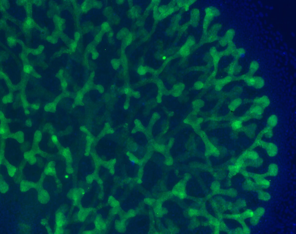
-
Application: ImmunocytochemistrySample Tested: Embryonic kidneySpecies: MouseVerified Customer | Posted 08/04/2018cultured embryonic kidney, stained with Calbindin and DapiFixation: 4% PFA, 30min RT Permeabilization: 100 % methanol -20°C, 20 min blocking buffer: 3% donkey serum antibody dilution: 1:200 in blocking buffer, incubation overnight 4°C

There are no reviews that match your criteria.
Protocols
Find general support by application which include: protocols, troubleshooting, illustrated assays, videos and webinars.
- Antigen Retrieval Protocol (PIER)
- Antigen Retrieval for Frozen Sections Protocol
- Appropriate Fixation of IHC/ICC Samples
- Cellular Response to Hypoxia Protocols
- Chromogenic IHC Staining of Formalin-Fixed Paraffin-Embedded (FFPE) Tissue Protocol
- Chromogenic Immunohistochemistry Staining of Frozen Tissue
- ClariTSA™ Fluorophore Kits
- Detection & Visualization of Antibody Binding
- Fluorescent IHC Staining of Frozen Tissue Protocol
- Graphic Protocol for Heat-induced Epitope Retrieval
- Graphic Protocol for the Preparation and Fluorescent IHC Staining of Frozen Tissue Sections
- Graphic Protocol for the Preparation and Fluorescent IHC Staining of Paraffin-embedded Tissue Sections
- Graphic Protocol for the Preparation of Gelatin-coated Slides for Histological Tissue Sections
- ICC Cell Smear Protocol for Suspension Cells
- ICC Immunocytochemistry Protocol Videos
- ICC for Adherent Cells
- IHC Sample Preparation (Frozen sections vs Paraffin)
- Immunocytochemistry (ICC) Protocol
- Immunocytochemistry Troubleshooting
- Immunofluorescence of Organoids Embedded in Cultrex Basement Membrane Extract
- Immunofluorescent IHC Staining of Formalin-Fixed Paraffin-Embedded (FFPE) Tissue Protocol
- Immunohistochemistry (IHC) and Immunocytochemistry (ICC) Protocols
- Immunohistochemistry Frozen Troubleshooting
- Immunohistochemistry Paraffin Troubleshooting
- Preparing Samples for IHC/ICC Experiments
- Preventing Non-Specific Staining (Non-Specific Binding)
- Primary Antibody Selection & Optimization
- Protocol for Heat-Induced Epitope Retrieval (HIER)
- Protocol for Making a 4% Formaldehyde Solution in PBS
- Protocol for VisUCyte™ HRP Polymer Detection Reagent
- Protocol for the Fluorescent ICC Staining of Cell Smears - Graphic
- Protocol for the Fluorescent ICC Staining of Cultured Cells on Coverslips - Graphic
- Protocol for the Preparation & Fixation of Cells on Coverslips
- Protocol for the Preparation and Chromogenic IHC Staining of Frozen Tissue Sections
- Protocol for the Preparation and Chromogenic IHC Staining of Frozen Tissue Sections - Graphic
- Protocol for the Preparation and Chromogenic IHC Staining of Paraffin-embedded Tissue Sections
- Protocol for the Preparation and Chromogenic IHC Staining of Paraffin-embedded Tissue Sections - Graphic
- Protocol for the Preparation and Fluorescent ICC Staining of Cells on Coverslips
- Protocol for the Preparation and Fluorescent ICC Staining of Non-adherent Cells
- Protocol for the Preparation and Fluorescent ICC Staining of Stem Cells on Coverslips
- Protocol for the Preparation and Fluorescent IHC Staining of Frozen Tissue Sections
- Protocol for the Preparation and Fluorescent IHC Staining of Paraffin-embedded Tissue Sections
- Protocol for the Preparation of Gelatin-coated Slides for Histological Tissue Sections
- Protocol for the Preparation of a Cell Smear for Non-adherent Cell ICC - Graphic
- R&D Systems Quality Control Western Blot Protocol
- TUNEL and Active Caspase-3 Detection by IHC/ICC Protocol
- The Importance of IHC/ICC Controls
- Troubleshooting Guide: Immunohistochemistry
- Troubleshooting Guide: Western Blot Figures
- Western Blot Conditions
- Western Blot Protocol
- Western Blot Protocol for Cell Lysates
- Western Blot Troubleshooting
- Western Blot Troubleshooting Guide
- View all Protocols, Troubleshooting, Illustrated assays and Webinars
FAQs for Calbindin D-28K Antibody - BSA Free
-
Q: I am doing research on Neuroscience and I would like to use some antibodies, especially in Western Blots, but I could do other assays: - alpha7 nicotinic cholinergic receptor; - parvalbumin; - calbindin D28K; The preferred reactivity is anti-human and anti-mouse. Do you have these antibodies? Are they in stock? And what about the prizes: How much are they? How much are the shipping costs? Could you make me any offer?
A:
This link will take you to all of our alpha7 nicotinic cholingergic receptor antibodies currently available for purchase. Please see the following link for our Calbinin D28K antibodies reactive against mouse and human. We currently do not have any Parvalbumin antibodies that have been tested in mouse. I would like to introduce you to our Innovators Reward Program. Try our primary antibodies in an untested species or application, without the financial risk of failure. The pricing and availability of our products depends on your country.