Caspase-9 Antibody - BSA Free
Novus Biologicals | Catalog # NB100-56118
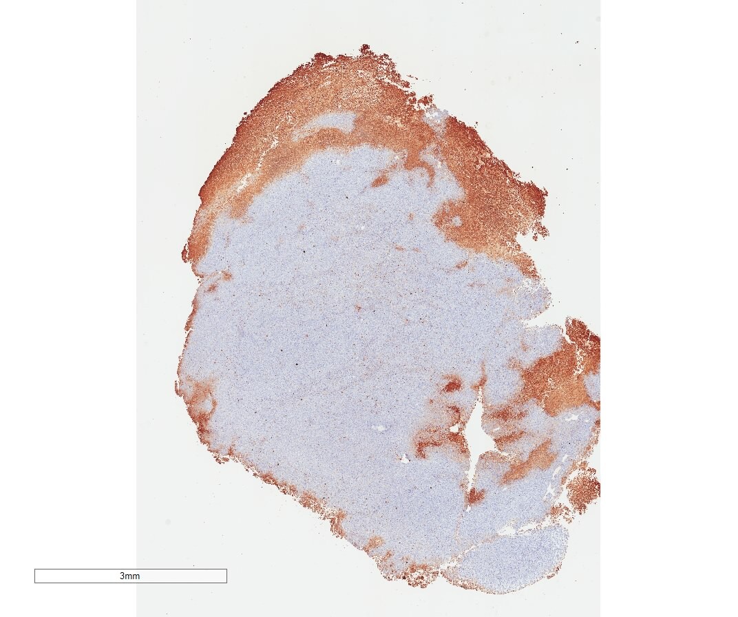
![Immunohistochemistry: Caspase-9 Antibody [NB100-56118]](https://resources.rndsystems.com/images/products/Caspase-9-Antibody-Immunohistochemistry-NB100-56118-img0014.jpg "Immunohistochemistry: Caspase-9 Antibody [NB100-56118]")
Key Product Details
Species Reactivity
Validated:
Cited:
Applications
Validated:
Cited:
Label
Antibody Source
Format
Product Specifications
Immunogen
Clonality
Host
Isotype
Scientific Data Images for Caspase-9 Antibody - BSA Free
Immunohistochemistry: Caspase-9 Antibody [NB100-56118]
Caspase-9-Antibody-Immunohistochemistry-NB100-56118-img0014.jpg![Immunohistochemistry: Caspase-9 Antibody [NB100-56118]](https://resources.rndsystems.com/images/products/Caspase-9-Antibody-Immunohistochemistry-NB100-56118-img0013.jpg "Immunohistochemistry: Caspase-9 Antibody [NB100-56118]")
Immunohistochemistry: Caspase-9 Antibody [NB100-56118]
Caspase-9-Antibody-Immunohistochemistry-NB100-56118-img0013.jpg![Immunohistochemistry-Paraffin: Caspase-9 Antibody [NB100-56118]](https://resources.rndsystems.com/images/products/Caspase-9-Antibody-Immunohistochemistry-Paraffin-NB100-56118-img0006.jpg "Immunohistochemistry-Paraffin: Caspase-9 Antibody [NB100-56118]")
Immunohistochemistry-Paraffin: Caspase-9 Antibody [NB100-56118]
Immunohistochemistry-Paraffin: Caspase-9 Antibody [NB100-56118] - Analysis of Capase-9 in mouse transplanted lung tumor section using anti-Caspase-9 antibody. Image from verified customer review.![Immunohistochemistry: Caspase-9 Antibody [NB100-56118]](https://resources.rndsystems.com/images/products/Caspase-9-Antibody-Immunohistochemistry-NB100-56118-img0012.jpg "Immunohistochemistry: Caspase-9 Antibody [NB100-56118]")
Immunohistochemistry: Caspase-9 Antibody [NB100-56118]
Caspase-9-Antibody-Immunohistochemistry-NB100-56118-img0012.jpg![Immunohistochemistry-Paraffin: Caspase-9 Antibody [NB100-56118]](https://resources.rndsystems.com/images/products/Caspase-9-Antibody-Immunohistochemistry-Paraffin-NB100-56118-img0007.jpg "Immunohistochemistry-Paraffin: Caspase-9 Antibody [NB100-56118]")
Immunohistochemistry-Paraffin: Caspase-9 Antibody [NB100-56118]
Immunohistochemistry-Paraffin: Caspase-9 Antibody [NB100-56118] - Analysis of Dog Brain sections stained for Active/Cleaved Caspase-9 expression at 1:2000. A and B. The pattern of Caspase-9 staining may vary between different types of neurons. Hematoxylin-eosin counterstain.![Immunohistochemistry-Paraffin: Caspase-9 Antibody [NB100-56118]](https://resources.rndsystems.com/images/products/Caspase-9-Antibody-Immunohistochemistry-Paraffin-NB100-56118-img0008.jpg "Immunohistochemistry-Paraffin: Caspase-9 Antibody [NB100-56118]")
Immunohistochemistry-Paraffin: Caspase-9 Antibody [NB100-56118]
Immunohistochemistry-Paraffin: Caspase-9 Antibody [NB100-56118] - Analysis of Dog Brain sections stained for cleaved Caspase-9 expression using this antibody at 1:2000. A) Section from a dog brain 2 hr after reperfusion injury. B) ection from a dog brain sham control (brain surgery but no injury). Hematoxylin-eosin counterstain.![Immunohistochemistry-Paraffin: Caspase-9 Antibody [NB100-56118]](https://resources.rndsystems.com/images/products/Caspase-9-Antibody-Immunohistochemistry-Paraffin-NB100-56118-img0009.jpg "Immunohistochemistry-Paraffin: Caspase-9 Antibody [NB100-56118]")
Immunohistochemistry-Paraffin: Caspase-9 Antibody [NB100-56118]
Immunohistochemistry-Paraffin: Caspase-9 Antibody [NB100-56118] - Analysis Human Brain sections stained for cleaved Caspase-9 expression using this antibody at 1:2000. A) Section from a patient 24 hr after head trauma. B) Control: section from a patient with no known neurological disease or head injury. Hematoxylin-eosin counterstain.
Immunocytochemistry/ Immunofluorescence: Caspase-9 Antibody [NB100-56118] -
Immunocytochemistry/ Immunofluorescence: Caspase-9 Antibody [NB100-56118] - Exacerbated apoptosis & altered inflammation in double-deficient Atg7 delta pan-Rip3−/− mice. (a) Depletion of Rip3 & pancreatic Atg7 (Atg7 delta pan-Rip3d/d) increased pancreatic caspase-3 in 12-week-old mice. Caspase-3 quantitation & representative IF microphotographs of Atg7 delta pan (n=5) & Atg7 delta pan-Rip3d/d (n=5) pancreatic tissue stained for DAPI (blue) & Caspase-3 (red) (anti-active caspase-3 ab2302, 1/50, scale bar=50 μm). (b) Depletion of Rip3 & pancreatic Atg7 (Atg7 delta pan-Rip3d/d) increased pancreatic Bax in 12-week-old mice. Bax quantitation & representative IF microphotographs of Atg7 delta pan (n=5) & Atg7 delta pan-Rip3d/d (n=5) pancreatic tissue stained for DAPI (blue) & Caspase-3 (red) (anti-Bax (sc-526, 1/50). (c) Depletion of Rip3 & pancreatic Atg7 (Atg7 delta pan-Rip3d/d) increased pancreatic caspase-9 in 12-week-old mice. Caspase-9 quantitation & representative IF microphotographs of Atg7 delta pan (n=5) & Atg7 delta pan-Rip3d/d (n=5) pancreatic tissue stained for DAPI (blue) & Caspase-9 (red) (anti-caspase-9 NB100-56118, 1/1000). (d) Reduced infiltration inflammation of macrophages (F4/80, NBP2-12506, 1/75) & early T-lymphocytes (MPO, ab9535, 1/50) in double-deficient Atg7 delta pan-Rip3d/d (n=10) mice compared with pancreatic Atg7 delta pan (n=7). Data are mean±S.E.M. for the numbers of animals as indicated in the graph, *P<0.05, **P<0.01, NS = no significance Image collected & cropped by CiteAb from the following publication (https://pubmed.ncbi.nlm.nih.gov/28703808), licensed under a CC-BY license. Not internally tested by Novus Biologicals.
Western Blot: Caspase-9 Antibody - BSA Free [NB100-56118] -
Nuclear translocation of TCA cycle enzymes prevents doxorubicin-mediated cellular damage.a–c Enzyme activity of IDH-2 (n = 4) (a), MDH-2 (n = 3) (b), and PDHC (n = 3) (c) in nuclei isolated from cells transduced with tetracycline-inducible NLS-IDH-2 constructs. Activity is represented as the rate in nM/min or delta OD/min, p value* <0.05, p value**** = 0.0001, and lysates were probed for PCNA to detect equal nuclear protein amounts used for the assay. d Reduced apoptotic cell death as measured by Annexin:V:FITC staining in cells expressing nuclear TCA cycle dehydrogenases prior to doxorubicin DRN treatment. N = 3 p value* <0.05 (e) Quantification of troponin I released from cardiomyocytes as a marker of cardiac injury, minimum n = 4 p value** <0.007, p value* <0.05. f Cell death as measured by Cell Titer GloTM in cells expressing nuclear dehydrogenases prior to DRN treatment. minimum n = 4 p value*** = 0.0001, p value**** <0.0001. NLS-EGFP-2A-NLS-mCherry was used as a control for the 2 A linker system. g DNA damage as assessed by ɣ-H2AX foci formation. Scale bar 20 um. h, i qRT‒PCR showing the expression of anti-apoptotic genes BCL2 (h) and MCL1 (i). n = 3 P value ** <0.005, *<0.05. j–l Protein levels of anti-apoptotic proteins (j, k along with reduced caspase 9 cleavage (l) in cells expressing nuclear IDH-2 compared to cells treated with doxorubicin. Image collected and cropped by CiteAb from the following open publication (https://pubmed.ncbi.nlm.nih.gov/37468519), licensed under a CC-BY license. Not internally tested by Novus Biologicals.Applications for Caspase-9 Antibody - BSA Free
Immunocytochemistry/ Immunofluorescence
Immunohistochemistry-Frozen
Immunohistochemistry-Paraffin
Immunoprecipitation
Western Blot
Reviewed Applications
Read 1 review rated 5 using NB100-56118 in the following applications:
Flow Cytometry Panel Builder
Bio-Techne Knows Flow Cytometry
Save time and reduce costly mistakes by quickly finding compatible reagents using the Panel Builder Tool.
Advanced Features
- Spectra Viewer - Custom analysis of spectra from multiple fluorochromes
- Spillover Popups - Visualize the spectra of individual fluorochromes
- Antigen Density Selector - Match fluorochrome brightness with antigen density

Formulation, Preparation, and Storage
Purification
Formulation
Format
Preservative
Concentration
Shipping
Stability & Storage
Background: Caspase-9
Alternate Names
Gene Symbol
UniProt
Additional Caspase-9 Products
Product Documents for Caspase-9 Antibody - BSA Free
Certificate of Analysis
To download a Certificate of Analysis, please enter a lot or batch number in the search box below.
Product Specific Notices for Caspase-9 Antibody - BSA Free
This product is for research use only and is not approved for use in humans or in clinical diagnosis. Primary Antibodies are guaranteed for 1 year from date of receipt.
Related Research Areas
Citations for Caspase-9 Antibody - BSA Free
 Powered by Bioz
Customer Reviews for Caspase-9 Antibody - BSA Free (1)
Have you used Caspase-9 Antibody - BSA Free?
Submit a review and receive an Amazon gift card!
$25/€18/£15/$25CAN/¥2500 Yen for a review with an image
$10/€7/£6/$10CAN/¥1110 Yen for a review without an image
Submit a review
Customer Images
-
Application: Immunohistochemistry-ParaffinSample Tested: Transplanted lung tumor sectionSpecies: MouseVerified Customer | Posted 01/08/2015Section of transplanted lung tumors from mouse

There are no reviews that match your criteria.
Protocols
Find general support by application which include: protocols, troubleshooting, illustrated assays, videos and webinars.
- 7-Amino Actinomycin D (7-AAD) Cell Viability Flow Cytometry Protocol
- Antigen Retrieval Protocol (PIER)
- Antigen Retrieval for Frozen Sections Protocol
- Appropriate Fixation of IHC/ICC Samples
- Cellular Response to Hypoxia Protocols
- Chromogenic IHC Staining of Formalin-Fixed Paraffin-Embedded (FFPE) Tissue Protocol
- Chromogenic Immunohistochemistry Staining of Frozen Tissue
- ClariTSA™ Fluorophore Kits
- Detection & Visualization of Antibody Binding
- Extracellular Membrane Flow Cytometry Protocol
- Flow Cytometry Protocol for Cell Surface Markers
- Flow Cytometry Protocol for Staining Membrane Associated Proteins
- Flow Cytometry Staining Protocols
- Flow Cytometry Troubleshooting Guide
- Fluorescent IHC Staining of Frozen Tissue Protocol
- Graphic Protocol for Heat-induced Epitope Retrieval
- Graphic Protocol for the Preparation and Fluorescent IHC Staining of Frozen Tissue Sections
- Graphic Protocol for the Preparation and Fluorescent IHC Staining of Paraffin-embedded Tissue Sections
- Graphic Protocol for the Preparation of Gelatin-coated Slides for Histological Tissue Sections
- ICC Cell Smear Protocol for Suspension Cells
- ICC Immunocytochemistry Protocol Videos
- ICC for Adherent Cells
- IHC Sample Preparation (Frozen sections vs Paraffin)
- Immunocytochemistry (ICC) Protocol
- Immunocytochemistry Troubleshooting
- Immunofluorescence of Organoids Embedded in Cultrex Basement Membrane Extract
- Immunofluorescent IHC Staining of Formalin-Fixed Paraffin-Embedded (FFPE) Tissue Protocol
- Immunohistochemistry (IHC) and Immunocytochemistry (ICC) Protocols
- Immunohistochemistry Frozen Troubleshooting
- Immunohistochemistry Paraffin Troubleshooting
- Immunoprecipitation Protocol
- Intracellular Flow Cytometry Protocol Using Alcohol (Methanol)
- Intracellular Flow Cytometry Protocol Using Detergents
- Intracellular Nuclear Staining Flow Cytometry Protocol Using Detergents
- Intracellular Staining Flow Cytometry Protocol Using Alcohol Permeabilization
- Intracellular Staining Flow Cytometry Protocol Using Detergents to Permeabilize Cells
- Preparing Samples for IHC/ICC Experiments
- Preventing Non-Specific Staining (Non-Specific Binding)
- Primary Antibody Selection & Optimization
- Propidium Iodide Cell Viability Flow Cytometry Protocol
- Protocol for Heat-Induced Epitope Retrieval (HIER)
- Protocol for Liperfluo
- Protocol for Making a 4% Formaldehyde Solution in PBS
- Protocol for VisUCyte™ HRP Polymer Detection Reagent
- Protocol for the Characterization of Human Th22 Cells
- Protocol for the Characterization of Human Th9 Cells
- Protocol for the Fluorescent ICC Staining of Cell Smears - Graphic
- Protocol for the Fluorescent ICC Staining of Cultured Cells on Coverslips - Graphic
- Protocol for the Preparation & Fixation of Cells on Coverslips
- Protocol for the Preparation and Chromogenic IHC Staining of Frozen Tissue Sections
- Protocol for the Preparation and Chromogenic IHC Staining of Frozen Tissue Sections - Graphic
- Protocol for the Preparation and Chromogenic IHC Staining of Paraffin-embedded Tissue Sections
- Protocol for the Preparation and Chromogenic IHC Staining of Paraffin-embedded Tissue Sections - Graphic
- Protocol for the Preparation and Fluorescent ICC Staining of Cells on Coverslips
- Protocol for the Preparation and Fluorescent ICC Staining of Non-adherent Cells
- Protocol for the Preparation and Fluorescent ICC Staining of Stem Cells on Coverslips
- Protocol for the Preparation and Fluorescent IHC Staining of Frozen Tissue Sections
- Protocol for the Preparation and Fluorescent IHC Staining of Paraffin-embedded Tissue Sections
- Protocol for the Preparation of Gelatin-coated Slides for Histological Tissue Sections
- Protocol for the Preparation of a Cell Smear for Non-adherent Cell ICC - Graphic
- Protocol: Annexin V and PI Staining by Flow Cytometry
- Protocol: Annexin V and PI Staining for Apoptosis by Flow Cytometry
- R&D Systems Quality Control Western Blot Protocol
- TUNEL and Active Caspase-3 Detection by IHC/ICC Protocol
- The Importance of IHC/ICC Controls
- Troubleshooting Guide: Fluorokine Flow Cytometry Kits
- Troubleshooting Guide: Immunohistochemistry
- Troubleshooting Guide: Western Blot Figures
- Western Blot Conditions
- Western Blot Protocol
- Western Blot Protocol for Cell Lysates
- Western Blot Troubleshooting
- Western Blot Troubleshooting Guide
- View all Protocols, Troubleshooting, Illustrated assays and Webinars
FAQs for Caspase-9 Antibody - BSA Free
-
Q: Could I use HeLa cell lysate as a positive control for Caspase 9 primary antibodies in WB?
A: After looking through our testing it does appear you should be fine using HeLa as a positive control cell lysate for Caspase 9 in Western Blot, as we have used this sample successfully in our QC tests and shown expression of Caspase 9.
Associated Pathways

