Caveolin-2 Antibody - BSA Free
Novus Biologicals | Catalog # NBP1-31116
![Western Blot: Caveolin-2 Antibody [NBP1-31116]](https://resources.rndsystems.com/images/products/Caveolin-2-Antibody-Western-Blot-NBP1-31116-img0014.jpg "Western Blot: Caveolin-2 Antibody [NBP1-31116]")
Loading...
Key Product Details
Species Reactivity
Validated:
Human, Mouse, Rat
Predicted:
Bovine (100%), Chimpanzee (100%), Feline (100%), Porcine (100%), Rabbit (100%), Rhesus Macaque (100%), Sheep (100%). Backed by our 100% Guarantee.
Applications
Validated:
Immunohistochemistry, Immunohistochemistry-Paraffin, Western Blot, Immunocytochemistry/ Immunofluorescence, Immunoprecipitation
Cited:
In vivo assay
Label
Unconjugated
Antibody Source
Polyclonal Rabbit IgG
Format
BSA Free
Loading...
Product Specifications
Immunogen
Carrier-protein conjugated synthetic peptide encompassing a sequence within the N-terminus region of human Caveolin-2. The exact sequence is proprietary.
Reactivity Notes
Cat (100%).
Localization
Golgi apparatus membrane, Single-pass membrane protein, Cell membrane, Single-pass membrane protein, Membrane, caveola, Single-pass membrane protein, Golgi apparatus membrane, Cell membrane, Membrane, caveola
Marker
Caveolae Marker
Clonality
Polyclonal
Host
Rabbit
Isotype
IgG
Theoretical MW
18 kDa.
Disclaimer note: The observed molecular weight of the protein may vary from the listed predicted molecular weight due to post translational modifications, post translation cleavages, relative charges, and other experimental factors.
Disclaimer note: The observed molecular weight of the protein may vary from the listed predicted molecular weight due to post translational modifications, post translation cleavages, relative charges, and other experimental factors.
Scientific Data Images for Caveolin-2 Antibody - BSA Free
Western Blot: Caveolin-2 Antibody [NBP1-31116]
Western Blot: Caveolin-2 Antibody [NBP1-31116] - Rat tissue extracts (50 ug) was separated by 15% SDS-PAGE, and the membrane was blotted with Caveolin 2 antibody diluted at 1:1000.![Immunohistochemistry-Paraffin: Caveolin-2 Antibody [NBP1-31116]](https://resources.rndsystems.com/images/products/Caveolin-2-Antibody-Immunohistochemistry-Paraffin-NBP1-31116-img0006.jpg "Immunohistochemistry-Paraffin: Caveolin-2 Antibody [NBP1-31116]")
Immunohistochemistry-Paraffin: Caveolin-2 Antibody [NBP1-31116]
Immunohistochemistry-Paraffin: Caveolin-2 Antibody [NBP1-31116] - FaDu xenograft, using Caveolin 2 antibody at 1:500 dilution. Antigen Retrieval: Trilogy™ (EDTA based, pH 8.0) buffer, 15min.![Western Blot: Caveolin-2 Antibody [NBP1-31116]](https://resources.rndsystems.com/images/products/Caveolin-2-Antibody-Western-Blot-NBP1-31116-img0008.jpg "Western Blot: Caveolin-2 Antibody [NBP1-31116]")
Western Blot: Caveolin-2 Antibody [NBP1-31116]
Western Blot: Caveolin-2 Antibody [NBP1-31116] - Sample (30 ug of whole cell lysate) A: A549 12% SDS PAGE; antibody diluted at 1:1000.![Western Blot: Caveolin-2 Antibody [NBP1-31116]](https://resources.rndsystems.com/images/products/Caveolin-2-Antibody-Western-Blot-NBP1-31116-img0013.jpg "Western Blot: Caveolin-2 Antibody [NBP1-31116]")
Western Blot: Caveolin-2 Antibody [NBP1-31116]
Western Blot: Caveolin-2 Antibody [NBP1-31116] - Whole cell extracts (30 ug) was separated by 15% SDS-PAGE, and the membrane was blotted with Caveolin 2 antibody diluted at 1:1000.![Immunoprecipitation: Caveolin-2 Antibody [NBP1-31116]](https://resources.rndsystems.com/images/products/Caveolin-2-Antibody-Immunoprecipitation-NBP1-31116-img0012.jpg "Immunoprecipitation: Caveolin-2 Antibody [NBP1-31116]")
Immunoprecipitation: Caveolin-2 Antibody [NBP1-31116]
Immunoprecipitation: Caveolin-2 Antibody [NBP1-31116] - IP samples: A549 whole cell extract A. 30 ug A549 whole cell extract B. Control with 4 ug of preimmune Rabbit IgG C. Immunoprecipitation of caveolin 2 protein by 4 ug Caveolin 2 antibody 15 % SDS-PAGE The immunoprecipitated caveolin 2 protein was detected by Caveolin 2 antibody diluted at 1:500. [EasyBlot anti-rabbit IgG was used as a secondary reagent]
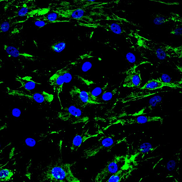
Immunocytochemistry/ Immunofluorescence: Caveolin-2 Antibody [NBP1-31116] -
Caveolin-2 antibody detects Caveolin-2 protein at membrane by immunofluorescent analysis.Sample: A431 cells were fixed in 4% paraformaldehyde at RT for 15 min.
Green: Caveolin-2 protein stained by Caveolin-2 antibody (NBP1-31116) diluted at 1:500.
Blue: Hoechst 33342 staining.

Western Blot: Caveolin-2 Antibody - BSA Free [NBP1-31116] -
The S100 protein family affects HNSCC metastasis and is regulated by CAV2.A The expression of S100 family proteins in CAV2-control and CAV2-knockdown (shCAV2-1, shCAV2-2) SCC15 and SCC25 HNSCC cell lines was evaluated by immunoblotting. B The expression of S100 family proteins in CAV2-control and CAV2-knockdown (shCAV2-1, shCAV2-2) SCC15 cell lines was evaluated by RT–qPCR. C Alterations in the invasive ability of SCC15 and SCC25 cells following siRNA interference of S100 protein family members. The invasive ability of SCC15 and SCC25 cells was detected using Transwell assays. *, **, *** and **** indicate p < 0.05, p < 0.01, p < 0.001 and p < 0.0001, respectively. Image collected and cropped by CiteAb from the following open publication (https://pubmed.ncbi.nlm.nih.gov/36114176), licensed under a CC-BY license. Not internally tested by Novus Biologicals.
Knockdown Validated: Caveolin-2 Antibody - BSA Free [NBP1-31116] -
CAV2 promotes HNSCC metastasis both in vitro and in vivo.Silencing CAV2 in SCC15 and SCC25 cells by transfection with two individual CAV2 shRNAs significantly decreased CAV2 expression, as detected by western blotting (A) and QT-PCR (B). C, D The silencing of CAV2 significantly inhibited the migration and invasion of SCC15 and SCC25 cells, as evaluated using Transwell assays. (Scale bar, 200 μm). E Images of the macroscopic lung tissues of tail vein-injected mice. F The lung metastasis nodules were counted, and the data from the shCAV2 and negative control groups are summarized. ‘****’ indicates ‘p < 0.0001’. G CAV2 IHC staining and H&E staining to assess lung metastasis in tail vein-injected mice. (Scale bars, 200 μm and 100 μm). Image collected and cropped by CiteAb from the following open publication (https://pubmed.ncbi.nlm.nih.gov/36114176), licensed under a CC-BY license. Not internally tested by Novus Biologicals.
Immunohistochemistry: Caveolin-2 Antibody - BSA Free [NBP1-31116] -
High expression of CAV2 is associated with the poor prognosis of HNSCC patients.A IHC analysis of CAV2 levels in 211 human HNSCC samples (scale bar, 100 μm or 400 μm). B Kaplan–Meier survival curve showing the correlation of overall survival and disease-free survival with the CAV2 IHC score. Image collected and cropped by CiteAb from the following open publication (https://pubmed.ncbi.nlm.nih.gov/36114176), licensed under a CC-BY license. Not internally tested by Novus Biologicals.
Immunohistochemistry: Caveolin-2 Antibody - BSA Free [NBP1-31116] -
CAV2 promotes HNSCC metastasis both in vitro and in vivo.Silencing CAV2 in SCC15 and SCC25 cells by transfection with two individual CAV2 shRNAs significantly decreased CAV2 expression, as detected by western blotting (A) and QT-PCR (B). C, D The silencing of CAV2 significantly inhibited the migration and invasion of SCC15 and SCC25 cells, as evaluated using Transwell assays. (Scale bar, 200 μm). E Images of the macroscopic lung tissues of tail vein-injected mice. F The lung metastasis nodules were counted, and the data from the shCAV2 and negative control groups are summarized. ‘****’ indicates ‘p < 0.0001’. G CAV2 IHC staining and H&E staining to assess lung metastasis in tail vein-injected mice. (Scale bars, 200 μm and 100 μm). Image collected and cropped by CiteAb from the following open publication (https://pubmed.ncbi.nlm.nih.gov/36114176), licensed under a CC-BY license. Not internally tested by Novus Biologicals.Applications for Caveolin-2 Antibody - BSA Free
Application
Recommended Usage
Immunocytochemistry/ Immunofluorescence
1:100-1:1000
Immunohistochemistry
1:100-1:1000
Immunohistochemistry-Paraffin
1:100-1:1000
Immunoprecipitation
1:100-1:500
Western Blot
1:500-1:3000
Reviewed Applications
Read 1 review rated 5 using NBP1-31116 in the following applications:
Formulation, Preparation, and Storage
Purification
Antigen Affinity-purified
Formulation
0.1M Tris, 0.1M Glycine, 10% Glycerol
Format
BSA Free
Preservative
0.01% Thimerosal
Concentration
Concentrations vary lot to lot. See vial label for concentration. If unlisted please contact technical services.
Shipping
The product is shipped with polar packs. Upon receipt, store it immediately at the temperature recommended below.
Stability & Storage
Aliquot and store at -20C or -80C. Avoid freeze-thaw cycles.
Background: Caveolin-2
Alternate Names
CAV2, Caveolin2
Gene Symbol
CAV2
Additional Caveolin-2 Products
Product Documents for Caveolin-2 Antibody - BSA Free
Certificate of Analysis
To download a Certificate of Analysis, please enter a lot or batch number in the search box below.
Product Specific Notices for Caveolin-2 Antibody - BSA Free
This product is for research use only and is not approved for use in humans or in clinical diagnosis. Primary Antibodies are guaranteed for 1 year from date of receipt.
⚠ WARNING: This product can expose you to chemicals including mercury, which is known to the State of California to cause reproductive toxicity with developmental effects. For more information go to www.P65Warnings.ca.gov.Related Research Areas
Citations for Caveolin-2 Antibody - BSA Free
 Powered by Bioz
Customer Reviews for Caveolin-2 Antibody - BSA Free (1)
5 out of 5
1 Customer Rating
Have you used Caveolin-2 Antibody - BSA Free?
Submit a review and receive an Amazon gift card!
$25/€18/£15/$25CAN/¥2500 Yen for a review with an image
$10/€7/£6/$10CAN/¥1110 Yen for a review without an image
Submit a review
Customer Images
Showing
1
-
1 of
1 review
Showing All
Filter By:
-
Application: ImmunocytochemistrySample Tested: abecular meshwork cellsSpecies: PigVerified Customer | Posted 11/28/2016Detection of caveolin2 in cultured pig trabecular meshwork cells using polyclonal Caveolin-2 Antibody (NBP1-31116) at 1:500 dilution overnight and donkey anti-rabbit Alexa 488 (1:1000) for 1 hour.

There are no reviews that match your criteria.
Protocols
Find general support by application which include: protocols, troubleshooting, illustrated assays, videos and webinars.
- Antigen Retrieval Protocol (PIER)
- Antigen Retrieval for Frozen Sections Protocol
- Appropriate Fixation of IHC/ICC Samples
- Cellular Response to Hypoxia Protocols
- Chromogenic IHC Staining of Formalin-Fixed Paraffin-Embedded (FFPE) Tissue Protocol
- Chromogenic Immunohistochemistry Staining of Frozen Tissue
- ClariTSA™ Fluorophore Kits
- Detection & Visualization of Antibody Binding
- Fluorescent IHC Staining of Frozen Tissue Protocol
- Graphic Protocol for Heat-induced Epitope Retrieval
- Graphic Protocol for the Preparation and Fluorescent IHC Staining of Frozen Tissue Sections
- Graphic Protocol for the Preparation and Fluorescent IHC Staining of Paraffin-embedded Tissue Sections
- Graphic Protocol for the Preparation of Gelatin-coated Slides for Histological Tissue Sections
- ICC Cell Smear Protocol for Suspension Cells
- ICC Immunocytochemistry Protocol Videos
- ICC for Adherent Cells
- IHC Sample Preparation (Frozen sections vs Paraffin)
- Immunocytochemistry (ICC) Protocol
- Immunocytochemistry Troubleshooting
- Immunofluorescence of Organoids Embedded in Cultrex Basement Membrane Extract
- Immunofluorescent IHC Staining of Formalin-Fixed Paraffin-Embedded (FFPE) Tissue Protocol
- Immunohistochemistry (IHC) and Immunocytochemistry (ICC) Protocols
- Immunohistochemistry Frozen Troubleshooting
- Immunohistochemistry Paraffin Troubleshooting
- Immunoprecipitation Protocol
- Preparing Samples for IHC/ICC Experiments
- Preventing Non-Specific Staining (Non-Specific Binding)
- Primary Antibody Selection & Optimization
- Protocol for Heat-Induced Epitope Retrieval (HIER)
- Protocol for Making a 4% Formaldehyde Solution in PBS
- Protocol for VisUCyte™ HRP Polymer Detection Reagent
- Protocol for the Fluorescent ICC Staining of Cell Smears - Graphic
- Protocol for the Fluorescent ICC Staining of Cultured Cells on Coverslips - Graphic
- Protocol for the Preparation & Fixation of Cells on Coverslips
- Protocol for the Preparation and Chromogenic IHC Staining of Frozen Tissue Sections
- Protocol for the Preparation and Chromogenic IHC Staining of Frozen Tissue Sections - Graphic
- Protocol for the Preparation and Chromogenic IHC Staining of Paraffin-embedded Tissue Sections
- Protocol for the Preparation and Chromogenic IHC Staining of Paraffin-embedded Tissue Sections - Graphic
- Protocol for the Preparation and Fluorescent ICC Staining of Cells on Coverslips
- Protocol for the Preparation and Fluorescent ICC Staining of Non-adherent Cells
- Protocol for the Preparation and Fluorescent ICC Staining of Stem Cells on Coverslips
- Protocol for the Preparation and Fluorescent IHC Staining of Frozen Tissue Sections
- Protocol for the Preparation and Fluorescent IHC Staining of Paraffin-embedded Tissue Sections
- Protocol for the Preparation of Gelatin-coated Slides for Histological Tissue Sections
- Protocol for the Preparation of a Cell Smear for Non-adherent Cell ICC - Graphic
- R&D Systems Quality Control Western Blot Protocol
- TUNEL and Active Caspase-3 Detection by IHC/ICC Protocol
- The Importance of IHC/ICC Controls
- Troubleshooting Guide: Immunohistochemistry
- Troubleshooting Guide: Western Blot Figures
- Western Blot Conditions
- Western Blot Protocol
- Western Blot Protocol for Cell Lysates
- Western Blot Troubleshooting
- Western Blot Troubleshooting Guide
- View all Protocols, Troubleshooting, Illustrated assays and Webinars
Loading...