CD11 Antibody (BLR107H)
Novus Biologicals | Catalog # NBP3-14697
Recombinant Monoclonal Antibody
Loading...
Key Product Details
Species Reactivity
Human
Applications
Multiplex Immunofluorescence, Immunohistochemistry, Western Blot, Immunocytochemistry/ Immunofluorescence, Immunoprecipitation
Label
Unconjugated
Antibody Source
Recombinant Monoclonal Rabbit Clone # BLR107H
Loading...
Product Specifications
Immunogen
residues 17-334 (ECD)
Clonality
Monoclonal
Host
Rabbit
Scientific Data Images for CD11 Antibody (BLR107H)
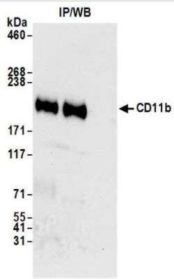
CD11 Antibody [NBP3-14697] - : Whole cell lysate (1.0 mgper IP reaction; 20% of IP loaded) from MUTZ-3 cellsprepared using NETN lysis buffer. Antibodies: Rabbit antiCD11b recombinant monoclonal antibody used for IP at 20 ul/mg lysate. CD11bwas also immunoprecipitated by rabbit anti-CD11bantibody. For blotting immunoprecipitatedCD11b, was used at 1:1000.Chemiluminescence with an exposure time of 10 seconds.
Applications for CD11 Antibody (BLR107H)
Application
Recommended Usage
Immunocytochemistry/ Immunofluorescence
Optimal dilutions of this antibody should be experimentally determined.
Immunohistochemistry
Optimal dilutions of this antibody should be experimentally determined.
Immunoprecipitation
Optimal dilutions of this antibody should be experimentally determined.
Multiplex Immunofluorescence
Optimal dilutions of this antibody should be experimentally determined.
Western Blot
Optimal dilutions of this antibody should be experimentally determined.
Formulation, Preparation, and Storage
Purification
>95%
Formulation
Borate Buffered Saline (BBS) pH 8.2 with 0.1% BSA and
Preservative
0.09% Sodium Azide
Concentration
Please see the vial label for concentration. If unlisted please contact technical services.
Shipping
The product is shipped with polar packs. Upon receipt, store it immediately at the temperature recommended below.
Stability & Storage
Store at 2 - 8 C / 1 year from date of receipt
Background: CD11
Alternate Names
antigen CD11b (p170), CD11 antigen-like family member B, CD11b, CD11b antigen, CD11B integrin, alpha M (complement component receptor 3, alpha; also known as CD11b(p170), macrophage antigen alpha polypeptide), Cell surface glycoprotein MAC-1 subunit alpha, CR-3 alpha chain, CR3AMGC117044, integrin alpha-M, integrin, alpha M (complement component 3 receptor 3 subunit), Leukocyte adhesion receptor MO1, MAC-1, MAC1A, macrophage antigen alpha polypeptide, MO1A, Neutrophil adherence receptor, neutrophil adherence receptor alpha-M subunit, SLEB6
Gene Symbol
ITGAM
Additional CD11 Products
Product Documents for CD11 Antibody (BLR107H)
Certificate of Analysis
To download a Certificate of Analysis, please enter a lot or batch number in the search box below.
Product Specific Notices for CD11 Antibody (BLR107H)
This product is for research use only and is not approved for use in humans or in clinical diagnosis. Primary Antibodies are guaranteed for 1 year from date of receipt.
Customer Reviews for CD11 Antibody (BLR107H)
There are currently no reviews for this product. Be the first to review CD11 Antibody (BLR107H) and earn rewards!
Have you used CD11 Antibody (BLR107H)?
Submit a review and receive an Amazon gift card!
$25/€18/£15/$25CAN/¥2500 Yen for a review with an image
$10/€7/£6/$10CAN/¥1110 Yen for a review without an image
Submit a review
Protocols
Find general support by application which include: protocols, troubleshooting, illustrated assays, videos and webinars.
- Antigen Retrieval Protocol (PIER)
- Antigen Retrieval for Frozen Sections Protocol
- Appropriate Fixation of IHC/ICC Samples
- Cellular Response to Hypoxia Protocols
- Chromogenic IHC Staining of Formalin-Fixed Paraffin-Embedded (FFPE) Tissue Protocol
- Chromogenic Immunohistochemistry Staining of Frozen Tissue
- ClariTSA™ Fluorophore Kits
- Detection & Visualization of Antibody Binding
- Fluorescent IHC Staining of Frozen Tissue Protocol
- Graphic Protocol for Heat-induced Epitope Retrieval
- Graphic Protocol for the Preparation and Fluorescent IHC Staining of Frozen Tissue Sections
- Graphic Protocol for the Preparation and Fluorescent IHC Staining of Paraffin-embedded Tissue Sections
- Graphic Protocol for the Preparation of Gelatin-coated Slides for Histological Tissue Sections
- ICC Cell Smear Protocol for Suspension Cells
- ICC Immunocytochemistry Protocol Videos
- ICC for Adherent Cells
- IHC Sample Preparation (Frozen sections vs Paraffin)
- Immunocytochemistry (ICC) Protocol
- Immunocytochemistry Troubleshooting
- Immunofluorescence of Organoids Embedded in Cultrex Basement Membrane Extract
- Immunofluorescent IHC Staining of Formalin-Fixed Paraffin-Embedded (FFPE) Tissue Protocol
- Immunohistochemistry (IHC) and Immunocytochemistry (ICC) Protocols
- Immunohistochemistry Frozen Troubleshooting
- Immunohistochemistry Paraffin Troubleshooting
- Immunoprecipitation Protocol
- Preparing Samples for IHC/ICC Experiments
- Preventing Non-Specific Staining (Non-Specific Binding)
- Primary Antibody Selection & Optimization
- Protocol for Heat-Induced Epitope Retrieval (HIER)
- Protocol for Making a 4% Formaldehyde Solution in PBS
- Protocol for VisUCyte™ HRP Polymer Detection Reagent
- Protocol for the Fluorescent ICC Staining of Cell Smears - Graphic
- Protocol for the Fluorescent ICC Staining of Cultured Cells on Coverslips - Graphic
- Protocol for the Preparation & Fixation of Cells on Coverslips
- Protocol for the Preparation and Chromogenic IHC Staining of Frozen Tissue Sections
- Protocol for the Preparation and Chromogenic IHC Staining of Frozen Tissue Sections - Graphic
- Protocol for the Preparation and Chromogenic IHC Staining of Paraffin-embedded Tissue Sections
- Protocol for the Preparation and Chromogenic IHC Staining of Paraffin-embedded Tissue Sections - Graphic
- Protocol for the Preparation and Fluorescent ICC Staining of Cells on Coverslips
- Protocol for the Preparation and Fluorescent ICC Staining of Non-adherent Cells
- Protocol for the Preparation and Fluorescent ICC Staining of Stem Cells on Coverslips
- Protocol for the Preparation and Fluorescent IHC Staining of Frozen Tissue Sections
- Protocol for the Preparation and Fluorescent IHC Staining of Paraffin-embedded Tissue Sections
- Protocol for the Preparation of Gelatin-coated Slides for Histological Tissue Sections
- Protocol for the Preparation of a Cell Smear for Non-adherent Cell ICC - Graphic
- R&D Systems Quality Control Western Blot Protocol
- TUNEL and Active Caspase-3 Detection by IHC/ICC Protocol
- The Importance of IHC/ICC Controls
- Troubleshooting Guide: Immunohistochemistry
- Troubleshooting Guide: Western Blot Figures
- Western Blot Conditions
- Western Blot Protocol
- Western Blot Protocol for Cell Lysates
- Western Blot Troubleshooting
- Western Blot Troubleshooting Guide
- View all Protocols, Troubleshooting, Illustrated assays and Webinars
Loading...