CD1a Antibody (NA1/34-HLK) - BSA Free
Novus Biologicals | Catalog # NB100-64852
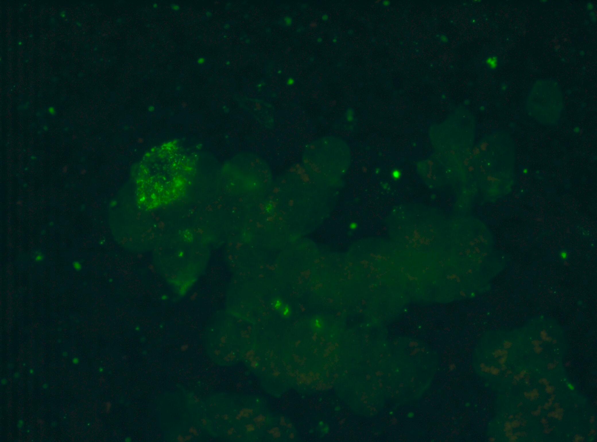
![Immunohistochemistry-Frozen: CD1a Antibody (NA1/34-HLK) - BSA Free [NB100-64852]](https://resources.rndsystems.com/images/products/CD1a-Antibody-NA1-34-HLK-Immunohistochemistry-Frozen-NB100-64852-img0007.jpg "Immunohistochemistry-Frozen: CD1a Antibody (NA1/34-HLK) - BSA Free [NB100-64852]")
Key Product Details
Species Reactivity
Validated:
Human
Cited:
Canine
Applications
Validated:
Immunohistochemistry, Immunohistochemistry-Frozen, Flow Cytometry, Immunofluorescence
Cited:
Flow Cytometry
Label
Unconjugated
Antibody Source
Monoclonal Mouse IgG2A Clone # NA1/34-HLK
Format
BSA Free
Loading...
Product Specifications
Immunogen
Human thymocytes
Reactivity Notes
Predicted cross-reactivities: Cynomolgus monkey, Dog
Specificity
NB100-64852 recognizes the human CD1a cell surface glycoprotein, a 49kD molecule expressed in association with beta 2 microglobulin. CD1a is expressed strongly by cortical thymocytes, and also by Langerhans cells and interdigitating cells.
Clonality
Monoclonal
Host
Mouse
Isotype
IgG2A
Scientific Data Images for CD1a Antibody (NA1/34-HLK) - BSA Free
Immunohistochemistry-Frozen: CD1a Antibody (NA1/34-HLK) - BSA Free [NB100-64852]
Immunohistochemistry-Frozen: CD1a Antibody (NA1/34-HLK) [NB100-64852] - Immunoperoxidase staining of human tonsil cryosection using Mouse anti Human CD1a antibody followed by HISTAR detection system. Medium power![Flow Cytometry: CD1a Antibody (NA1/34-HLK) - BSA Free [NB100-64852]](https://resources.rndsystems.com/images/products/CD1a-Antibody-NA1-34-HLK-Flow-Cytometry-NB100-64852-img0005.jpg "Flow Cytometry: CD1a Antibody (NA1/34-HLK) - BSA Free [NB100-64852]")
Flow Cytometry: CD1a Antibody (NA1/34-HLK) - BSA Free [NB100-64852]
Flow Cytometry: CD1a Antibody (NA1/34-HLK) [NB100-64852] - Analysis using the FITC conjugate of NB100-64852. Staining of MOLT 4 cells.![Immunohistochemistry-Frozen: CD1a Antibody (NA1/34-HLK) - BSA Free [NB100-64852]](https://resources.rndsystems.com/images/products/CD1a-Antibody-NA1-34-HLK-Immunohistochemistry-Frozen-NB100-64852-img0006.jpg "Immunohistochemistry-Frozen: CD1a Antibody (NA1/34-HLK) - BSA Free [NB100-64852]")
Immunohistochemistry-Frozen: CD1a Antibody (NA1/34-HLK) - BSA Free [NB100-64852]
Immunohistochemistry-Frozen: CD1a Antibody (NA1/34-HLK) [NB100-64852] - mmunoperoxidase staining of human tonsil cryosection using Mouse anti Human CD1a antibody followed by HISTAR detection system. Low power![Flow Cytometry: CD1a Antibody (NA1/34-HLK) - BSA Free [NB100-64852]](https://resources.rndsystems.com/images/products/CD1a-Antibody-NA1-34-HLK-Flow-Cytometry-NB100-64852-img0004.jpg "Flow Cytometry: CD1a Antibody (NA1/34-HLK) - BSA Free [NB100-64852]")
Flow Cytometry: CD1a Antibody (NA1/34-HLK) - BSA Free [NB100-64852]
Flow Cytometry: CD1a Antibody (NA1/34-HLK) [NB100-64852] - Analysis using the Biotin conjugate of NB100-64852. Staining of MOLT 4 cells with CD1a antibody.Applications for CD1a Antibody (NA1/34-HLK) - BSA Free
Application
Recommended Usage
Flow Cytometry
1:50-1:100
Immunohistochemistry
1:10-1:500
Immunohistochemistry-Frozen
1:10-1:500
Reviewed Applications
Read 1 review rated 5 using NB100-64852 in the following applications:
Flow Cytometry Panel Builder
Bio-Techne Knows Flow Cytometry
Save time and reduce costly mistakes by quickly finding compatible reagents using the Panel Builder Tool.
Advanced Features
- Spectra Viewer - Custom analysis of spectra from multiple fluorochromes
- Spillover Popups - Visualize the spectra of individual fluorochromes
- Antigen Density Selector - Match fluorochrome brightness with antigen density

Formulation, Preparation, and Storage
Purification
Protein A purified
Formulation
PBS
Format
BSA Free
Preservative
0.09% Sodium Azide
Concentration
1.0 mg/ml
Shipping
The product is shipped with polar packs. Upon receipt, store it immediately at the temperature recommended below.
Stability & Storage
Store at 4C short term. Aliquot and store at -20C long term. Avoid freeze-thaw cycles.
Background: CD1a
Long Name
T Cell Surface Glycoprotein CD1a
Alternate Names
CD1a, FCB6, HTA1, R4, T6
Gene Symbol
CD1A
UniProt
Additional CD1a Products
Product Documents for CD1a Antibody (NA1/34-HLK) - BSA Free
Certificate of Analysis
To download a Certificate of Analysis, please enter a lot or batch number in the search box below.
Product Specific Notices for CD1a Antibody (NA1/34-HLK) - BSA Free
This product is for research use only and is not approved for use in humans or in clinical diagnosis. Primary Antibodies are guaranteed for 1 year from date of receipt.
Citations for CD1a Antibody (NA1/34-HLK) - BSA Free
 Powered by Bioz
Customer Reviews for CD1a Antibody (NA1/34-HLK) - BSA Free (1)
5 out of 5
1 Customer Rating
Have you used CD1a Antibody (NA1/34-HLK) - BSA Free?
Submit a review and receive an Amazon gift card!
$25/€18/£15/$25CAN/¥2500 Yen for a review with an image
$10/€7/£6/$10CAN/¥1110 Yen for a review without an image
Submit a review
Customer Images
Showing
1
-
1 of
1 review
Showing All
Filter By:
-
Application: ImmunocytochemistrySample Tested: Dog blood cells on slideSpecies: CanineVerified Customer | Posted 06/13/2017ICC on canine blood cells.

There are no reviews that match your criteria.
Protocols
Find general support by application which include: protocols, troubleshooting, illustrated assays, videos and webinars.
- 7-Amino Actinomycin D (7-AAD) Cell Viability Flow Cytometry Protocol
- Antigen Retrieval Protocol (PIER)
- Antigen Retrieval for Frozen Sections Protocol
- Appropriate Fixation of IHC/ICC Samples
- Cellular Response to Hypoxia Protocols
- Chromogenic IHC Staining of Formalin-Fixed Paraffin-Embedded (FFPE) Tissue Protocol
- Chromogenic Immunohistochemistry Staining of Frozen Tissue
- ClariTSA™ Fluorophore Kits
- Detection & Visualization of Antibody Binding
- Extracellular Membrane Flow Cytometry Protocol
- Flow Cytometry Protocol for Cell Surface Markers
- Flow Cytometry Protocol for Staining Membrane Associated Proteins
- Flow Cytometry Staining Protocols
- Flow Cytometry Troubleshooting Guide
- Fluorescent IHC Staining of Frozen Tissue Protocol
- Graphic Protocol for Heat-induced Epitope Retrieval
- Graphic Protocol for the Preparation and Fluorescent IHC Staining of Frozen Tissue Sections
- Graphic Protocol for the Preparation and Fluorescent IHC Staining of Paraffin-embedded Tissue Sections
- Graphic Protocol for the Preparation of Gelatin-coated Slides for Histological Tissue Sections
- ICC Cell Smear Protocol for Suspension Cells
- ICC Immunocytochemistry Protocol Videos
- ICC for Adherent Cells
- IHC Sample Preparation (Frozen sections vs Paraffin)
- Immunocytochemistry (ICC) Protocol
- Immunocytochemistry Troubleshooting
- Immunofluorescence of Organoids Embedded in Cultrex Basement Membrane Extract
- Immunofluorescent IHC Staining of Formalin-Fixed Paraffin-Embedded (FFPE) Tissue Protocol
- Immunohistochemistry (IHC) and Immunocytochemistry (ICC) Protocols
- Immunohistochemistry Frozen Troubleshooting
- Immunohistochemistry Paraffin Troubleshooting
- Intracellular Flow Cytometry Protocol Using Alcohol (Methanol)
- Intracellular Flow Cytometry Protocol Using Detergents
- Intracellular Nuclear Staining Flow Cytometry Protocol Using Detergents
- Intracellular Staining Flow Cytometry Protocol Using Alcohol Permeabilization
- Intracellular Staining Flow Cytometry Protocol Using Detergents to Permeabilize Cells
- Preparing Samples for IHC/ICC Experiments
- Preventing Non-Specific Staining (Non-Specific Binding)
- Primary Antibody Selection & Optimization
- Propidium Iodide Cell Viability Flow Cytometry Protocol
- Protocol for Heat-Induced Epitope Retrieval (HIER)
- Protocol for Liperfluo
- Protocol for Making a 4% Formaldehyde Solution in PBS
- Protocol for VisUCyte™ HRP Polymer Detection Reagent
- Protocol for the Characterization of Human Th22 Cells
- Protocol for the Characterization of Human Th9 Cells
- Protocol for the Fluorescent ICC Staining of Cell Smears - Graphic
- Protocol for the Fluorescent ICC Staining of Cultured Cells on Coverslips - Graphic
- Protocol for the Preparation & Fixation of Cells on Coverslips
- Protocol for the Preparation and Chromogenic IHC Staining of Frozen Tissue Sections
- Protocol for the Preparation and Chromogenic IHC Staining of Frozen Tissue Sections - Graphic
- Protocol for the Preparation and Chromogenic IHC Staining of Paraffin-embedded Tissue Sections
- Protocol for the Preparation and Chromogenic IHC Staining of Paraffin-embedded Tissue Sections - Graphic
- Protocol for the Preparation and Fluorescent ICC Staining of Cells on Coverslips
- Protocol for the Preparation and Fluorescent ICC Staining of Non-adherent Cells
- Protocol for the Preparation and Fluorescent ICC Staining of Stem Cells on Coverslips
- Protocol for the Preparation and Fluorescent IHC Staining of Frozen Tissue Sections
- Protocol for the Preparation and Fluorescent IHC Staining of Paraffin-embedded Tissue Sections
- Protocol for the Preparation of Gelatin-coated Slides for Histological Tissue Sections
- Protocol for the Preparation of a Cell Smear for Non-adherent Cell ICC - Graphic
- Protocol: Annexin V and PI Staining by Flow Cytometry
- Protocol: Annexin V and PI Staining for Apoptosis by Flow Cytometry
- TUNEL and Active Caspase-3 Detection by IHC/ICC Protocol
- The Importance of IHC/ICC Controls
- Troubleshooting Guide: Fluorokine Flow Cytometry Kits
- Troubleshooting Guide: Immunohistochemistry
- View all Protocols, Troubleshooting, Illustrated assays and Webinars
Loading...
Associated Pathways
