![Immunocytochemistry/ Immunofluorescence: CD37 Antibody (2B8) - BSA Free [NBP1-28869]](https://resources.rndsystems.com/images/products/CD37-Antibody-2B8-Immunocytochemistry-NBP1-28869-img0003.jpg "Immunocytochemistry/ Immunofluorescence: CD37 Antibody (2B8) - BSA Free [NBP1-28869]")
Loading...
Key Product Details
Species Reactivity
Human, Mouse
Applications
Immunohistochemistry, Immunohistochemistry-Paraffin, ELISA, Flow Cytometry, Immunocytochemistry/ Immunofluorescence
Label
Unconjugated
Antibody Source
Monoclonal Mouse IgG1 Clone # 2B8
Loading...
Product Specifications
Immunogen
Purified recombinant fragment of CD37 expressed in E. Coli.
Reactivity Notes
Mouse blocking reagent may be needed for IHC and ICC experiments to reduce high background signal. You can find these reagents under catalog numbers PK-2200-NB and MP-2400-NB. Please contact Technical Support if you have any Please note that this antibody is reactive to Mouse and derived from the same host, Mouse. Mouse-On-questions.
Clonality
Monoclonal
Host
Mouse
Isotype
IgG1
Theoretical MW
31.7 kDa.
Disclaimer note: The observed molecular weight of the protein may vary from the listed predicted molecular weight due to post translational modifications, post translation cleavages, relative charges, and other experimental factors.
Disclaimer note: The observed molecular weight of the protein may vary from the listed predicted molecular weight due to post translational modifications, post translation cleavages, relative charges, and other experimental factors.
Scientific Data Images for CD37 Antibody (2B8)
Immunocytochemistry/ Immunofluorescence: CD37 Antibody (2B8) - BSA Free [NBP1-28869]
Immunocytochemistry/Immunofluorescence: CD37 Antibody (2B8) [NBP1-28869] - Confocal immunofluorescence analysis of methanol-fixed BCBL-1 (left) and L1210 (right) cells using CD37 mouse mAb(green), showing membrane localization. Blue: DRAQ5 fluorescent DNA dye.![Immunohistochemistry-Paraffin: CD37 Antibody (2B8) - BSA Free [NBP1-28869]](https://resources.rndsystems.com/images/products/CD37-Antibody-2B8-Immunohistochemistry-Paraffin-NBP1-28869-img0004.jpg "Immunohistochemistry-Paraffin: CD37 Antibody (2B8) - BSA Free [NBP1-28869]")
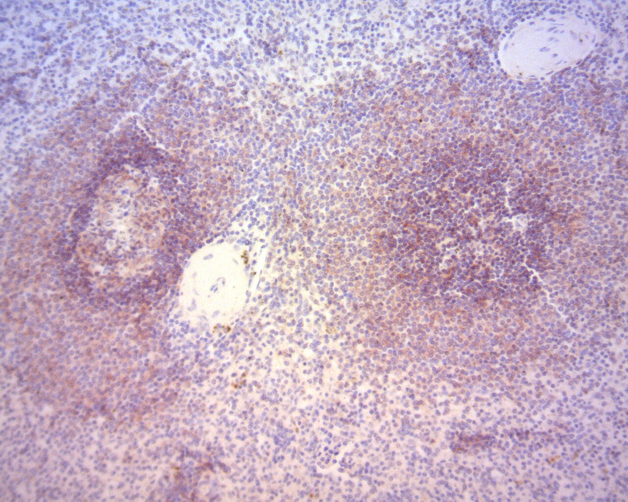
Immunohistochemistry-Paraffin: CD37 Antibody (2B8) - BSA Free [NBP1-28869]
Immunohistochemistry-Paraffin: CD37 Antibody (2B8) [NBP1-28869] - Immunohistochemistry staining of CD37 (NBP1-28869: 2B8) on human paraffin-embedded spleen tissues, developed with NovaRed HRP substrate. Epitope retrieval: HIER Citrate. This image was submitted via customer review.![Flow Cytometry: CD37 Antibody (2B8) - BSA Free [NBP1-28869]](https://resources.rndsystems.com/images/products/CD37-Antibody-2B8-Flow-Cytometry-NBP1-28869-img0005.jpg "Flow Cytometry: CD37 Antibody (2B8) - BSA Free [NBP1-28869]")
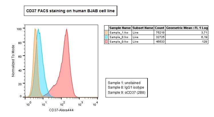
Flow Cytometry: CD37 Antibody (2B8) - BSA Free [NBP1-28869]
Flow Cytometry: CD37 Antibody (2B8) [NBP1-28869] - CD37 FACS staining on BJAB cells (1:200), stained with GaM IgG1-Alexa488 secondary antibody. This image was submtted via customer review.Applications for CD37 Antibody (2B8)
Application
Recommended Usage
ELISA
1:10000
Immunocytochemistry/ Immunofluorescence
1:200-1:1000
Application Notes
Flow Cytometry and Immunohistochemistry Paraffin reported in verified customer reviews.
Reviewed Applications
Read 2 reviews rated 5 using NBP1-28869 in the following applications:
Flow Cytometry Panel Builder
Bio-Techne Knows Flow Cytometry
Save time and reduce costly mistakes by quickly finding compatible reagents using the Panel Builder Tool.
Advanced Features
- Spectra Viewer - Custom analysis of spectra from multiple fluorochromes
- Spillover Popups - Visualize the spectra of individual fluorochromes
- Antigen Density Selector - Match fluorochrome brightness with antigen density

Formulation, Preparation, and Storage
Purification
Ascites
Formulation
Ascites
Preservative
0.03% Sodium Azide
Concentration
This product is unpurified. The exact concentration of antibody is not quantifiable.
Shipping
The product is shipped with polar packs. Upon receipt, store it immediately at the temperature recommended below.
Stability & Storage
Store at 4C short term. Aliquot and store at -20C long term. Avoid freeze-thaw cycles.
Background: CD37
Alternate Names
CD37, TSPAN26
Gene Symbol
CD37
Additional CD37 Products
Product Documents for CD37 Antibody (2B8)
Certificate of Analysis
To download a Certificate of Analysis, please enter a lot or batch number in the search box below.
Product Specific Notices for CD37 Antibody (2B8)
This product is for research use only and is not approved for use in humans or in clinical diagnosis. Primary Antibodies are guaranteed for 1 year from date of receipt.
Related Research Areas
Customer Reviews for CD37 Antibody (2B8) (2)
5 out of 5
2 Customer Ratings
Have you used CD37 Antibody (2B8)?
Submit a review and receive an Amazon gift card!
$25/€18/£15/$25CAN/¥2500 Yen for a review with an image
$10/€7/£6/$10CAN/¥1110 Yen for a review without an image
Submit a review
Customer Images
Showing
1
-
2 of
2 reviews
Showing All
Filter By:
-
Application: Flow CytometrySample Tested: BJAB human Burkitt's lymphoma cell lineSpecies: HumanVerified Customer | Posted 03/13/2018CD37 FACS staining on BJAB cells (1:200), stained with GaM IgG1-Alexa488 secondary antibody.

-
Application: Immunohistochemistry-ParaffinSample Tested: Human paraffin embedded spleen tissueSpecies: HumanVerified Customer | Posted 03/13/2018Immunohistochemistry staining of CD37 (NBP1-28869: 2B8) on human paraffin-embedded spleen tissues, developed with NovaRed HRP substrate. Epitope retrieval: HIER Citrate.Epitope retrieval: HIER Citrate
There are no reviews that match your criteria.
Protocols
Find general support by application which include: protocols, troubleshooting, illustrated assays, videos and webinars.
- 7-Amino Actinomycin D (7-AAD) Cell Viability Flow Cytometry Protocol
- Antigen Retrieval Protocol (PIER)
- Antigen Retrieval for Frozen Sections Protocol
- Appropriate Fixation of IHC/ICC Samples
- Cellular Response to Hypoxia Protocols
- Chromogenic IHC Staining of Formalin-Fixed Paraffin-Embedded (FFPE) Tissue Protocol
- Chromogenic Immunohistochemistry Staining of Frozen Tissue
- ClariTSA™ Fluorophore Kits
- Detection & Visualization of Antibody Binding
- ELISA Sample Preparation & Collection Guide
- ELISA Troubleshooting Guide
- Extracellular Membrane Flow Cytometry Protocol
- Flow Cytometry Protocol for Cell Surface Markers
- Flow Cytometry Protocol for Staining Membrane Associated Proteins
- Flow Cytometry Staining Protocols
- Flow Cytometry Troubleshooting Guide
- Fluorescent IHC Staining of Frozen Tissue Protocol
- Graphic Protocol for Heat-induced Epitope Retrieval
- Graphic Protocol for the Preparation and Fluorescent IHC Staining of Frozen Tissue Sections
- Graphic Protocol for the Preparation and Fluorescent IHC Staining of Paraffin-embedded Tissue Sections
- Graphic Protocol for the Preparation of Gelatin-coated Slides for Histological Tissue Sections
- How to Run an R&D Systems DuoSet ELISA
- How to Run an R&D Systems Quantikine ELISA
- How to Run an R&D Systems Quantikine™ QuicKit™ ELISA
- ICC Cell Smear Protocol for Suspension Cells
- ICC Immunocytochemistry Protocol Videos
- ICC for Adherent Cells
- IHC Sample Preparation (Frozen sections vs Paraffin)
- Immunocytochemistry (ICC) Protocol
- Immunocytochemistry Troubleshooting
- Immunofluorescence of Organoids Embedded in Cultrex Basement Membrane Extract
- Immunofluorescent IHC Staining of Formalin-Fixed Paraffin-Embedded (FFPE) Tissue Protocol
- Immunohistochemistry (IHC) and Immunocytochemistry (ICC) Protocols
- Immunohistochemistry Frozen Troubleshooting
- Immunohistochemistry Paraffin Troubleshooting
- Intracellular Flow Cytometry Protocol Using Alcohol (Methanol)
- Intracellular Flow Cytometry Protocol Using Detergents
- Intracellular Nuclear Staining Flow Cytometry Protocol Using Detergents
- Intracellular Staining Flow Cytometry Protocol Using Alcohol Permeabilization
- Intracellular Staining Flow Cytometry Protocol Using Detergents to Permeabilize Cells
- Preparing Samples for IHC/ICC Experiments
- Preventing Non-Specific Staining (Non-Specific Binding)
- Primary Antibody Selection & Optimization
- Propidium Iodide Cell Viability Flow Cytometry Protocol
- Protocol for Heat-Induced Epitope Retrieval (HIER)
- Protocol for Liperfluo
- Protocol for Making a 4% Formaldehyde Solution in PBS
- Protocol for VisUCyte™ HRP Polymer Detection Reagent
- Protocol for the Characterization of Human Th22 Cells
- Protocol for the Characterization of Human Th9 Cells
- Protocol for the Fluorescent ICC Staining of Cell Smears - Graphic
- Protocol for the Fluorescent ICC Staining of Cultured Cells on Coverslips - Graphic
- Protocol for the Preparation & Fixation of Cells on Coverslips
- Protocol for the Preparation and Chromogenic IHC Staining of Frozen Tissue Sections
- Protocol for the Preparation and Chromogenic IHC Staining of Frozen Tissue Sections - Graphic
- Protocol for the Preparation and Chromogenic IHC Staining of Paraffin-embedded Tissue Sections
- Protocol for the Preparation and Chromogenic IHC Staining of Paraffin-embedded Tissue Sections - Graphic
- Protocol for the Preparation and Fluorescent ICC Staining of Cells on Coverslips
- Protocol for the Preparation and Fluorescent ICC Staining of Non-adherent Cells
- Protocol for the Preparation and Fluorescent ICC Staining of Stem Cells on Coverslips
- Protocol for the Preparation and Fluorescent IHC Staining of Frozen Tissue Sections
- Protocol for the Preparation and Fluorescent IHC Staining of Paraffin-embedded Tissue Sections
- Protocol for the Preparation of Gelatin-coated Slides for Histological Tissue Sections
- Protocol for the Preparation of a Cell Smear for Non-adherent Cell ICC - Graphic
- Protocol: Annexin V and PI Staining by Flow Cytometry
- Protocol: Annexin V and PI Staining for Apoptosis by Flow Cytometry
- Quantikine HS ELISA Kit Assay Principle, Alkaline Phosphatase
- Quantikine HS ELISA Kit Principle, Streptavidin-HRP Polymer
- Sandwich ELISA (Colorimetric) – Biotin/Streptavidin Detection Protocol
- Sandwich ELISA (Colorimetric) – Direct Detection Protocol
- TUNEL and Active Caspase-3 Detection by IHC/ICC Protocol
- The Importance of IHC/ICC Controls
- Troubleshooting Guide: ELISA
- Troubleshooting Guide: Fluorokine Flow Cytometry Kits
- Troubleshooting Guide: Immunohistochemistry
- View all Protocols, Troubleshooting, Illustrated assays and Webinars
Loading...