CD5 Antibody (C5/473 + CD5/54/F6) - Azide and BSA Free
Novus Biologicals | Catalog # NBP2-34583
Key Product Details
Species Reactivity
Human, Rat, Bovine, Rabbit
Applications
Immunohistochemistry, Immunohistochemistry-Paraffin, ELISA
Label
Unconjugated
Antibody Source
Monoclonal Mouse IgG1 Clone # C5/473 + CD5/54/F6
Format
Azide and BSA Free
Loading...
Product Specifications
Immunogen
Human CD5 recombinant protein (C5/473); A synthetic peptide from the intracellular region of human CD5 (CD5/54/F6)
Localization
Cell surface
Marker
Mantle Cell Lymphoma Marker
Specificity
Recognizes a 67kDa transmembrane protein, which is identified as CD5. The CD5 antigen is found on 95% of thymocytes and 72% of peripheral blood lymphocytes. In lymph nodes, the main reactivity is observed in T cell areas. Anti-CD5 is a pan T-cell marker that also reacts with a range of neoplastic B-cells, e.g. chronic lymphocytic leukemia/small lymphocytic lymphoma (CLL/SLL), mantle cell lymphoma, and a subset (~10%) of diffuse large B-cell lymphoma. CD5 aberrant expression is useful in making a diagnosis of mature T-cell neoplasms. Anti-CD5 detection is diagnostic in CLL/SLL within a panel of other B-cell markers, especially one that includes anti-CD23. Anti-CD5 is also very useful in differentiating among mature small lymphoid cell malignancies. In addition, anti-CD5 can be used in distinguishing thymic carcinoma (+) from thymoma (-). Anti-CD5 does not react with granulocytes or monocytes.
Clonality
Monoclonal
Host
Mouse
Isotype
IgG1
Theoretical MW
67 kDa.
Disclaimer note: The observed molecular weight of the protein may vary from the listed predicted molecular weight due to post translational modifications, post translation cleavages, relative charges, and other experimental factors.
Disclaimer note: The observed molecular weight of the protein may vary from the listed predicted molecular weight due to post translational modifications, post translation cleavages, relative charges, and other experimental factors.
Description
1.0 mg/ml of antibody purified from Bioreactor Concentrate by Protein A/G. Prepared in 10mM PBS WITHOUT BSA & azide. Also available at 200 ug/ml WITH BSA & azide (NBP2-34316).
Antibody with azide - store at 2 to 8C. Antibody without azide - store at -20 to -80C.
Antibody with azide - store at 2 to 8C. Antibody without azide - store at -20 to -80C.
Scientific Data Images for CD5 Antibody (C5/473 + CD5/54/F6) - Azide and BSA Free
![Immunohistochemistry-Paraffin: CD5 Antibody (C5/473 + CD5/54/F6) - Azide and BSA Free [NBP2-34583]](https://resources.rndsystems.com/images/products/CD5-Antibody-C5-473-+-CD5-54-F6-Azide-and-BSA-Free-Immunohistochemistry-Paraffin-NBP2-34583-img0002.jpg "Immunohistochemistry-Paraffin: CD5 Antibody (C5/473 + CD5/54/F6) - Azide and BSA Free [NBP2-34583]")
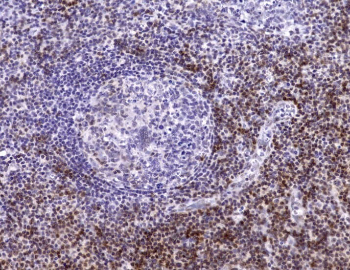
Immunohistochemistry-Paraffin: CD5 Antibody (C5/473 + CD5/54/F6) - Azide and BSA Free [NBP2-34583]
Immunohistochemistry-Paraffin: CD5 Antibody (C5/473 + CD5/54/F6) - Azide and BSA Free [NBP2-34583] - CD5 CODEX stain of human tonsil tissue. Image from verified customer review.![Immunohistochemistry-Paraffin: CD5 Antibody (C5/473 + CD5/54/F6) - Azide and BSA Free [NBP2-34583]](https://resources.rndsystems.com/images/products/CD5-Antibody-C5-473-+-CD5-54-F6-Azide-and-BSA-Free-Immunohistochemistry-Paraffin-NBP2-34583-img0001.jpg "Immunohistochemistry-Paraffin: CD5 Antibody (C5/473 + CD5/54/F6) - Azide and BSA Free [NBP2-34583]")
Immunohistochemistry-Paraffin: CD5 Antibody (C5/473 + CD5/54/F6) - Azide and BSA Free [NBP2-34583]
Immunohistochemistry-Paraffin: CD5 Antibody (C5/473 + CD5/54/F6) - Azide and BSA Free [NBP2-34583] - Formalin-fixed paraffin-embedded human tonsil stained with CD5 Monoclonal antibody (C5/473+CD5/54/F6)Applications for CD5 Antibody (C5/473 + CD5/54/F6) - Azide and BSA Free
Application
Recommended Usage
ELISA
1-2 ug/ml
Application Notes
ELISA: For coating use Ab at 1-2ug/ml, order Ab without BSA.
Immunohistochemistry (Formalin-fixed): 1-2ug/ml for 30 minutes at RT. Staining of formalin-fixed tissues requires heating tissue sections in 1mM EDTA, pH 7.5-8.5, for 45 min at 95C followed by cooling at RT for 20 minutes.
Optimal dilution for a specific application should be determined.
Immunohistochemistry (Formalin-fixed): 1-2ug/ml for 30 minutes at RT. Staining of formalin-fixed tissues requires heating tissue sections in 1mM EDTA, pH 7.5-8.5, for 45 min at 95C followed by cooling at RT for 20 minutes.
Optimal dilution for a specific application should be determined.
Reviewed Applications
Read 1 review rated 5 using NBP2-34583 in the following applications:
Formulation, Preparation, and Storage
Purification
Protein A or G purified
Formulation
10 mM PBS
Format
Azide and BSA Free
Preservative
No Preservative
Concentration
1.0 mg/ml
Shipping
The product is shipped with polar packs. Upon receipt, store it immediately at the temperature recommended below.
Stability & Storage
Store at -20 to -80C. Avoid freeze-thaw cycles.
Background: CD5
Alternate Names
CD5
Gene Symbol
CD5
Additional CD5 Products
Product Documents for CD5 Antibody (C5/473 + CD5/54/F6) - Azide and BSA Free
Certificate of Analysis
To download a Certificate of Analysis, please enter a lot or batch number in the search box below.
Product Specific Notices for CD5 Antibody (C5/473 + CD5/54/F6) - Azide and BSA Free
This product is for research use only and is not approved for use in humans or in clinical diagnosis. Primary Antibodies are guaranteed for 1 year from date of receipt.
Customer Reviews for CD5 Antibody (C5/473 + CD5/54/F6) - Azide and BSA Free (1)
5 out of 5
1 Customer Rating
Have you used CD5 Antibody (C5/473 + CD5/54/F6) - Azide and BSA Free?
Submit a review and receive an Amazon gift card!
$25/€18/£15/$25CAN/¥2500 Yen for a review with an image
$10/€7/£6/$10CAN/¥1110 Yen for a review without an image
Submit a review
Customer Images
Showing
1
-
1 of
1 review
Showing All
Filter By:
-
Application: Immunohistochemistry-ParaffinSample Tested: Human Tonsil tissueSpecies: HumanVerified Customer | Posted 10/25/2022CD5 CODEX stain of human tonsil tissue

There are no reviews that match your criteria.
Protocols
Find general support by application which include: protocols, troubleshooting, illustrated assays, videos and webinars.
- Antigen Retrieval Protocol (PIER)
- Antigen Retrieval for Frozen Sections Protocol
- Appropriate Fixation of IHC/ICC Samples
- Cellular Response to Hypoxia Protocols
- Chromogenic IHC Staining of Formalin-Fixed Paraffin-Embedded (FFPE) Tissue Protocol
- Chromogenic Immunohistochemistry Staining of Frozen Tissue
- ClariTSA™ Fluorophore Kits
- Detection & Visualization of Antibody Binding
- ELISA Sample Preparation & Collection Guide
- ELISA Troubleshooting Guide
- Fluorescent IHC Staining of Frozen Tissue Protocol
- Graphic Protocol for Heat-induced Epitope Retrieval
- Graphic Protocol for the Preparation and Fluorescent IHC Staining of Frozen Tissue Sections
- Graphic Protocol for the Preparation and Fluorescent IHC Staining of Paraffin-embedded Tissue Sections
- Graphic Protocol for the Preparation of Gelatin-coated Slides for Histological Tissue Sections
- How to Run an R&D Systems DuoSet ELISA
- How to Run an R&D Systems Quantikine ELISA
- How to Run an R&D Systems Quantikine™ QuicKit™ ELISA
- IHC Sample Preparation (Frozen sections vs Paraffin)
- Immunofluorescent IHC Staining of Formalin-Fixed Paraffin-Embedded (FFPE) Tissue Protocol
- Immunohistochemistry (IHC) and Immunocytochemistry (ICC) Protocols
- Immunohistochemistry Frozen Troubleshooting
- Immunohistochemistry Paraffin Troubleshooting
- Preparing Samples for IHC/ICC Experiments
- Preventing Non-Specific Staining (Non-Specific Binding)
- Primary Antibody Selection & Optimization
- Protocol for Heat-Induced Epitope Retrieval (HIER)
- Protocol for Making a 4% Formaldehyde Solution in PBS
- Protocol for VisUCyte™ HRP Polymer Detection Reagent
- Protocol for the Preparation & Fixation of Cells on Coverslips
- Protocol for the Preparation and Chromogenic IHC Staining of Frozen Tissue Sections
- Protocol for the Preparation and Chromogenic IHC Staining of Frozen Tissue Sections - Graphic
- Protocol for the Preparation and Chromogenic IHC Staining of Paraffin-embedded Tissue Sections
- Protocol for the Preparation and Chromogenic IHC Staining of Paraffin-embedded Tissue Sections - Graphic
- Protocol for the Preparation and Fluorescent IHC Staining of Frozen Tissue Sections
- Protocol for the Preparation and Fluorescent IHC Staining of Paraffin-embedded Tissue Sections
- Protocol for the Preparation of Gelatin-coated Slides for Histological Tissue Sections
- Quantikine HS ELISA Kit Assay Principle, Alkaline Phosphatase
- Quantikine HS ELISA Kit Principle, Streptavidin-HRP Polymer
- Sandwich ELISA (Colorimetric) – Biotin/Streptavidin Detection Protocol
- Sandwich ELISA (Colorimetric) – Direct Detection Protocol
- TUNEL and Active Caspase-3 Detection by IHC/ICC Protocol
- The Importance of IHC/ICC Controls
- Troubleshooting Guide: ELISA
- Troubleshooting Guide: Immunohistochemistry
- View all Protocols, Troubleshooting, Illustrated assays and Webinars
Loading...
Associated Pathways
