CD63 Antibody (MEM-259) - BSA Free
Novus Biologicals | Catalog # NB100-77913
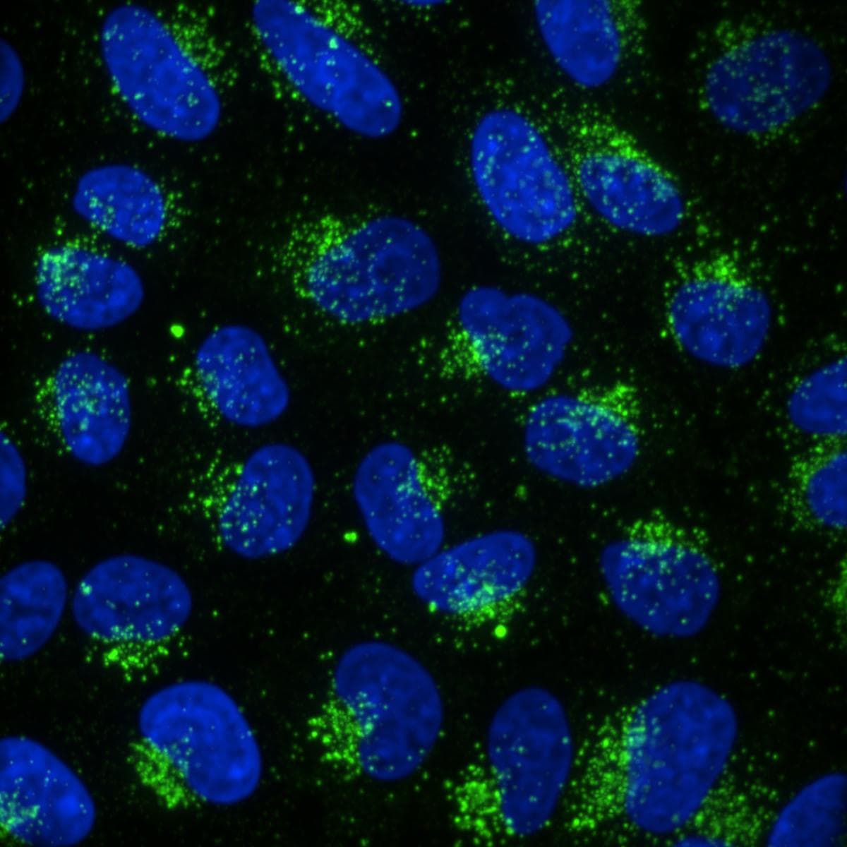
![Immunocytochemistry/ Immunofluorescence: CD63 Antibody (MEM-259) - BSA Free [NB100-77913]](https://resources.rndsystems.com/images/products/CD63-Antibody-MEM-259-Immunocytochemistry-Immunofluorescence-NB100-77913-img0015.jpg "Immunocytochemistry/ Immunofluorescence: CD63 Antibody (MEM-259) - BSA Free [NB100-77913]")
Key Product Details
Species Reactivity
Validated:
Human, Mouse, Rat
Cited:
Human, Rat
Applications
Validated:
Immunohistochemistry, Immunohistochemistry-Paraffin, Western Blot, Flow Cytometry, Immunocytochemistry/ Immunofluorescence, Immunoprecipitation, CyTOF-ready
Cited:
Immunohistochemistry, Western Blot, Flow Cytometry, Immunocytochemistry/ Immunofluorescence, IF/IHC
Label
Unconjugated
Antibody Source
Monoclonal Mouse IgG1 Clone # MEM-259
Format
BSA Free
Loading...
Product Specifications
Immunogen
HPB-ALL T cell line
Reactivity Notes
Use in Mouse and Rat reported in scientific literature (PMID:32111836).
Marker
Late Endosomes Marker
Specificity
The antibody MEM-259 reacts with an extracellular/luminal epitope of CD63 (LAMP-3), a 40-60 kDa tetraspan glycoprotein expressed by granulocytes, platelets, T-cells, monocytes/macrophages and endothelial cells. Cell surface exposition of CD63 is usually activation-dependent.
Clonality
Monoclonal
Host
Mouse
Isotype
IgG1
Theoretical MW
60 kDa.
Disclaimer note: The observed molecular weight of the protein may vary from the listed predicted molecular weight due to post translational modifications, post translation cleavages, relative charges, and other experimental factors.
Disclaimer note: The observed molecular weight of the protein may vary from the listed predicted molecular weight due to post translational modifications, post translation cleavages, relative charges, and other experimental factors.
Scientific Data Images for CD63 Antibody (MEM-259) - BSA Free
Immunocytochemistry/ Immunofluorescence: CD63 Antibody (MEM-259) - BSA Free [NB100-77913]
Immunocytochemistry/Immunofluorescence: CD63 Antibody (MEM-259) [NB100-77913] - Human HeLa cells. ICC/IF image submitted by a verified customer review.![Western Blot: CD63 Antibody (MEM-259)BSA Free [NB100-77913]](https://resources.rndsystems.com/images/products/CD63-Antibody-MEM-259-Western-Blot-NB100-77913-img0016.jpg "Western Blot: CD63 Antibody (MEM-259)BSA Free [NB100-77913]")
Western Blot: CD63 Antibody (MEM-259)BSA Free [NB100-77913]
CD63-Antibody-MEM-259-Western-Blot-NB100-77913-img0016.jpg![Immunohistochemistry-Paraffin: CD63 Antibody (MEM-259) - BSA Free [NB100-77913]](https://resources.rndsystems.com/images/products/CD63-Antibody-MEM-259-Immunohistochemistry-Paraffin-NB100-77913-img0018.jpg "Immunohistochemistry-Paraffin: CD63 Antibody (MEM-259) - BSA Free [NB100-77913]")
Immunohistochemistry-Paraffin: CD63 Antibody (MEM-259) - BSA Free [NB100-77913]
Immunohistochemistry-Paraffin: CD63 Antibody (MEM-259) [NB100-77913] - Staining of human spleen (paraffin sections) using anti-CD63 (MEM-259).![Flow Cytometry: CD63 Antibody (MEM-259) - BSA Free [NB100-77913]](https://resources.rndsystems.com/images/products/CD63-Antibody-MEM-259-Flow-Cytometry-NB100-77913-img0014.jpg "Flow Cytometry: CD63 Antibody (MEM-259) - BSA Free [NB100-77913]")
Flow Cytometry: CD63 Antibody (MEM-259) - BSA Free [NB100-77913]
Flow Cytometry: CD63 Antibody (MEM-259) [NB100-77913] - Flow Cytometry: CD63 Antibody (MEM-259) [FITC] [NB500-483] - Analysis of IgE-activated peripheral blood stained with anti-human CD63 (MEM-259) PE-CyTM5. Image using the FITC form of this antibody.![Immunocytochemistry/ Immunofluorescence: CD63 Antibody (MEM-259) - BSA Free [NB100-77913]](https://resources.rndsystems.com/images/products/CD63-Antibody-MEM-259-Immunocytochemistry-Immunofluorescence-NB100-77913-img0001.jpg "Immunocytochemistry/ Immunofluorescence: CD63 Antibody (MEM-259) - BSA Free [NB100-77913]")
Immunocytochemistry/ Immunofluorescence: CD63 Antibody (MEM-259) - BSA Free [NB100-77913]
Immunocytochemistry/Immunofluorescence: CD63 Antibody (MEM-259) [NB100-77913] - Staining of human skin fibroblasts with anti-CD63 (MEM-259; green) after co-incubation of living cells with human Transferrin - Dyomics 547 (red); cell nuclei stained with DAPI (blue).![Immunocytochemistry/ Immunofluorescence: CD63 Antibody (MEM-259) - BSA Free [NB100-77913]](https://resources.rndsystems.com/images/products/CD63-Antibody-MEM-259-Immunocytochemistry-Immunofluorescence-NB100-77913-img0002.jpg "Immunocytochemistry/ Immunofluorescence: CD63 Antibody (MEM-259) - BSA Free [NB100-77913]")
Immunocytochemistry/ Immunofluorescence: CD63 Antibody (MEM-259) - BSA Free [NB100-77913]
Immunocytochemistry/Immunofluorescence: CD63 Antibody (MEM-259) [NB100-77913] - Staining of CD63 in human primary fibroblasts using anti-CD63 (MEM-259; green). Actin cytoskeleton was decorated by phalloidin (red) and cell nuclei stained with DAPI (blue).![Immunocytochemistry/ Immunofluorescence: CD63 Antibody (MEM-259) - BSA Free [NB100-77913]](https://resources.rndsystems.com/images/products/CD63-Antibody-MEM-259-Immunocytochemistry-Immunofluorescence-NB100-77913-img0003.jpg "Immunocytochemistry/ Immunofluorescence: CD63 Antibody (MEM-259) - BSA Free [NB100-77913]")
Immunocytochemistry/ Immunofluorescence: CD63 Antibody (MEM-259) - BSA Free [NB100-77913]
Immunocytochemistry/Immunofluorescence: CD63 Antibody (MEM-259) [NB100-77913] - Staining of CD63 in human HeLa cell line using anti-CD63 (MEM-259; green). Actin cytoskeleton was decorated by phalloidin (red) and cell nuclei stained with DAPI (blue).![Flow Cytometry: CD63 Antibody (MEM-259) - BSA Free [NB100-77913]](https://resources.rndsystems.com/images/products/CD63-Antibody-MEM-259-Flow-Cytometry-NB100-77913-img0006.jpg "Flow Cytometry: CD63 Antibody (MEM-259) - BSA Free [NB100-77913]")
Flow Cytometry: CD63 Antibody (MEM-259) - BSA Free [NB100-77913]
Flow Cytometry: CD63 Antibody (MEM-259) [NB100-77913] - Analysis using the FITC conjugate of NB100-77913. Staining of peripheral blood lymphocytes from a patient with allergy to bee venom after stimulation with bee venom, stained with anti-human CD63 (MEM-259) FITC.![Flow Cytometry: CD63 Antibody (MEM-259) - BSA Free [NB100-77913]](https://resources.rndsystems.com/images/products/CD63-Antibody-MEM-259-Flow-Cytometry-NB100-77913-img0009.jpg "Flow Cytometry: CD63 Antibody (MEM-259) - BSA Free [NB100-77913]")
Flow Cytometry: CD63 Antibody (MEM-259) - BSA Free [NB100-77913]
Flow Cytometry: CD63 Antibody (MEM-259) [NB100-77913] - Analysis of IgE-activated peripheral blood stained with anti-human CD63 (MEM-259) PE.![Flow Cytometry: CD63 Antibody (MEM-259) - BSA Free [NB100-77913]](https://resources.rndsystems.com/images/products/CD63-Antibody-MEM-259-Flow-Cytometry-NB100-77913-img0010.jpg "Flow Cytometry: CD63 Antibody (MEM-259) - BSA Free [NB100-77913]")
Flow Cytometry: CD63 Antibody (MEM-259) - BSA Free [NB100-77913]
Flow Cytometry: CD63 Antibody (MEM-259) [NB100-77913] - Analysis of IgE-activated peripheral blood stained with anti-human CD63 (MEM-259) purified, GAM-APC.![Flow Cytometry: CD63 Antibody (MEM-259) - BSA Free [NB100-77913]](https://resources.rndsystems.com/images/products/CD63-Antibody-MEM-259-Flow-Cytometry-NB100-77913-img0011.jpg "Flow Cytometry: CD63 Antibody (MEM-259) - BSA Free [NB100-77913]")
Flow Cytometry: CD63 Antibody (MEM-259) - BSA Free [NB100-77913]
Flow Cytometry: CD63 Antibody (MEM-259) [NB100-77913] - Analysis of IgE-activated peripheral blood stained with anti-human CD63 (MEM-259) PerCP-CyTM5.5.![Flow Cytometry: CD63 Antibody (MEM-259) - BSA Free [NB100-77913]](https://resources.rndsystems.com/images/products/CD63-Antibody-MEM-259-Flow-Cytometry-NB100-77913-img0012.jpg "Flow Cytometry: CD63 Antibody (MEM-259) - BSA Free [NB100-77913]")
Flow Cytometry: CD63 Antibody (MEM-259) - BSA Free [NB100-77913]
Flow Cytometry: CD63 Antibody (MEM-259) [NB100-77913] - Analysis of IgE-activated peripheral blood stained with anti-human CD63 (MEM-259) PerCP.![Flow Cytometry: CD63 Antibody (MEM-259) - BSA Free [NB100-77913]](https://resources.rndsystems.com/images/products/CD63-Antibody-MEM-259-Flow-Cytometry-NB100-77913-img0013.jpg "Flow Cytometry: CD63 Antibody (MEM-259) - BSA Free [NB100-77913]")
Flow Cytometry: CD63 Antibody (MEM-259) - BSA Free [NB100-77913]
Flow Cytometry: CD63 Antibody (MEM-259) [NB100-77913] - Flow Cytometry: CD63 Antibody (MEM-259) [FITC] [NB500-483] - Analysis of IgE-activated peripheral blood stained with anti-human CD63 (MEM-259) Alexa Fluor® 488. Image using the FITC form of this antibody. - BSA Free [NB100-77913] -")
Western Blot: CD63 Antibody (MEM-259) - BSA Free [NB100-77913] -
Characterization & internalization of M2-Exos. (A) TEM image of M2-Exos. Scale bar: 200 nm. (B) DLS measurement of M2-Exos size. (C) Western blotting assay of exosomal markers in THP-1-M2 cellular lysate & M2-Exos preparation. (D,F) Fluorescence images of 786-O & ACHN cells treated with or without PKH67-labeled M2-Exos (green). Scale bar: 50 μm. (E,G) Three-dimensional confocal reconstruction of 786-O & ACHN cells treated with PKH67-labeled M2-Exos (green). (H) Fluorescence staining analyzing the internalization of M2-Exos by 786-O cells over 12 h. Scale bar: 10 μm. CT: control. Image collected & cropped by CiteAb from the following publication (https://pubmed.ncbi.nlm.nih.gov/35328425), licensed under a CC-BY license. Not internally tested by Novus Biologicals. - BSA Free [NB100-77913] -")
Western Blot: CD63 Antibody (MEM-259) - BSA Free [NB100-77913] -
Western Blot: CD63 Antibody (MEM-259) - BSA Free [NB100-77913] - Cytokine & checkpoint molecule arrays from plasma exosomes showed a decreased concentration of IFN-gamma, IL-10, IL-13, B7-1, B7-2, & ICOSL in GBM patients in comparison to normal donors. (A) Quantification of IFN-gamma, IL-10, & IL-13 in plasma exosomes from normal donors & grade 4 GBM patients' plasma exosomes (mean ± SEM, n = 4/group, *P < 0.05). (B) Quantification of B7-1, B7-2, & ICOSL in plasma exosomes from normal donors & grade 4 GBM patients' plasma exosomes (mean ± SEM, n = 4/group). (C) Quantification of the immunosuppressive checkpoint & T cell costimulatory homolog protein PD-L1 in plasma exosomes from normal donors & grade 4 GBM patients' plasma exosomes (mean ± SEM, n = 4/group). Interestingly, PD-L1 is found at similar levels in both normal donor & glioma patient plasma exosomes & these findings were confirmed by western blot. Western Blot analysis also shows that exosomal markers Flotillin-1 & CD63 are found universally in plasma exosomes from normal donors (n = 3), glioblastoma (grade 4) patients (n = 4), & grade 3 glioma patients (n = 3), though CD63 expression may be slightly reduced in glioma patients. Image collected & cropped by CiteAb from the following publication (https://pubmed.ncbi.nlm.nih.gov/31380286), licensed under a CC-BY license. Not internally tested by Novus Biologicals.Applications for CD63 Antibody (MEM-259) - BSA Free
Application
Recommended Usage
Flow Cytometry
2 ug/ml
Immunocytochemistry/ Immunofluorescence
1:10 - 1:2000
Immunohistochemistry
10 ug/mL
Immunohistochemistry-Paraffin
10 ug/ml
Immunoprecipitation
1:10 - 1:500
Western Blot
1:100 - 1:2000
Application Notes
This antibody is CyTOF ready. WB reactivity reported in (PMID: 25034888). Clone MEM-259 has been used for extracellular vesicle flow cytometry
Reviewed Applications
Read 1 review rated 5 using NB100-77913 in the following applications:
Flow Cytometry Panel Builder
Bio-Techne Knows Flow Cytometry
Save time and reduce costly mistakes by quickly finding compatible reagents using the Panel Builder Tool.
Advanced Features
- Spectra Viewer - Custom analysis of spectra from multiple fluorochromes
- Spillover Popups - Visualize the spectra of individual fluorochromes
- Antigen Density Selector - Match fluorochrome brightness with antigen density

Formulation, Preparation, and Storage
Purification
Protein A purified
Formulation
Phosphate buffered saline (PBS), pH 7.4
Format
BSA Free
Preservative
15mM Sodium Azide
Concentration
1.0 mg/ml
Shipping
The product is shipped with polar packs. Upon receipt, store it immediately at the temperature recommended below.
Stability & Storage
Store at 4C. Do not freeze.
Background: CD63
References
1. Pols, M. S., & Klumperman, J. (2009). Trafficking and function of the tetraspanin CD63. Experimental cell research. https://doi.org/10.1016/j.yexcr.2008.09.020
2. Metzelaar, M. J., Wijngaard, P. L., Peters, P. J., Sixma, J. J., Nieuwenhuis, H. K., & Clevers, H. C. (1991). CD63 antigen. A novel lysosomal membrane glycoprotein, cloned by a screening procedure for intracellular antigens in eukaryotic cells. The Journal of biological chemistry.
3. Horejsi, V., & Vlcek, C. (1991). Novel structurally distinct family of leucocyte surface glycoproteins including CD9, CD37, CD53 and CD63. FEBS letters. https://doi.org/10.1016/0014-5793(91)80988-f
4. Eckfeld, C., HauBler, D., Schoeps, B., Hermann, C. D., & Kruger, A. (2019). Functional disparities within the TIMP family in cancer: hints from molecular divergence. Cancer metastasis reviews. https://doi.org/10.1007/s10555-019-09812-6
5. Hoffmann, H. J., Santos, A. F., Mayorga, C., Nopp, A., Eberlein, B., Ferrer, M., Rouzaire, P., Ebo, D. G., Sabato, V., Sanz, M. L., Pecaric-Petkovic, T., Patil, S. U., Hausmann, O. V., Shreffler, W. G., Korosec, P., & Knol, E. F. (2015). The clinical utility of basophil activation testing in diagnosis and monitoring of allergic disease. Allergy. https://doi.org/10.1111/all.12698
6. Dell'Angelica, E. C., Shotelersuk, V., Aguilar, R. C., Gahl, W. A., & Bonifacino, J. S. (1999). Altered trafficking of lysosomal proteins in Hermansky-Pudlak syndrome due to mutations in the beta 3A subunit of the AP-3 adaptor. Molecular cell. https://doi.org/10.1016/s1097-2765(00)80170-7
Alternate Names
CD63, Granulophysin, Lamp-3, ME491, OMA81H, Tspan30
Entrez Gene IDs
967 (Human)
Gene Symbol
CD63
UniProt
Additional CD63 Products
Product Documents for CD63 Antibody (MEM-259) - BSA Free
Certificate of Analysis
To download a Certificate of Analysis, please enter a lot or batch number in the search box below.
Product Specific Notices for CD63 Antibody (MEM-259) - BSA Free
This product is for research use only and is not approved for use in humans or in clinical diagnosis. Primary Antibodies are guaranteed for 1 year from date of receipt.
Related Research Areas
Citations for CD63 Antibody (MEM-259) - BSA Free
 Powered by Bioz
Customer Reviews for CD63 Antibody (MEM-259) - BSA Free (1)
5 out of 5
1 Customer Rating
Have you used CD63 Antibody (MEM-259) - BSA Free?
Submit a review and receive an Amazon gift card!
$25/€18/£15/$25CAN/¥2500 Yen for a review with an image
$10/€7/£6/$10CAN/¥1110 Yen for a review without an image
Submit a review
Customer Images
Showing
1
-
1 of
1 review
Showing All
Filter By:
-
Application: ImmunofluorescenceSample Tested: hela cellSpecies: HumanVerified Customer | Posted 10/23/2019

There are no reviews that match your criteria.
Protocols
Find general support by application which include: protocols, troubleshooting, illustrated assays, videos and webinars.
- 7-Amino Actinomycin D (7-AAD) Cell Viability Flow Cytometry Protocol
- Antigen Retrieval Protocol (PIER)
- Antigen Retrieval for Frozen Sections Protocol
- Appropriate Fixation of IHC/ICC Samples
- Cellular Response to Hypoxia Protocols
- Chromogenic IHC Staining of Formalin-Fixed Paraffin-Embedded (FFPE) Tissue Protocol
- Chromogenic Immunohistochemistry Staining of Frozen Tissue
- ClariTSA™ Fluorophore Kits
- Detection & Visualization of Antibody Binding
- Extracellular Membrane Flow Cytometry Protocol
- Flow Cytometry Protocol for Cell Surface Markers
- Flow Cytometry Protocol for Staining Membrane Associated Proteins
- Flow Cytometry Staining Protocols
- Flow Cytometry Troubleshooting Guide
- Fluorescent IHC Staining of Frozen Tissue Protocol
- Graphic Protocol for Heat-induced Epitope Retrieval
- Graphic Protocol for the Preparation and Fluorescent IHC Staining of Frozen Tissue Sections
- Graphic Protocol for the Preparation and Fluorescent IHC Staining of Paraffin-embedded Tissue Sections
- Graphic Protocol for the Preparation of Gelatin-coated Slides for Histological Tissue Sections
- ICC Cell Smear Protocol for Suspension Cells
- ICC Immunocytochemistry Protocol Videos
- ICC for Adherent Cells
- IHC Sample Preparation (Frozen sections vs Paraffin)
- Immunocytochemistry (ICC) Protocol
- Immunocytochemistry Troubleshooting
- Immunofluorescence of Organoids Embedded in Cultrex Basement Membrane Extract
- Immunofluorescent IHC Staining of Formalin-Fixed Paraffin-Embedded (FFPE) Tissue Protocol
- Immunohistochemistry (IHC) and Immunocytochemistry (ICC) Protocols
- Immunohistochemistry Frozen Troubleshooting
- Immunohistochemistry Paraffin Troubleshooting
- Immunoprecipitation Protocol
- Intracellular Flow Cytometry Protocol Using Alcohol (Methanol)
- Intracellular Flow Cytometry Protocol Using Detergents
- Intracellular Nuclear Staining Flow Cytometry Protocol Using Detergents
- Intracellular Staining Flow Cytometry Protocol Using Alcohol Permeabilization
- Intracellular Staining Flow Cytometry Protocol Using Detergents to Permeabilize Cells
- Preparing Samples for IHC/ICC Experiments
- Preventing Non-Specific Staining (Non-Specific Binding)
- Primary Antibody Selection & Optimization
- Propidium Iodide Cell Viability Flow Cytometry Protocol
- Protocol for Heat-Induced Epitope Retrieval (HIER)
- Protocol for Liperfluo
- Protocol for Making a 4% Formaldehyde Solution in PBS
- Protocol for VisUCyte™ HRP Polymer Detection Reagent
- Protocol for the Characterization of Human Th22 Cells
- Protocol for the Characterization of Human Th9 Cells
- Protocol for the Fluorescent ICC Staining of Cell Smears - Graphic
- Protocol for the Fluorescent ICC Staining of Cultured Cells on Coverslips - Graphic
- Protocol for the Preparation & Fixation of Cells on Coverslips
- Protocol for the Preparation and Chromogenic IHC Staining of Frozen Tissue Sections
- Protocol for the Preparation and Chromogenic IHC Staining of Frozen Tissue Sections - Graphic
- Protocol for the Preparation and Chromogenic IHC Staining of Paraffin-embedded Tissue Sections
- Protocol for the Preparation and Chromogenic IHC Staining of Paraffin-embedded Tissue Sections - Graphic
- Protocol for the Preparation and Fluorescent ICC Staining of Cells on Coverslips
- Protocol for the Preparation and Fluorescent ICC Staining of Non-adherent Cells
- Protocol for the Preparation and Fluorescent ICC Staining of Stem Cells on Coverslips
- Protocol for the Preparation and Fluorescent IHC Staining of Frozen Tissue Sections
- Protocol for the Preparation and Fluorescent IHC Staining of Paraffin-embedded Tissue Sections
- Protocol for the Preparation of Gelatin-coated Slides for Histological Tissue Sections
- Protocol for the Preparation of a Cell Smear for Non-adherent Cell ICC - Graphic
- Protocol: Annexin V and PI Staining by Flow Cytometry
- Protocol: Annexin V and PI Staining for Apoptosis by Flow Cytometry
- R&D Systems Quality Control Western Blot Protocol
- TUNEL and Active Caspase-3 Detection by IHC/ICC Protocol
- The Importance of IHC/ICC Controls
- Troubleshooting Guide: Fluorokine Flow Cytometry Kits
- Troubleshooting Guide: Immunohistochemistry
- Troubleshooting Guide: Western Blot Figures
- Western Blot Conditions
- Western Blot Protocol
- Western Blot Protocol for Cell Lysates
- Western Blot Troubleshooting
- Western Blot Troubleshooting Guide
- View all Protocols, Troubleshooting, Illustrated assays and Webinars
FAQs for CD63 Antibody (MEM-259) - BSA Free
Showing
1
-
1 of
1 FAQ
Showing All
-
Q: Hi, does this antibody work in western blots under reducing conditions? Thanks!
A: In regards to your inquiry about our CD63 antibody, we do recommend running this antibody in a Western Blot under reducing conditions.
Loading...