CD63 Antibody (MX-49.129.5)
Novus Biologicals | Catalog # NBP2-32830
Key Product Details
Species Reactivity
Validated:
Human, Mouse
Cited:
Human, Mouse
Applications
Validated:
Immunohistochemistry, Immunohistochemistry-Paraffin, Western Blot, Flow Cytometry, Immunocytochemistry/ Immunofluorescence
Cited:
Immunohistochemistry-Paraffin, Western Blot, Flow Cytometry, Immunocytochemistry/ Immunofluorescence, IF/IHC
Label
Unconjugated
Antibody Source
Monoclonal Mouse IgG1 kappa Clone # MX-49.129.5
Loading...
Product Specifications
Immunogen
Full length CD63 of human origin (Uniprot: P08962 )
Localization
Cell Surface & Cytoplasmic
Marker
Late Endosomes Marker
Specificity
This monoclonal antibody recognizes protein of 26kDa-60kDa, which is identified as CD63. Its epitope is different from that of monoclonal antibody LAMP3/529. The tetraspanins are integral membrane proteins expressed on cell surface and granular membranes of hematopoietic cells and are components of multi-molecular complexes with specific integrins. The tetraspanin CD63 is a lysosomal membrane glycoprotein that translocates to the plasma membrane after platelet activation. CD63 is expressed on activated platelets, monocytes and macrophages, and is weakly expressed on granulocytes, T cell and B cells. It is located on the basophilic granule membranes and on the plasma membranes of lymphocytes and granulocytes. CD63 is a member of the TM4 superfamily of leukocyte glycoproteins that includes CD9, CD37 and CD53, which contain four transmembrane regions. CD63 may play a role in phagocytic and intracellular lysosome-phagosome fusion events. CD63 deficiency is associated with Hermansky-Pudlak syndrome and is strongly expressed during the early stages of melanoma progression.
Clonality
Monoclonal
Host
Mouse
Isotype
IgG1 kappa
Description
200ug/ml of antibody purified from Bioreactor Concentrate by Protein A or G. Prepared in 10 mM PBS with 0.05% BSA & 0.05% azide. Also available WITHOUT BSA & azide at 1.0 mg/ml. (NBP2-34689)
Antibody with azide - store at 2 to 8C. Antibody without azide - store at -20 to -80 C.
Antibody with azide - store at 2 to 8C. Antibody without azide - store at -20 to -80 C.
Scientific Data Images for CD63 Antibody (MX-49.129.5)
![Western Blot: CD63 Antibody (MX-49.129.5) [NBP2-32830]](https://resources.rndsystems.com/images/products/CD63-Antibody-MX-49-129-5-Western-Blot-NBP2-32830-img0020.jpg "Western Blot: CD63 Antibody (MX-49.129.5) [NBP2-32830]")
Western Blot: CD63 Antibody (MX-49.129.5) [NBP2-32830]
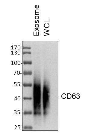
Western Blot: CD63 Antibody (MX-49.129.5) [NBP2-32830] - Exosome or whole cell lysates (WCL) sample from MDA-MB-231 cells was loaded with 10 ug/lane. 10% SDS-PAGE. CD63 antibody (NBP2-32830) was used for primary antibody: 1:500, 4C, overnight. Image from verified customer review.![Immunocytochemistry/ Immunofluorescence: CD63 Antibody (MX-49.129.5) [NBP2-32830]](https://resources.rndsystems.com/images/products/CD63-Antibody-MX-49-129-5-Immunocytochemistry-Immunofluorescence-NBP2-32830-img0011.jpg "Immunocytochemistry/ Immunofluorescence: CD63 Antibody (MX-49.129.5) [NBP2-32830]")
Immunocytochemistry/ Immunofluorescence: CD63 Antibody (MX-49.129.5) [NBP2-32830]
Immunocytochemistry/Immunofluorescence: CD63 Antibody (MX-49.129.5) [NBP2-32830] - Immunofluorescence: - Azide and BSA Free [NBP2-34689] - IF staining of HeLa cells using AF488 labeled CD63 Monoclonal Antibody (MX-49. 129.5) (green). F-actin filaments are labeled with dylight 554 phalloidin (red) Nuclei stained with DAPI (blue).![Immunohistochemistry-Paraffin: CD63 Antibody (MX-49.129.5) [NBP2-32830]](https://resources.rndsystems.com/images/products/CD63-Antibody-MX-49-129-5-Immunohistochemistry-Paraffin-NBP2-32830-img0014.jpg "Immunohistochemistry-Paraffin: CD63 Antibody (MX-49.129.5) [NBP2-32830]")
Immunohistochemistry-Paraffin: CD63 Antibody (MX-49.129.5) [NBP2-32830]
Immunohistochemistry-Paraffin: CD63 Antibody (MX-49.129.5) [NBP2-32830] - Formalin-fixed, paraffin-embedded Mouse spleen stained with CD63 Antibody (MX-49.129.5)![Flow Cytometry: CD63 Antibody (MX-49.129.5) [NBP2-32830]](https://resources.rndsystems.com/images/products/CD63-Antibody-MX-49-129-5-Flow-Cytometry-NBP2-32830-img0019.jpg "Flow Cytometry: CD63 Antibody (MX-49.129.5) [NBP2-32830]")
Flow Cytometry: CD63 Antibody (MX-49.129.5) [NBP2-32830]
Flow Cytometry: CD63 Antibody (MX-49.129.5) [NBP2-32830] - Flow cytometric analysis of bead-bound exosomes derived from MCF-7 cells. CD63 Mouse Monoclonal antibody (MX-49.129.5) followed by goat anti-mouse IgG-CF568 (orange); unstained exosomes (gray).![Western Blot: CD63 Antibody (MX-49.129.5) [NBP2-32830]](https://resources.rndsystems.com/images/products/CD63-Antibody-MX-49-129-5-Western-Blot-NBP2-32830-img0008.jpg "Western Blot: CD63 Antibody (MX-49.129.5) [NBP2-32830]")
Western Blot: CD63 Antibody (MX-49.129.5) [NBP2-32830]
Western Blot: CD63 Antibody (MX-49.129.5) [NBP2-32830] - Western Blot: CD63 Antibody (MX-49.129.5) - Azide and BSA Free [NBP2-34689] - Western blot of human spleen Lysate with CD63 Monoclonal Antibody (IMX-49. 129.5)![Immunohistochemistry-Paraffin: CD63 Antibody (MX-49.129.5) [NBP2-32830]](https://resources.rndsystems.com/images/products/CD63-Antibody-MX-49-129-5-Immunohistochemistry-Paraffin-NBP2-32830-img0009.jpg "Immunohistochemistry-Paraffin: CD63 Antibody (MX-49.129.5) [NBP2-32830]")
Immunohistochemistry-Paraffin: CD63 Antibody (MX-49.129.5) [NBP2-32830]
Immunohistochemistry-Paraffin: CD63 Antibody (MX-49.129.5) [NBP2-32830] - Immunohistochemistry-Paraffin: CD63 Antibody (MX-49.129.5) - Azide and BSA Free [NBP2-34689] - Formalin-fixed, paraffin-embedded human prostate carcinoma stained with CD63 Monoclonal Antibody (MX-49. 129.5)![Immunohistochemistry-Paraffin: CD63 Antibody (MX-49.129.5) [NBP2-32830]](https://resources.rndsystems.com/images/products/CD63-Antibody-MX-49-129-5-Immunohistochemistry-Paraffin-NBP2-32830-img0013.jpg "Immunohistochemistry-Paraffin: CD63 Antibody (MX-49.129.5) [NBP2-32830]")
Immunohistochemistry-Paraffin: CD63 Antibody (MX-49.129.5) [NBP2-32830]
Immunohistochemistry-Paraffin: CD63 Antibody (MX-49.129.5) [NBP2-32830] - Formalin-fixed, paraffin-embedded human melanoma stained with CD63 Antibody (MX-49.129.5)![Flow Cytometry: CD63 Antibody (MX-49.129.5) [NBP2-32830]](https://resources.rndsystems.com/images/products/CD63-Antibody-MX-49-129-5-Flow-Cytometry-NBP2-32830-img0010.jpg "Flow Cytometry: CD63 Antibody (MX-49.129.5) [NBP2-32830]")
Flow Cytometry: CD63 Antibody (MX-49.129.5) [NBP2-32830]
Flow Cytometry: CD63 Antibody (MX-49.129.5) [NBP2-32830] - Flow Cytometry: CD63 Antibody (MX-49.129.5) - Azide and BSA Free [NBP2-34689] - Flow Cytometry of NIH/3T3 Cells. Black: Cells alone; Green: Isotype Control; Red; PE-labeled CD63 Monoclonal Antibody (MX-49. 129.5)![Flow Cytometry: CD63 Antibody (MX-49.129.5) [NBP2-32830]](https://resources.rndsystems.com/images/products/CD63-Antibody-MX-49-129-5-Flow-Cytometry-NBP2-32830-img0015.jpg "Flow Cytometry: CD63 Antibody (MX-49.129.5) [NBP2-32830]")
Flow Cytometry: CD63 Antibody (MX-49.129.5) [NBP2-32830]
Flow Cytometry: CD63 Antibody (MX-49.129.5) [NBP2-32830] - Flow Cytometric staining of CD63 on human PBMC cells. Black: cells alone; Green: Isotype Control; Red: PE-labeled CD63 Monoclonal Antibody (MX-49.129.5).![Flow Cytometry: CD63 Antibody (MX-49.129.5) [NBP2-32830]](https://resources.rndsystems.com/images/products/CD63-Antibody-MX-49-129-5-Flow-Cytometry-NBP2-32830-img0016.jpg "Flow Cytometry: CD63 Antibody (MX-49.129.5) [NBP2-32830]")
Flow Cytometry: CD63 Antibody (MX-49.129.5) [NBP2-32830]
Flow Cytometry: CD63 Antibody (MX-49.129.5) [NBP2-32830] - Flow cytometry of MCF-7 cells unstained (gray) or stained with CF488A-labeled CD63 monoclonal antibody (MX-49.129.5) (green).![Flow Cytometry: CD63 Antibody (MX-49.129.5) [NBP2-32830]](https://resources.rndsystems.com/images/products/CD63-Antibody-MX-49-129-5-Flow-Cytometry-NBP2-32830-img0017.jpg "Flow Cytometry: CD63 Antibody (MX-49.129.5) [NBP2-32830]")
Flow Cytometry: CD63 Antibody (MX-49.129.5) [NBP2-32830]
Flow Cytometry: CD63 Antibody (MX-49.129.5) [NBP2-32830] - Flow cytometry of bead-bound exosomes derived from MCF-7 cells. Unstained exosomes (gray) or stained with CF488A-labeled CD63 monoclonal antibody (MX-49.129.5) (green)![Flow Cytometry: CD63 Antibody (MX-49.129.5) [NBP2-32830]](https://resources.rndsystems.com/images/products/CD63-Antibody-MX-49-129-5-Flow-Cytometry-NBP2-32830-img0018.jpg "Flow Cytometry: CD63 Antibody (MX-49.129.5) [NBP2-32830]")
Flow Cytometry: CD63 Antibody (MX-49.129.5) [NBP2-32830]
Flow Cytometry: CD63 Antibody (MX-49.129.5) [NBP2-32830] - Flow cytometric analysis of MCF-7 cells. CD63 Mouse Monoclonal antibody (MX-49.129.5) followed by goat anti-mouse IgG-CF568 (orange); unstained cells (gray). [NBP2-32830] -")
Western Blot: CD63 Antibody (MX-49.129.5) [NBP2-32830] -
Characterization of urine and urinary exosomes isolated by BEST of PE and HC. (A) Representative nanoparticle tracking analysis for untreated urine and isolated urine from PE and HC, depicting particle diameter and the number of particles. (B) and (C) Comparisons of particle concentration and particle size between PE and HC. (D) Western blots of TSG101 and CD63 protein extracted from isolated exosomes. (E) and (F) Quantitative analysis of exosome marker TSG101 and CD63 protein. All data are presented as mean \documentclass[12pt]{minimal}\usepackage{amsmath}\usepackage{wasysym} \usepackage{amsfonts} \usepackage{amssymb} \usepackage{amsbsy}\usepackage{mathrsfs}\usepackage{upgreek}\setlength{\oddsidemargin}{-69pt}\begin{document}$$\:\pm\:$$\end{document} standard error and analyzed by paired t-test and independent two-tailed t-test. BEST, biologically intact exosome separation technology; PE, patients with preeclampsia; HC, healthy controls. Image collected and cropped by CiteAb from the following open publication (https://pubmed.ncbi.nlm.nih.gov/39406891), licensed under a CC-BY license. Not internally tested by Novus Biologicals.Applications for CD63 Antibody (MX-49.129.5)
Application
Recommended Usage
Flow Cytometry
0.5-1 ug/million cells
Immunocytochemistry/ Immunofluorescence
0.5-1 ug/ml
Immunohistochemistry-Paraffin
1-2 ug/ml
Western Blot
0.5-1 ug/ml
Application Notes
Immunohistochemistry (Formalin-fixed): 1-2ug/ml for 30 minutes at RT. Staining of formalin-fixed tissues requires heating tissue sections in 10mM Tris with 1mM EDTA, pH 9.0, for 45 min at 95C followed by cooling at RT for 20 minutes.
Optimal dilution for a specific application should be determined.
Optimal dilution for a specific application should be determined.
Reviewed Applications
Read 2 reviews rated 5 using NBP2-32830 in the following applications:
Flow Cytometry Panel Builder
Bio-Techne Knows Flow Cytometry
Save time and reduce costly mistakes by quickly finding compatible reagents using the Panel Builder Tool.
Advanced Features
- Spectra Viewer - Custom analysis of spectra from multiple fluorochromes
- Spillover Popups - Visualize the spectra of individual fluorochromes
- Antigen Density Selector - Match fluorochrome brightness with antigen density

Formulation, Preparation, and Storage
Purification
Protein A or G purified
Formulation
10 mM PBS with 0.05% BSA
Preservative
0.05% Sodium Azide
Concentration
0.2 mg/ml
Shipping
The product is shipped with polar packs. Upon receipt, store it immediately at the temperature recommended below.
Stability & Storage
Store at 4C.
Background: CD63
References
1. Pols, M. S., & Klumperman, J. (2009). Trafficking and function of the tetraspanin CD63. Experimental cell research. https://doi.org/10.1016/j.yexcr.2008.09.020
2. Metzelaar, M. J., Wijngaard, P. L., Peters, P. J., Sixma, J. J., Nieuwenhuis, H. K., & Clevers, H. C. (1991). CD63 antigen. A novel lysosomal membrane glycoprotein, cloned by a screening procedure for intracellular antigens in eukaryotic cells. The Journal of biological chemistry.
3. Horejsi, V., & Vlcek, C. (1991). Novel structurally distinct family of leucocyte surface glycoproteins including CD9, CD37, CD53 and CD63. FEBS letters. https://doi.org/10.1016/0014-5793(91)80988-f
4. Eckfeld, C., HauBler, D., Schoeps, B., Hermann, C. D., & Kruger, A. (2019). Functional disparities within the TIMP family in cancer: hints from molecular divergence. Cancer metastasis reviews. https://doi.org/10.1007/s10555-019-09812-6
5. Hoffmann, H. J., Santos, A. F., Mayorga, C., Nopp, A., Eberlein, B., Ferrer, M., Rouzaire, P., Ebo, D. G., Sabato, V., Sanz, M. L., Pecaric-Petkovic, T., Patil, S. U., Hausmann, O. V., Shreffler, W. G., Korosec, P., & Knol, E. F. (2015). The clinical utility of basophil activation testing in diagnosis and monitoring of allergic disease. Allergy. https://doi.org/10.1111/all.12698
6. Dell'Angelica, E. C., Shotelersuk, V., Aguilar, R. C., Gahl, W. A., & Bonifacino, J. S. (1999). Altered trafficking of lysosomal proteins in Hermansky-Pudlak syndrome due to mutations in the beta 3A subunit of the AP-3 adaptor. Molecular cell. https://doi.org/10.1016/s1097-2765(00)80170-7
Additional CD63 Products
Product Documents for CD63 Antibody (MX-49.129.5)
Certificate of Analysis
To download a Certificate of Analysis, please enter a lot or batch number in the search box below.
Product Specific Notices for CD63 Antibody (MX-49.129.5)
This product is for research use only and is not approved for use in humans or in clinical diagnosis. Primary Antibodies are guaranteed for 1 year from date of receipt.
Related Research Areas
Citations for CD63 Antibody (MX-49.129.5)
 Powered by Bioz
Customer Reviews for CD63 Antibody (MX-49.129.5) (2)
5 out of 5
2 Customer Ratings
Have you used CD63 Antibody (MX-49.129.5)?
Submit a review and receive an Amazon gift card!
$25/€18/£15/$25CAN/¥2500 Yen for a review with an image
$10/€7/£6/$10CAN/¥1110 Yen for a review without an image
Submit a review
Customer Images

Showing
1
-
2 of
2 reviews
Showing All
Filter By:
-
Application: Western BlotSample Tested: MDA-MB-231Species: HumanVerified Customer | Posted 10/20/2022Western Blot: Exosome or whole cell lysates (WCL) sample from MDA-MB-231 cells was loaded with 10 ug/lane. 10% SDS-PAGE. CD63 Antibody (NBP2-32830) was used for primary antibody: 1:500, 4℃, overnight.

-
Application: Western BlotSample Tested: 293T cell lysateSpecies: HumanVerified Customer | Posted 08/03/2022CD63 overexpression cells1:500 in TBST
There are no reviews that match your criteria.
Protocols
Find general support by application which include: protocols, troubleshooting, illustrated assays, videos and webinars.
- 7-Amino Actinomycin D (7-AAD) Cell Viability Flow Cytometry Protocol
- Antigen Retrieval Protocol (PIER)
- Antigen Retrieval for Frozen Sections Protocol
- Appropriate Fixation of IHC/ICC Samples
- Cellular Response to Hypoxia Protocols
- Chromogenic IHC Staining of Formalin-Fixed Paraffin-Embedded (FFPE) Tissue Protocol
- Chromogenic Immunohistochemistry Staining of Frozen Tissue
- ClariTSA™ Fluorophore Kits
- Detection & Visualization of Antibody Binding
- Extracellular Membrane Flow Cytometry Protocol
- Flow Cytometry Protocol for Cell Surface Markers
- Flow Cytometry Protocol for Staining Membrane Associated Proteins
- Flow Cytometry Staining Protocols
- Flow Cytometry Troubleshooting Guide
- Fluorescent IHC Staining of Frozen Tissue Protocol
- Graphic Protocol for Heat-induced Epitope Retrieval
- Graphic Protocol for the Preparation and Fluorescent IHC Staining of Frozen Tissue Sections
- Graphic Protocol for the Preparation and Fluorescent IHC Staining of Paraffin-embedded Tissue Sections
- Graphic Protocol for the Preparation of Gelatin-coated Slides for Histological Tissue Sections
- ICC Cell Smear Protocol for Suspension Cells
- ICC Immunocytochemistry Protocol Videos
- ICC for Adherent Cells
- IHC Sample Preparation (Frozen sections vs Paraffin)
- Immunocytochemistry (ICC) Protocol
- Immunocytochemistry Troubleshooting
- Immunofluorescence of Organoids Embedded in Cultrex Basement Membrane Extract
- Immunofluorescent IHC Staining of Formalin-Fixed Paraffin-Embedded (FFPE) Tissue Protocol
- Immunohistochemistry (IHC) and Immunocytochemistry (ICC) Protocols
- Immunohistochemistry Frozen Troubleshooting
- Immunohistochemistry Paraffin Troubleshooting
- Intracellular Flow Cytometry Protocol Using Alcohol (Methanol)
- Intracellular Flow Cytometry Protocol Using Detergents
- Intracellular Nuclear Staining Flow Cytometry Protocol Using Detergents
- Intracellular Staining Flow Cytometry Protocol Using Alcohol Permeabilization
- Intracellular Staining Flow Cytometry Protocol Using Detergents to Permeabilize Cells
- Preparing Samples for IHC/ICC Experiments
- Preventing Non-Specific Staining (Non-Specific Binding)
- Primary Antibody Selection & Optimization
- Propidium Iodide Cell Viability Flow Cytometry Protocol
- Protocol for Heat-Induced Epitope Retrieval (HIER)
- Protocol for Liperfluo
- Protocol for Making a 4% Formaldehyde Solution in PBS
- Protocol for VisUCyte™ HRP Polymer Detection Reagent
- Protocol for the Characterization of Human Th22 Cells
- Protocol for the Characterization of Human Th9 Cells
- Protocol for the Fluorescent ICC Staining of Cell Smears - Graphic
- Protocol for the Fluorescent ICC Staining of Cultured Cells on Coverslips - Graphic
- Protocol for the Preparation & Fixation of Cells on Coverslips
- Protocol for the Preparation and Chromogenic IHC Staining of Frozen Tissue Sections
- Protocol for the Preparation and Chromogenic IHC Staining of Frozen Tissue Sections - Graphic
- Protocol for the Preparation and Chromogenic IHC Staining of Paraffin-embedded Tissue Sections
- Protocol for the Preparation and Chromogenic IHC Staining of Paraffin-embedded Tissue Sections - Graphic
- Protocol for the Preparation and Fluorescent ICC Staining of Cells on Coverslips
- Protocol for the Preparation and Fluorescent ICC Staining of Non-adherent Cells
- Protocol for the Preparation and Fluorescent ICC Staining of Stem Cells on Coverslips
- Protocol for the Preparation and Fluorescent IHC Staining of Frozen Tissue Sections
- Protocol for the Preparation and Fluorescent IHC Staining of Paraffin-embedded Tissue Sections
- Protocol for the Preparation of Gelatin-coated Slides for Histological Tissue Sections
- Protocol for the Preparation of a Cell Smear for Non-adherent Cell ICC - Graphic
- Protocol: Annexin V and PI Staining by Flow Cytometry
- Protocol: Annexin V and PI Staining for Apoptosis by Flow Cytometry
- R&D Systems Quality Control Western Blot Protocol
- TUNEL and Active Caspase-3 Detection by IHC/ICC Protocol
- The Importance of IHC/ICC Controls
- Troubleshooting Guide: Fluorokine Flow Cytometry Kits
- Troubleshooting Guide: Immunohistochemistry
- Troubleshooting Guide: Western Blot Figures
- Western Blot Conditions
- Western Blot Protocol
- Western Blot Protocol for Cell Lysates
- Western Blot Troubleshooting
- Western Blot Troubleshooting Guide
- View all Protocols, Troubleshooting, Illustrated assays and Webinars
FAQs for CD63 Antibody (MX-49.129.5)
Showing
1
-
1 of
1 FAQ
Showing All
-
Q: I am wondering whether the CD62 MX-49.129.5 monoclonal antibody (NBP2-32830) is working for western blot under reducing conditions?
A: One specific condition isn't recommended, so the CD63 antibody should work in both reducing and non reducing conditions.
Loading...