CD79A Antibody (HM57) - BSA Free
Novus Biologicals | Catalog # NB100-64347
Clone HM57 was used by HLDA to establish CD designation.
![Immunohistochemistry: CD79A Antibody (HM57) - BSA Free [NB100-64347]](https://resources.rndsystems.com/images/products/CD79A-Antibody-HM57-Immunohistochemistry-NB100-64347-img0005.jpg "Immunohistochemistry: CD79A Antibody (HM57) - BSA Free [NB100-64347]")
Key Product Details
Species Reactivity
Validated:
Human
Cited:
Human, Avian - Chicken, Bovine, Canine
Applications
Validated:
Immunohistochemistry, Immunohistochemistry-Paraffin, Immunohistochemistry-Frozen, Flow Cytometry
Cited:
Immunohistochemistry-Paraffin, IF/IHC
Label
Unconjugated
Antibody Source
Monoclonal Mouse IgG1 Clone # HM57
Format
BSA Free
Loading...
Product Specifications
Immunogen
Synthetic peptide corresponding to 202-216 amino acid sequence of human mb-1
Reactivity Notes
Predicted cross-reactivities: Equine, Monkey, Guinea Pig, Goat, Fallow deer, Ferret, Dog, Bovine, American Bison, Porcine, Rabbit, Mouse, Rat, Red deer
Please note that this antibody is reactive to Mouse and derived from the same host, Mouse. Additional Mouse on Mouse blocking steps may be required for IHC and ICC experiments. Please contact Technical Support for more information.
Please note that this antibody is reactive to Mouse and derived from the same host, Mouse. Additional Mouse on Mouse blocking steps may be required for IHC and ICC experiments. Please contact Technical Support for more information.
Specificity
NB100-64347 recognizes an epitope within the cytoplasmic domain of CD79a. CD79a, also known as mb-1, is a 45kD protein that is expressed by B lymphocytes during differentiation from early pre-B cell stage through to plasma cells. The CD79a molecule associates with CD79b (B29) to form a heterodimer that is non-covalently linked to surface immunoglobulin, forming the B-cell receptor (BCR) complex. The CD79a/CD79b heterodimers are also necessary for intracellular signalling following antigen-binding to surface immunoglobulin. Clone HM57 has been reported to work in western blotting applications.
Clonality
Monoclonal
Host
Mouse
Isotype
IgG1
Scientific Data Images for CD79A Antibody (HM57) - BSA Free
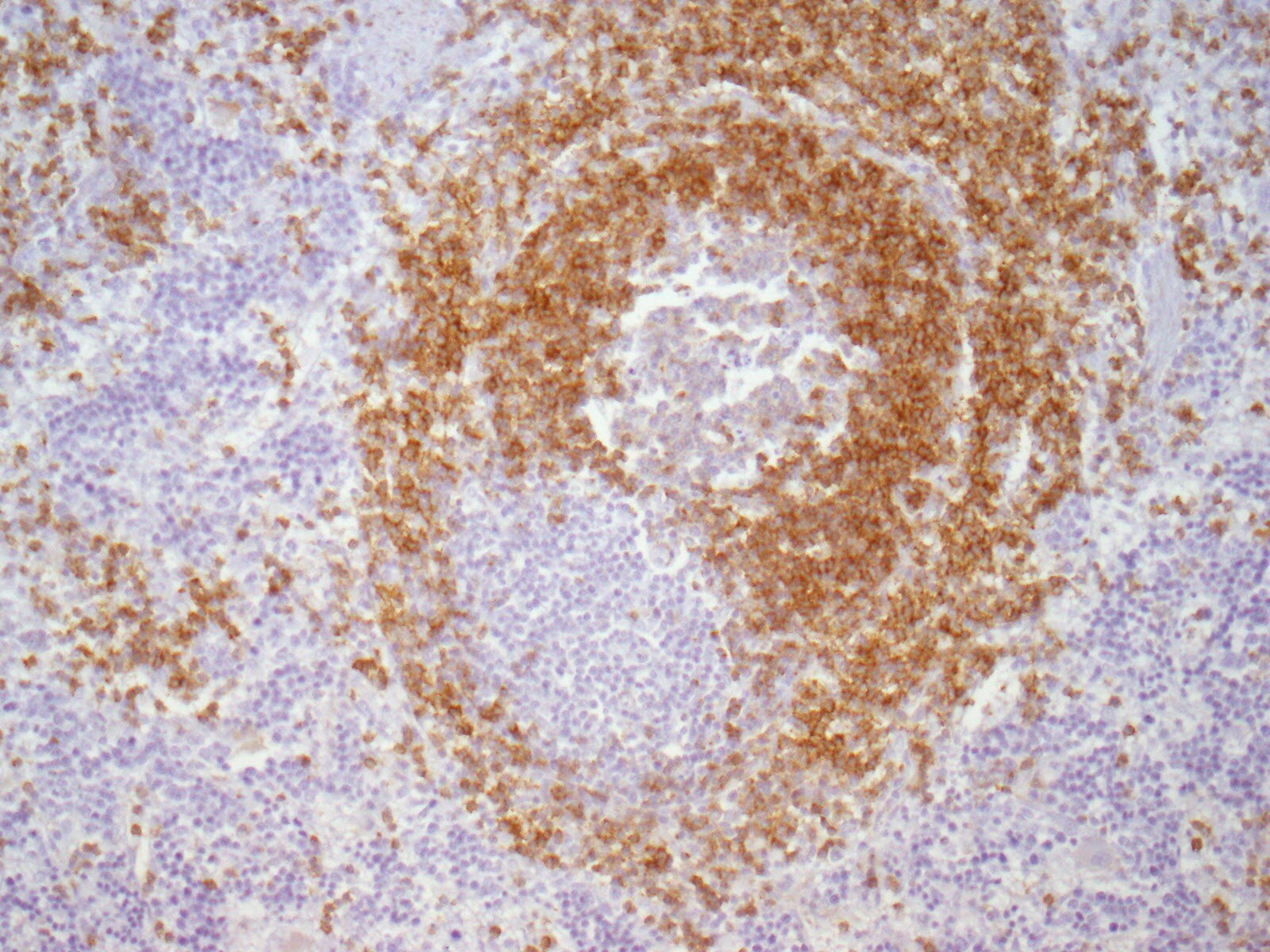
Immunohistochemistry: CD79A Antibody (HM57) - BSA Free [NB100-64347]
Immunohistochemistry: CD79A Antibody (HM57) [NB100-64347] - Paraffin-embedded alcohol fixed Rat Spleen tissue (20x). Antigen retrieval pH6. Dilution: 1:500 and ON incubation at 4C. This image was submitted via customer revies.![Flow Cytometry: CD79A Antibody (HM57) - BSA Free [NB100-64347]](https://resources.rndsystems.com/images/products/CD79A-Antibody-HM57-Flow-Cytometry-NB100-64347-img0003.jpg "Flow Cytometry: CD79A Antibody (HM57) - BSA Free [NB100-64347]")
Flow Cytometry: CD79A Antibody (HM57) - BSA Free [NB100-64347]
Flow Cytometry: CD79A Antibody (HM57) [NB100-64347] - Using the Allophycocyanin direct conjugate An intracellular stain was performed on Daudi cells with CD79A (HM 57) antibody NB100-64347APC (blue) and a matched isotype control NBP1-97005APC (orange). Cells were fixed with 4% PFA and then permeablized with 0.1% saponin. Cells were incubated in an antibody dilution of 0.5 ug/mL for 30 minutes at room temperature. Both antibodies were conjugated to Allophycocyanin.![Immunohistochemistry-Paraffin: CD79A Antibody (HM57) - BSA Free [NB100-64347]](https://resources.rndsystems.com/images/products/CD79A-Antibody-HM57-Immunohistochemistry-Paraffin-NB100-64347-img0002.jpg "Immunohistochemistry-Paraffin: CD79A Antibody (HM57) - BSA Free [NB100-64347]")
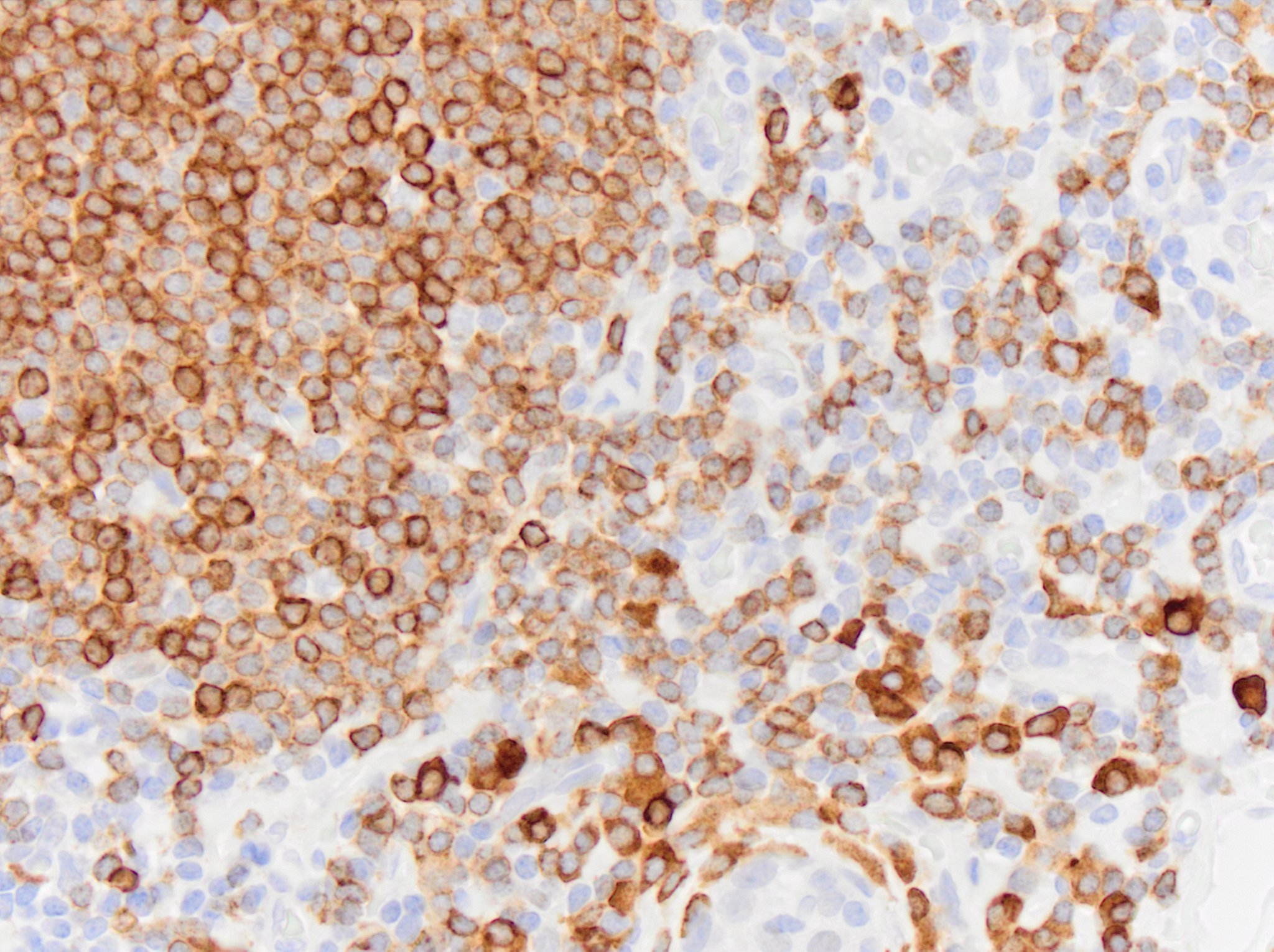
Immunohistochemistry-Paraffin: CD79A Antibody (HM57) - BSA Free [NB100-64347]
Immunohistochemistry-Paraffin: CD79A Antibody (HM57) [NB100-64347] - Embedded human tonsil.Applications for CD79A Antibody (HM57) - BSA Free
Application
Recommended Usage
Flow Cytometry
1:50-1:100
Immunohistochemistry
1:10-1:500
Immunohistochemistry-Frozen
1:10-1:500
Immunohistochemistry-Paraffin
1:100-1:500
Application Notes
For Flow Cytometry: Use 10 ul of the suggested working dilution to label 10^6 cells in 100 ul. For IHC: heat treated antigen retrieval using sodium citrate buffer pH 6.0 is recommended for this purpose. Although not tested this antibody may be useful in Western Blot. NB100-64347 has been used successfully in Immunofluorescence reported by a by a customer review.
Reviewed Applications
Read 6 reviews rated 3.5 using NB100-64347 in the following applications:
Flow Cytometry Panel Builder
Bio-Techne Knows Flow Cytometry
Save time and reduce costly mistakes by quickly finding compatible reagents using the Panel Builder Tool.
Advanced Features
- Spectra Viewer - Custom analysis of spectra from multiple fluorochromes
- Spillover Popups - Visualize the spectra of individual fluorochromes
- Antigen Density Selector - Match fluorochrome brightness with antigen density

Formulation, Preparation, and Storage
Purification
Protein A purified
Formulation
PBS
Format
BSA Free
Preservative
0.09% Sodium Azide
Concentration
1.0 mg/ml
Shipping
The product is shipped with polar packs. Upon receipt, store it immediately at the temperature recommended below.
Stability & Storage
Store at 4C short term. Aliquot and store at -20C long term. Avoid freeze-thaw cycles.
Background: CD79A
Alternate Names
CD79A, MB-1
Gene Symbol
CD79A
UniProt
Additional CD79A Products
Product Documents for CD79A Antibody (HM57) - BSA Free
Certificate of Analysis
To download a Certificate of Analysis, please enter a lot or batch number in the search box below.
Product Specific Notices for CD79A Antibody (HM57) - BSA Free
This product is for research use only and is not approved for use in humans or in clinical diagnosis. Primary Antibodies are guaranteed for 1 year from date of receipt.
Related Research Areas
Citations for CD79A Antibody (HM57) - BSA Free
 Powered by Bioz
Customer Reviews for CD79A Antibody (HM57) - BSA Free (6)
3.5 out of 5
6 Customer Ratings
Have you used CD79A Antibody (HM57) - BSA Free?
Submit a review and receive an Amazon gift card!
$25/€18/£15/$25CAN/¥2500 Yen for a review with an image
$10/€7/£6/$10CAN/¥1110 Yen for a review without an image
Submit a review
Customer Images
Showing
1
-
5 of
6 reviews
Showing All
Filter By:
-
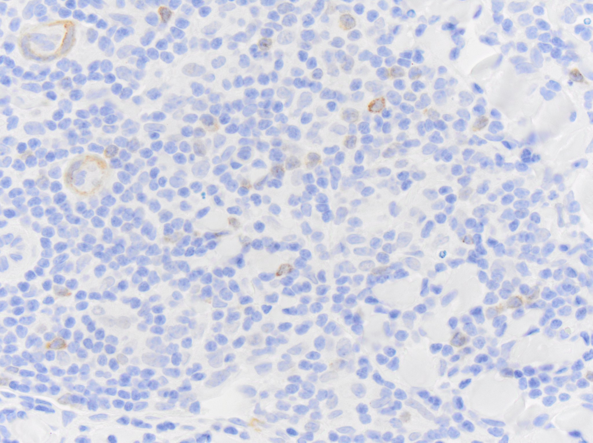
Application: Immunohistochemistry-ParaffinSample Tested: TonsilSpecies: HumanVerified Customer | Posted 10/13/2023CD79a NB100-64347 immunoreactivity in human tonsil. The antibody was diluted to 0.5ug/mL and was incubated with sections for 30min at room temperature. Secondary was Horse Anti-Mouse HRP polymer.Reacts with smooth muscle

-
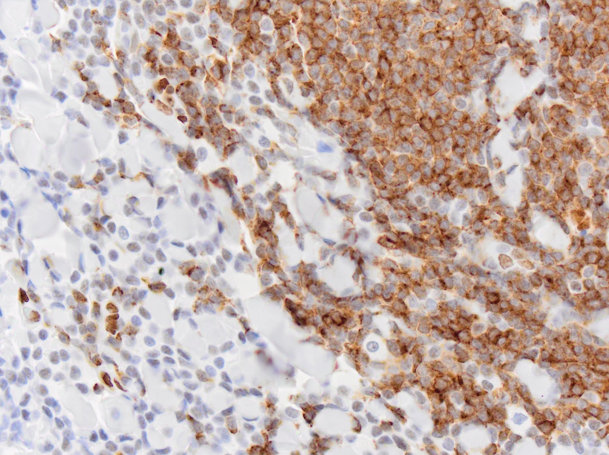
Application: Immunohistochemistry-ParaffinSample Tested: SkinSpecies: EquineVerified Customer | Posted 10/13/2023CD79a NB100-64347 immunoreactivity in horse skin (lymphoma). The antibody was diluted to 0.5ug/mL and was incubated with tissue section for 30min at room temperature. Secondary was Horse Anti-Mouse HRP polymer.Reacted strongly with smooth muscle as well as immune cells.
-
Application: Immunohistochemistry-ParaffinSample Tested: SkinSpecies: FelineVerified Customer | Posted 10/13/2023CD79a NB100-64347 immunoreactivity in cat skin (lymphoma). Antibody was diluted to 0.5ug/mL and was left on tissue sections for 30m at room temperature. Secondary was Horse Anti-Mouse HRP polymer.Reacted with muscle and nucleus. Attempts to limit this labeling resulted in a loss of suspected genuine immunoreactivity as well.
Bio-Techne ResponseThis review was submitted through the legacy Novus Innovators Program, reflecting a new species or application tested on a primary antibody.
-
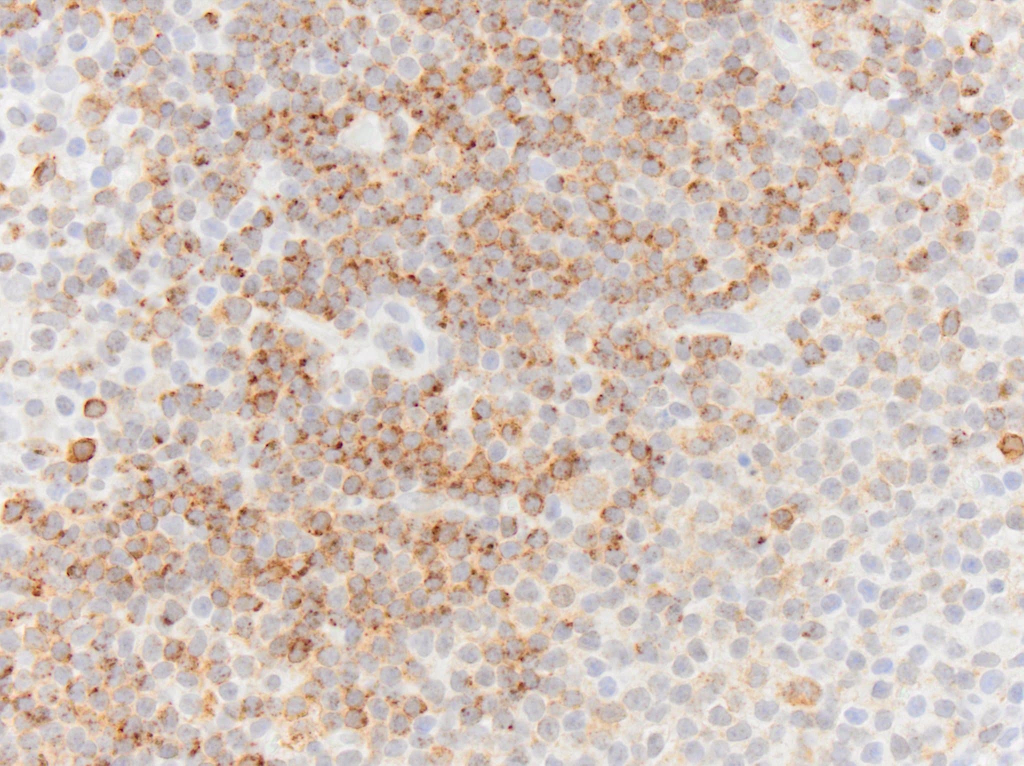
Application: Immunohistochemistry-ParaffinSample Tested: cynomolgus monkey lymph node and Lymph NodeSpecies: CanineVerified Customer | Posted 10/13/2023CD79a NB100-64347 immunoreactivity in dog lymph node. Antibody was diluted to 0.5ug/mL and was left on tissue sections for 30m at room temperature. Secondary was Horse Anti-Mouse HRP polymer.Reacts with smooth muscle. Attempts to dilute the antibody to eliminate muscle labeling resulted in a loss of signal in immune cells.
-
Application: Immunohistochemistry-ParaffinSample Tested: Spleen tissueSpecies: RatVerified Customer | Posted 02/17/2018IHC: Rat spleen tissue (20X). CD79a dilution 1:500 incubation ON at 4°CIHC performed on paraffin-embedded alcohol fixed tissue. Antigen retrieval pH6. Dilution: 1:500 and ON incubation at 4°C.
-
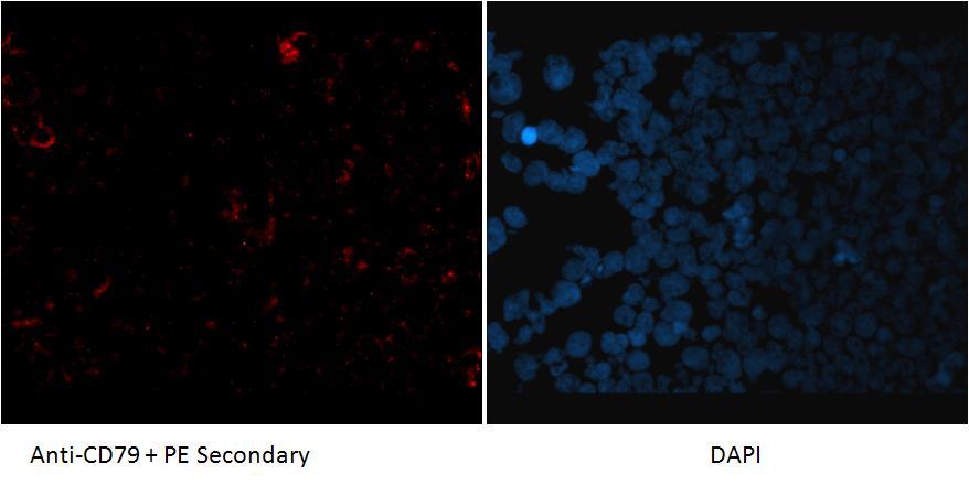
Application: ImmunofluorescenceSample Tested: Dog LymphomaSpecies: OtherVerified Customer | Posted 08/15/2012
There are no reviews that match your criteria.
Protocols
Find general support by application which include: protocols, troubleshooting, illustrated assays, videos and webinars.
- 7-Amino Actinomycin D (7-AAD) Cell Viability Flow Cytometry Protocol
- Antigen Retrieval Protocol (PIER)
- Antigen Retrieval for Frozen Sections Protocol
- Appropriate Fixation of IHC/ICC Samples
- Cellular Response to Hypoxia Protocols
- Chromogenic IHC Staining of Formalin-Fixed Paraffin-Embedded (FFPE) Tissue Protocol
- Chromogenic Immunohistochemistry Staining of Frozen Tissue
- ClariTSA™ Fluorophore Kits
- Detection & Visualization of Antibody Binding
- Extracellular Membrane Flow Cytometry Protocol
- Flow Cytometry Protocol for Cell Surface Markers
- Flow Cytometry Protocol for Staining Membrane Associated Proteins
- Flow Cytometry Staining Protocols
- Flow Cytometry Troubleshooting Guide
- Fluorescent IHC Staining of Frozen Tissue Protocol
- Graphic Protocol for Heat-induced Epitope Retrieval
- Graphic Protocol for the Preparation and Fluorescent IHC Staining of Frozen Tissue Sections
- Graphic Protocol for the Preparation and Fluorescent IHC Staining of Paraffin-embedded Tissue Sections
- Graphic Protocol for the Preparation of Gelatin-coated Slides for Histological Tissue Sections
- IHC Sample Preparation (Frozen sections vs Paraffin)
- Immunofluorescent IHC Staining of Formalin-Fixed Paraffin-Embedded (FFPE) Tissue Protocol
- Immunohistochemistry (IHC) and Immunocytochemistry (ICC) Protocols
- Immunohistochemistry Frozen Troubleshooting
- Immunohistochemistry Paraffin Troubleshooting
- Intracellular Flow Cytometry Protocol Using Alcohol (Methanol)
- Intracellular Flow Cytometry Protocol Using Detergents
- Intracellular Nuclear Staining Flow Cytometry Protocol Using Detergents
- Intracellular Staining Flow Cytometry Protocol Using Alcohol Permeabilization
- Intracellular Staining Flow Cytometry Protocol Using Detergents to Permeabilize Cells
- Preparing Samples for IHC/ICC Experiments
- Preventing Non-Specific Staining (Non-Specific Binding)
- Primary Antibody Selection & Optimization
- Propidium Iodide Cell Viability Flow Cytometry Protocol
- Protocol for Heat-Induced Epitope Retrieval (HIER)
- Protocol for Liperfluo
- Protocol for Making a 4% Formaldehyde Solution in PBS
- Protocol for VisUCyte™ HRP Polymer Detection Reagent
- Protocol for the Characterization of Human Th22 Cells
- Protocol for the Characterization of Human Th9 Cells
- Protocol for the Preparation & Fixation of Cells on Coverslips
- Protocol for the Preparation and Chromogenic IHC Staining of Frozen Tissue Sections
- Protocol for the Preparation and Chromogenic IHC Staining of Frozen Tissue Sections - Graphic
- Protocol for the Preparation and Chromogenic IHC Staining of Paraffin-embedded Tissue Sections
- Protocol for the Preparation and Chromogenic IHC Staining of Paraffin-embedded Tissue Sections - Graphic
- Protocol for the Preparation and Fluorescent IHC Staining of Frozen Tissue Sections
- Protocol for the Preparation and Fluorescent IHC Staining of Paraffin-embedded Tissue Sections
- Protocol for the Preparation of Gelatin-coated Slides for Histological Tissue Sections
- Protocol: Annexin V and PI Staining by Flow Cytometry
- Protocol: Annexin V and PI Staining for Apoptosis by Flow Cytometry
- TUNEL and Active Caspase-3 Detection by IHC/ICC Protocol
- The Importance of IHC/ICC Controls
- Troubleshooting Guide: Fluorokine Flow Cytometry Kits
- Troubleshooting Guide: Immunohistochemistry
- View all Protocols, Troubleshooting, Illustrated assays and Webinars

