CD9 Antibody (MEM-61) - BSA Free
Novus Biologicals | Catalog # NB500-327
Clone MEM-61 was used by HLDA to establish CD designation.
![Flow Cytometry: CD9 Antibody (MEM-61) - BSA Free [NB500-327]](https://resources.rndsystems.com/images/products/CD9-Antibody-MEM-61-Flow-Cytometry-NB500-327-img0008.jpg "Flow Cytometry: CD9 Antibody (MEM-61) - BSA Free [NB500-327]")
Key Product Details
Species Reactivity
Validated:
Human
Cited:
Human
Applications
Validated:
Immunohistochemistry, Immunohistochemistry-Paraffin, Western Blot, ELISA, Flow Cytometry, In vitro assay, CyTOF-ready
Cited:
Western Blot, ELISA, Flow Cytometry, Immunocytochemistry/ Immunofluorescence, In vitro assay, CyTOF-ready
Label
Unconjugated
Antibody Source
Monoclonal Mouse IgG1 Clone # MEM-61
Format
BSA Free
Loading...
Product Specifications
Immunogen
Pre-B cell line NALM-6
Specificity
The antibody MEM-61 recognizes an epitope on second extracellular domain (EC2) of CD9 antigen, a 24 kDa transmembrane protein expressed on platelets, monocytes, pre-B lymphocytes, granulocytes and activated T lymphocytes. HLDA VI; WS Code P P-15
Clonality
Monoclonal
Host
Mouse
Isotype
IgG1
Theoretical MW
24 kDa.
Disclaimer note: The observed molecular weight of the protein may vary from the listed predicted molecular weight due to post translational modifications, post translation cleavages, relative charges, and other experimental factors.
Disclaimer note: The observed molecular weight of the protein may vary from the listed predicted molecular weight due to post translational modifications, post translation cleavages, relative charges, and other experimental factors.
Scientific Data Images for CD9 Antibody (MEM-61) - BSA Free
Flow Cytometry: CD9 Antibody (MEM-61) - BSA Free [NB500-327]
Flow Cytometry: CD9 Antibody (MEM-61) [NB500-327] - Staining of human peripheral blood with anti-CD9 (MEM-61) Alexa Fluor® 647.![Flow Cytometry: CD9 Antibody (MEM-61) - BSA Free [NB500-327]](https://resources.rndsystems.com/images/products/CD9-Antibody-MEM-61-Flow-Cytometry-NB500-327-img0001.jpg "Flow Cytometry: CD9 Antibody (MEM-61) - BSA Free [NB500-327]")
Flow Cytometry: CD9 Antibody (MEM-61) - BSA Free [NB500-327]
Flow Cytometry: CD9 Antibody (MEM-61) [NB500-327] - Surface staining of NALM-6 human pre-B cell leukemia cell line Total viable cells were used for analysis.![Flow Cytometry: CD9 Antibody (MEM-61) - BSA Free [NB500-327]](https://resources.rndsystems.com/images/products/CD9-Antibody-MEM-61-Flow-Cytometry-NB500-327-img0002.jpg "Flow Cytometry: CD9 Antibody (MEM-61) - BSA Free [NB500-327]")
Flow Cytometry: CD9 Antibody (MEM-61) - BSA Free [NB500-327]
Flow Cytometry: CD9 Antibody (MEM-61) [NB500-327] - Surface staining (mass cytometry) of PBMC after Ficoll-Paque separation with anti-human CD9 (MEM-61) Dy161.![Flow Cytometry: CD9 Antibody (MEM-61) - BSA Free [NB500-327]](https://resources.rndsystems.com/images/products/CD9-Antibody-MEM-61-Flow-Cytometry-NB500-327-img0003.jpg "Flow Cytometry: CD9 Antibody (MEM-61) - BSA Free [NB500-327]")
Flow Cytometry: CD9 Antibody (MEM-61) - BSA Free [NB500-327]
Flow Cytometry: CD9 Antibody (MEM-61) [NB500-327] - Surface staining of human peripheral blood with anti-CD9 (MEM-61) biotin / streptavidin-APC.![Flow Cytometry: CD9 Antibody (MEM-61) - BSA Free [NB500-327]](https://resources.rndsystems.com/images/products/CD9-Antibody-MEM-61-Flow-Cytometry-NB500-327-img0004.jpg "Flow Cytometry: CD9 Antibody (MEM-61) - BSA Free [NB500-327]")
Flow Cytometry: CD9 Antibody (MEM-61) - BSA Free [NB500-327]
Flow Cytometry: CD9 Antibody (MEM-61) [NB500-327] - Surface staining of human peripheral blood with anti-CD9 (MEM-61) APC-CyTM7.![Flow Cytometry: CD9 Antibody (MEM-61) - BSA Free [NB500-327]](https://resources.rndsystems.com/images/products/CD9-Antibody-MEM-61-Flow-Cytometry-NB500-327-img0005.jpg "Flow Cytometry: CD9 Antibody (MEM-61) - BSA Free [NB500-327]")
Flow Cytometry: CD9 Antibody (MEM-61) - BSA Free [NB500-327]
Flow Cytometry: CD9 Antibody (MEM-61) [NB500-327] - Surface staining of human peripheral blood with anti-CD9 (MEM-61) Pacific BlueTM.![Flow Cytometry: CD9 Antibody (MEM-61) - BSA Free [NB500-327]](https://resources.rndsystems.com/images/products/CD9-Antibody-MEM-61-Flow-Cytometry-NB500-327-img0006.jpg "Flow Cytometry: CD9 Antibody (MEM-61) - BSA Free [NB500-327]")
Flow Cytometry: CD9 Antibody (MEM-61) - BSA Free [NB500-327]
Flow Cytometry: CD9 Antibody (MEM-61) [NB500-327] - Staining of human peripheral blood with anti-CD9 (MEM-61) APC.![Flow Cytometry: CD9 Antibody (MEM-61) - BSA Free [NB500-327]](https://resources.rndsystems.com/images/products/CD9-Antibody-MEM-61-Flow-Cytometry-NB500-327-img0007.jpg "Flow Cytometry: CD9 Antibody (MEM-61) - BSA Free [NB500-327]")
Flow Cytometry: CD9 Antibody (MEM-61) - BSA Free [NB500-327]
Flow Cytometry: CD9 Antibody (MEM-61) [NB500-327] - Staining of human peripheral blood with anti-CD9 (MEM-61) PE.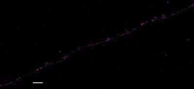
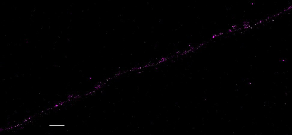
CyTOF-ready: CD9 Antibody (MEM-61) [NB500-327] - Analysis of CD9 antibody on human tissue. Scale, 50um. Image from verified customer review.
Applications for CD9 Antibody (MEM-61) - BSA Free
Application
Recommended Usage
Flow Cytometry
1-4 ug/ml
Immunohistochemistry
1:10-1:500
Immunohistochemistry-Paraffin
20 ug/ml
Western Blot
2-4 ug/ml
Application Notes
Western Blot -The antibody MEM-61 induces FcR-dependent platelet aggregation. This antibody is CyTOF ready.Use in In vitro reported in scientific literature (PMID: 28823976). Use in ELISA reported in secitific publication PMID: 32393780. Clone MEM-61 has been used for extracellular vesicle flow cytometry.
Reviewed Applications
Read 1 review rated 4 using NB500-327 in the following applications:
Flow Cytometry Panel Builder
Bio-Techne Knows Flow Cytometry
Save time and reduce costly mistakes by quickly finding compatible reagents using the Panel Builder Tool.
Advanced Features
- Spectra Viewer - Custom analysis of spectra from multiple fluorochromes
- Spillover Popups - Visualize the spectra of individual fluorochromes
- Antigen Density Selector - Match fluorochrome brightness with antigen density

Formulation, Preparation, and Storage
Purification
Protein A purified
Formulation
Phosphate buffered saline (PBS), pH 7.4
Format
BSA Free
Preservative
15mM Sodium Azide
Concentration
1 mg/ml
Shipping
The product is shipped with polar packs. Upon receipt, store it immediately at the temperature recommended below.
Stability & Storage
Store at 4C. Do not freeze.
Background: CD9
Alternate Names
CD9, DRAP-27, MIC3, p24, TSPAN29
Gene Symbol
CD9
Additional CD9 Products
Product Documents for CD9 Antibody (MEM-61) - BSA Free
Certificate of Analysis
To download a Certificate of Analysis, please enter a lot or batch number in the search box below.
Product Specific Notices for CD9 Antibody (MEM-61) - BSA Free
This product is for research use only and is not approved for use in humans or in clinical diagnosis. Primary Antibodies are guaranteed for 1 year from date of receipt.
Related Research Areas
Citations for CD9 Antibody (MEM-61) - BSA Free
 Powered by Bioz
Customer Reviews for CD9 Antibody (MEM-61) - BSA Free (1)
4 out of 5
1 Customer Rating
Have you used CD9 Antibody (MEM-61) - BSA Free?
Submit a review and receive an Amazon gift card!
$25/€18/£15/$25CAN/¥2500 Yen for a review with an image
$10/€7/£6/$10CAN/¥1110 Yen for a review without an image
Submit a review
Customer Images
Showing
1
-
1 of
1 review
Showing All
Filter By:
-
Application: CyTOFSample Tested: Human tissue originSpecies: HumanVerified Customer | Posted 01/04/2022Scale, 50um.

There are no reviews that match your criteria.
Protocols
Find general support by application which include: protocols, troubleshooting, illustrated assays, videos and webinars.
- 7-Amino Actinomycin D (7-AAD) Cell Viability Flow Cytometry Protocol
- Antigen Retrieval Protocol (PIER)
- Antigen Retrieval for Frozen Sections Protocol
- Appropriate Fixation of IHC/ICC Samples
- Cellular Response to Hypoxia Protocols
- Chromogenic IHC Staining of Formalin-Fixed Paraffin-Embedded (FFPE) Tissue Protocol
- Chromogenic Immunohistochemistry Staining of Frozen Tissue
- ClariTSA™ Fluorophore Kits
- Detection & Visualization of Antibody Binding
- ELISA Sample Preparation & Collection Guide
- ELISA Troubleshooting Guide
- Extracellular Membrane Flow Cytometry Protocol
- Flow Cytometry Protocol for Cell Surface Markers
- Flow Cytometry Protocol for Staining Membrane Associated Proteins
- Flow Cytometry Staining Protocols
- Flow Cytometry Troubleshooting Guide
- Fluorescent IHC Staining of Frozen Tissue Protocol
- Graphic Protocol for Heat-induced Epitope Retrieval
- Graphic Protocol for the Preparation and Fluorescent IHC Staining of Frozen Tissue Sections
- Graphic Protocol for the Preparation and Fluorescent IHC Staining of Paraffin-embedded Tissue Sections
- Graphic Protocol for the Preparation of Gelatin-coated Slides for Histological Tissue Sections
- How to Run an R&D Systems DuoSet ELISA
- How to Run an R&D Systems Quantikine ELISA
- How to Run an R&D Systems Quantikine™ QuicKit™ ELISA
- IHC Sample Preparation (Frozen sections vs Paraffin)
- Immunofluorescent IHC Staining of Formalin-Fixed Paraffin-Embedded (FFPE) Tissue Protocol
- Immunohistochemistry (IHC) and Immunocytochemistry (ICC) Protocols
- Immunohistochemistry Frozen Troubleshooting
- Immunohistochemistry Paraffin Troubleshooting
- Intracellular Flow Cytometry Protocol Using Alcohol (Methanol)
- Intracellular Flow Cytometry Protocol Using Detergents
- Intracellular Nuclear Staining Flow Cytometry Protocol Using Detergents
- Intracellular Staining Flow Cytometry Protocol Using Alcohol Permeabilization
- Intracellular Staining Flow Cytometry Protocol Using Detergents to Permeabilize Cells
- Preparing Samples for IHC/ICC Experiments
- Preventing Non-Specific Staining (Non-Specific Binding)
- Primary Antibody Selection & Optimization
- Propidium Iodide Cell Viability Flow Cytometry Protocol
- Protocol for Heat-Induced Epitope Retrieval (HIER)
- Protocol for Liperfluo
- Protocol for Making a 4% Formaldehyde Solution in PBS
- Protocol for VisUCyte™ HRP Polymer Detection Reagent
- Protocol for the Characterization of Human Th22 Cells
- Protocol for the Characterization of Human Th9 Cells
- Protocol for the Preparation & Fixation of Cells on Coverslips
- Protocol for the Preparation and Chromogenic IHC Staining of Frozen Tissue Sections
- Protocol for the Preparation and Chromogenic IHC Staining of Frozen Tissue Sections - Graphic
- Protocol for the Preparation and Chromogenic IHC Staining of Paraffin-embedded Tissue Sections
- Protocol for the Preparation and Chromogenic IHC Staining of Paraffin-embedded Tissue Sections - Graphic
- Protocol for the Preparation and Fluorescent IHC Staining of Frozen Tissue Sections
- Protocol for the Preparation and Fluorescent IHC Staining of Paraffin-embedded Tissue Sections
- Protocol for the Preparation of Gelatin-coated Slides for Histological Tissue Sections
- Protocol: Annexin V and PI Staining by Flow Cytometry
- Protocol: Annexin V and PI Staining for Apoptosis by Flow Cytometry
- Quantikine HS ELISA Kit Assay Principle, Alkaline Phosphatase
- Quantikine HS ELISA Kit Principle, Streptavidin-HRP Polymer
- R&D Systems Quality Control Western Blot Protocol
- Sandwich ELISA (Colorimetric) – Biotin/Streptavidin Detection Protocol
- Sandwich ELISA (Colorimetric) – Direct Detection Protocol
- TUNEL and Active Caspase-3 Detection by IHC/ICC Protocol
- The Importance of IHC/ICC Controls
- Troubleshooting Guide: ELISA
- Troubleshooting Guide: Fluorokine Flow Cytometry Kits
- Troubleshooting Guide: Immunohistochemistry
- Troubleshooting Guide: Western Blot Figures
- Western Blot Conditions
- Western Blot Protocol
- Western Blot Protocol for Cell Lysates
- Western Blot Troubleshooting
- Western Blot Troubleshooting Guide
- View all Protocols, Troubleshooting, Illustrated assays and Webinars
Loading...