![Western Blot: CDC42EP1 Antibody [NBP1-88379]](https://resources.rndsystems.com/images/products/CDC42EP1-Antibody-Western-Blot-NBP1-88379-img0005.jpg "Western Blot: CDC42EP1 Antibody [NBP1-88379]")
Loading...
Key Product Details
Validated by
Knockout/Knockdown
Species Reactivity
Human
Applications
Immunohistochemistry, Immunohistochemistry-Paraffin, Immunocytochemistry/ Immunofluorescence, Immunoprecipitation, Knockdown Validated
Label
Unconjugated
Antibody Source
Polyclonal Rabbit IgG
Format
BSA Free
Loading...
Product Specifications
Immunogen
This antibody was developed against Recombinant Protein corresponding to amino acids: SGFCTISRLPRSEKPHDRDRDGSFPSEPGLRRSDSLLSFRLDLDLGPSLLSELLGVMSLPEAPAAETPAPAANPPAPTANPTGPAANPPATTANPPAPAANPSAPAATPTGPAANPPAPAASSTPHGHCPNGV
Clonality
Polyclonal
Host
Rabbit
Isotype
IgG
Scientific Data Images for CDC42EP1 Antibody - BSA Free
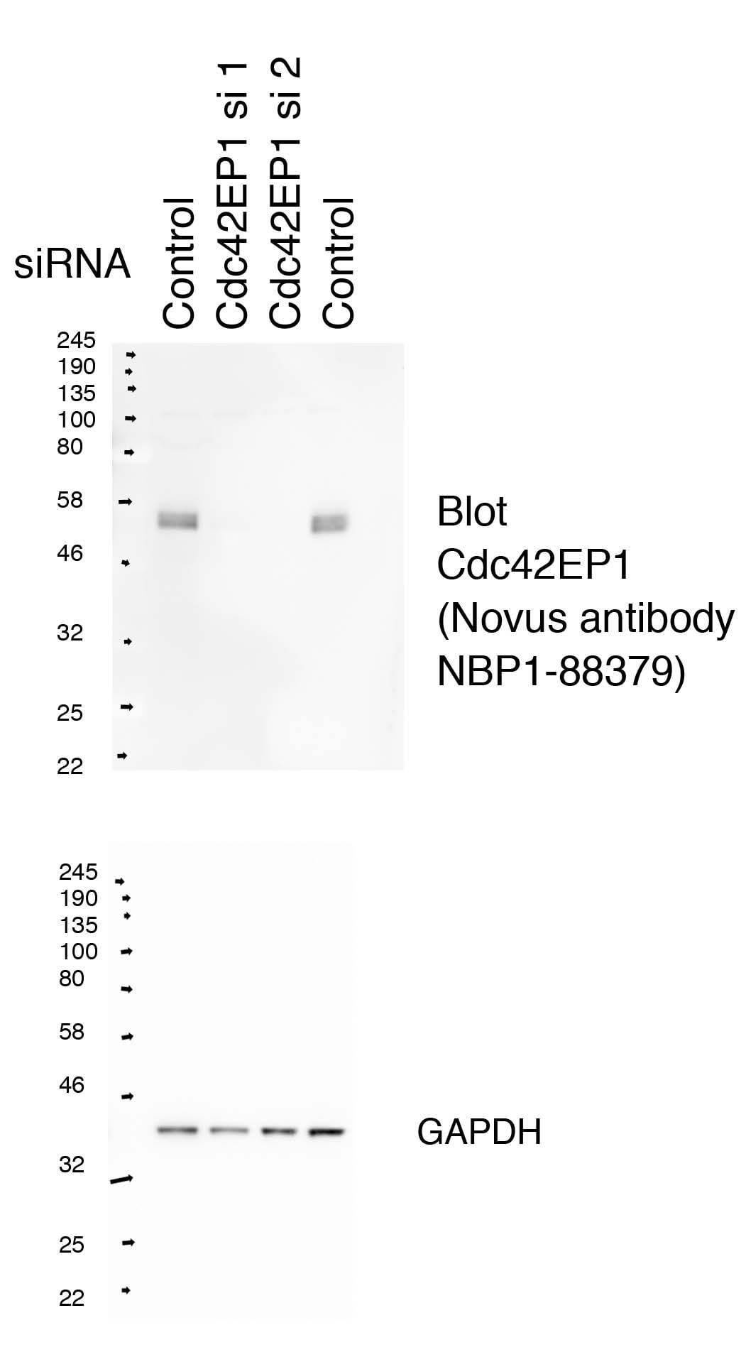
Western Blot: CDC42EP1 Antibody [NBP1-88379]
Western Blot: CDC42EP1 Antibody [NBP1-88379] - HeLa whole cell lysate. Lanes 1 and 4: HeLa cells transfected with control non-targeting siRNA. Lanes 2 and 3: HeLa cells transfected with two different siRNA-targeting Cdc42EP1. Blot probed with cdc42ep1 antibody and reprobed with GAPDH as loading control. Image from verified customer review.![Immunohistochemistry-Paraffin: CDC42EP1 Antibody [NBP1-88379]](https://resources.rndsystems.com/images/products/CDC42EP1-Antibody-Immunohistochemistry-Paraffin-NBP1-88379-img0002.jpg "Immunohistochemistry-Paraffin: CDC42EP1 Antibody [NBP1-88379]")
Immunohistochemistry-Paraffin: CDC42EP1 Antibody [NBP1-88379]
Immunohistochemistry-Paraffin: CDC42EP1 Antibody [NBP1-88379] - Staining of human stomach shows strong cytoplasmic positivity in glandular cells.![Immunoprecipitation: CDC42EP1 Antibody [NBP1-88379]](https://resources.rndsystems.com/images/products/CDC42EP1-Antibody-Immunoprecipitation-NBP1-88379-img0006.jpg "Immunoprecipitation: CDC42EP1 Antibody [NBP1-88379]")
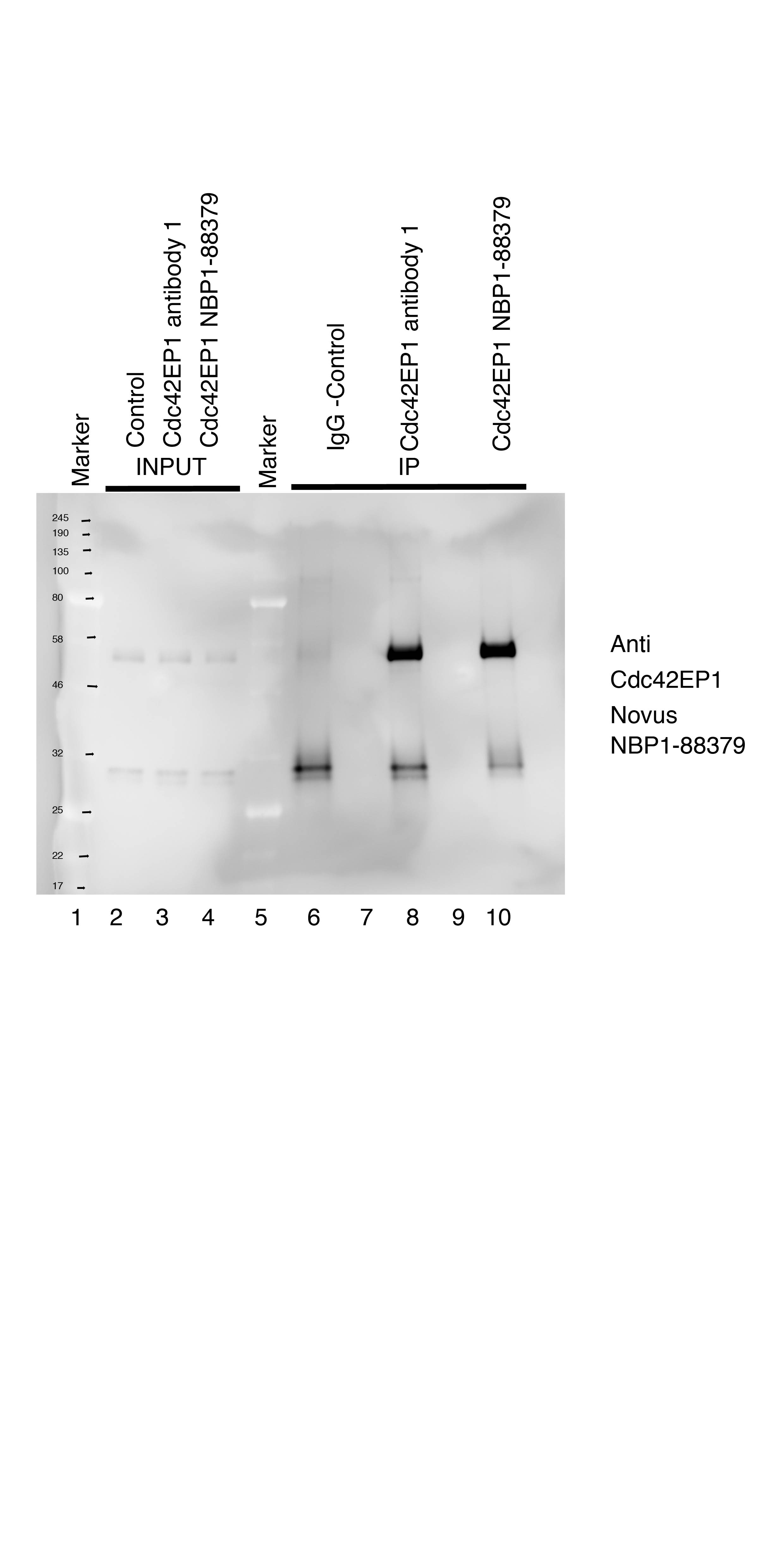
Immunoprecipitation: CDC42EP1 Antibody [NBP1-88379]
Immunoprecipitation: CDC42EP1 Antibody [NBP1-88379] - Lanes 1 and 5: Marker. Lanes 2-4 HeLa cell input controls. Lane 6: IP using 2 mg of Control Rabbit IgG cell lysate. Lane 8: IP using 2 mg Cdc42EP1 cell lysate and Betheyl antibody A302-381A. Lane 10: IP using 2 mg Cdc42EP1 cell lysate and Novus antibody NBP1-88379. Image from verified customer review.![CDC42EP1 Antibody - BSA Free Immunocytochemistry/ Immunofluorescence: CDC42EP1 Antibody [NBP1-88379]](https://resources.rndsystems.com/images/products/nbp1-88379_-immunocytochemistry-immunofluorescence-639174076711077770.jpg "Immunocytochemistry/ Immunofluorescence: CDC42EP1 Antibody [NBP1-88379]")
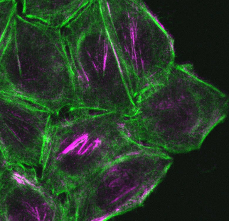
Immunocytochemistry/ Immunofluorescence: CDC42EP1 Antibody [NBP1-88379]
Staining of human cell line A-431 shows localization to plasma membrane & actin filaments.Applications for CDC42EP1 Antibody - BSA Free
Application
Recommended Usage
Immunocytochemistry/ Immunofluorescence
0.25-2 ug/ml
Immunohistochemistry
1:50 - 1:200
Immunohistochemistry-Paraffin
1:50 - 1:200
Immunoprecipitation
Validated from verified customer review
Application Notes
For IHC-Paraffin, HIER pH 6 antigen retrieval is recommended. ICC/IF fixation Permeabilization, Use PFA/Triton X-100.
Reviewed Applications
Read 3 reviews rated 5 using NBP1-88379 in the following applications:
Formulation, Preparation, and Storage
Purification
Affinity purified
Formulation
PBS (pH 7.2) and 40% Glycerol
Format
BSA Free
Preservative
0.02% Sodium Azide
Concentration
Concentrations vary lot to lot. See vial label for concentration. If unlisted please contact technical services.
Shipping
The product is shipped with polar packs. Upon receipt, store it immediately at the temperature recommended below.
Stability & Storage
Store at 4C short term. Aliquot and store at -20C long term. Avoid freeze-thaw cycles.
Background: CDC42EP1
Alternate Names
Binder of Rho GTPases 5, Borg5, BORG5cdc42 effector protein 1, CDC42 effector protein (Rho GTPase binding) 1, CEP155 kDa bone marrow stromal/endothelial cell protein, MSE55MGC15316, serum constituent protein, Serum protein MSE55
Gene Symbol
CDC42EP1
Additional CDC42EP1 Products
Product Documents for CDC42EP1 Antibody - BSA Free
Certificate of Analysis
To download a Certificate of Analysis, please enter a lot or batch number in the search box below.
Product Specific Notices for CDC42EP1 Antibody - BSA Free
This product is for research use only and is not approved for use in humans or in clinical diagnosis. Primary Antibodies are guaranteed for 1 year from date of receipt.
Customer Reviews for CDC42EP1 Antibody - BSA Free (3)
5 out of 5
3 Customer Ratings
Have you used CDC42EP1 Antibody - BSA Free?
Submit a review and receive an Amazon gift card!
$25/€18/£15/$25CAN/¥2500 Yen for a review with an image
$10/€7/£6/$10CAN/¥1110 Yen for a review without an image
Submit a review
Customer Images
Showing
1
-
3 of
3 reviews
Showing All
Filter By:
-
Application: ImmunocytochemistrySample Tested: hela cellSpecies: HumanVerified Customer | Posted 10/25/2018Confocal Imunofluorescent images of HeLa cells fixed with PFA and stained with Cdc42EP1 antibody anti 1:200 magenta & phalloidin-Alexa 488, green to visualize F-actin. Stains sub nuclear filaments some coloclisation to actin filamentsCells fixed with 4% PFA for 20 minutes room temperature. Fixed cells incubated with with 0.5% triton x100 in 1xPBS for 5 minutes room. Washed twice in blocking buffer (1% BSA 1xPBS, 20mM glycine). Washed once with blocking buffer left for 1hour room temperature. incubated with Anti Cdc42EP1 antibody in blocking buffer 1:200 for 20 hours at 4 degrees. Washed 3 x 5 minutes in blocking buffer Incubated with Anti rabbit-Alexa 594 conjugated antibody (Jackson immunoscience) for 2 hours room temperature. Washed 3 x 5 minutes in blocking buffer Washed 1 x 5 minutes in PBS Imaged with 60X oil objective on Nikon Eclipse Ti Inverted Fast confocal - Equipped with a Yokogawa CSU-X1 spinning disk unit and okogawa CSU-1 disk head and Andor Neo sCMOS camera.

-
Application: ImmunoprecipitationSample Tested: hela cellSpecies: HumanVerified Customer | Posted 08/20/20181&5-marker.2-4 HeLa cell input controls.6 IP using 2mg of Control Rabbit IgG/mg cell lysate. 8 IP using 2mg/mg of cell of Cdc42EP1 Ab from bethyl A302-381A. 8 IP using 2mg of Cdc42EP1 ab NBP1-88379/mg of cell.Vortex tube of mag protein G beads (NEB) Take 30 μl put in tube Add 1 ml of 1XPBS to wash Put on mag rack discard PBS Suspend beads in 60 μl of 1XPBS Add between 2mg of primary IP antibody to beads Put on rotating wheel at 4°C for 20h Lysis of cells Put all microfuge tubes that you are going to use on ice Cool centrifuge to 4°C Take HeLa cells grown on 10cm dishes to about 70% confluence Aspirate media wash twice with 10ml cold 1xPBS Add 1 ml of lysis buffer + 1xphosphatase + 1xprotease inhibtors (Roche) to each 10cm plate Scrape cells off plate Syringe 7-8 times with 25 G needle Transfer to microfuge tube on ice pre clear lysate by adding 60 μl of mag protein G beads (NEB) to each 1 ml of lysate Leave on ice for 10 min inverting several times. Spin tubes in cool centrifuge max speed 13K rpm for 5 min Put tubes in mag rack pipette off supernatant Save 50 μl of one of the samples to use as an input lane add 50 μl of 3X sample buffer +0.15M DTT to this boil for 5 min store -20°C save as input -load 20ul of this on gel Add 1ml of pre cleared lysate to each of the protein G –Antibody conjugates prepared day before Put on rotating wheel in cold room for 2h Put tubes on mag rack Wash once with 1ml cold 0.5% triton x100 PBS Put beads on rack remove liquid Wash 3X more with 1ml cold 1XPBS Suspend beads in 80 μl of 3X of 3X sample buffer +0.3MDTT boil 10 min Put on mag rack Transfer liquid to new tube Load 15 μl on gel along with 20 μl of inputs Gel run out on 10% SDS PAGE gel in MOPS buffer Transfered 100V 2hours 4 degrees nitrocellulose membrane blocked 5% milk PBST 1 hour incubated overnight with Cdc42EP1 antibody NBP1-88379 1:2000 in 5% milk PBST Washed 3X PBST Incubated with secondary anti rabbit HRP con light chain specific antibody 1:5000 (Jackson immunoresearch 211-032-171) in for 1hour 5% milk PBST Washed 3X PBST washed once 1X PBS ECL prime kit GE healthcare used for detection of chemilumincence
-
Application: Western BlotSample Tested: Hela whole cell lysateSpecies: HumanVerified Customer | Posted 08/03/2018Lane 1&4 HeLa cells transfected with control non targeting siRNA. Lane 2 and 3 HeLa cells transfected with two different siRNA targeting Cdc42EP1. Blot probed cdc42ep1 antibody and reprobed with GAPDH as loading control.For siRNA blot two different siRNAs were transfected at 40nM, targeting cdc42ep1 or non targeting control siRNA cells were lysed after 72 hours post tranfection Protein lysates were lysed in 1.5X Lammeli sample buffer boiled at 100 degrees for 5 minutes 10ug of protein loaded on 10% SDS PAGE gel run in MOPS buffer I used the antibody at 1:2000 in 5% milk TBST incubation overnight 4 degrees antibody gives a band at -55kD that reduces in RNAi Membranes were reprobed with GAPDH antibody as a loading control
There are no reviews that match your criteria.
Protocols
Find general support by application which include: protocols, troubleshooting, illustrated assays, videos and webinars.
- Antigen Retrieval Protocol (PIER)
- Antigen Retrieval for Frozen Sections Protocol
- Appropriate Fixation of IHC/ICC Samples
- Cellular Response to Hypoxia Protocols
- Chromogenic IHC Staining of Formalin-Fixed Paraffin-Embedded (FFPE) Tissue Protocol
- Chromogenic Immunohistochemistry Staining of Frozen Tissue
- ClariTSA™ Fluorophore Kits
- Detection & Visualization of Antibody Binding
- Fluorescent IHC Staining of Frozen Tissue Protocol
- Graphic Protocol for Heat-induced Epitope Retrieval
- Graphic Protocol for the Preparation and Fluorescent IHC Staining of Frozen Tissue Sections
- Graphic Protocol for the Preparation and Fluorescent IHC Staining of Paraffin-embedded Tissue Sections
- Graphic Protocol for the Preparation of Gelatin-coated Slides for Histological Tissue Sections
- ICC Cell Smear Protocol for Suspension Cells
- ICC Immunocytochemistry Protocol Videos
- ICC for Adherent Cells
- IHC Sample Preparation (Frozen sections vs Paraffin)
- Immunocytochemistry (ICC) Protocol
- Immunocytochemistry Troubleshooting
- Immunofluorescence of Organoids Embedded in Cultrex Basement Membrane Extract
- Immunofluorescent IHC Staining of Formalin-Fixed Paraffin-Embedded (FFPE) Tissue Protocol
- Immunohistochemistry (IHC) and Immunocytochemistry (ICC) Protocols
- Immunohistochemistry Frozen Troubleshooting
- Immunohistochemistry Paraffin Troubleshooting
- Immunoprecipitation Protocol
- Preparing Samples for IHC/ICC Experiments
- Preventing Non-Specific Staining (Non-Specific Binding)
- Primary Antibody Selection & Optimization
- Protocol for Heat-Induced Epitope Retrieval (HIER)
- Protocol for Making a 4% Formaldehyde Solution in PBS
- Protocol for VisUCyte™ HRP Polymer Detection Reagent
- Protocol for the Fluorescent ICC Staining of Cell Smears - Graphic
- Protocol for the Fluorescent ICC Staining of Cultured Cells on Coverslips - Graphic
- Protocol for the Preparation & Fixation of Cells on Coverslips
- Protocol for the Preparation and Chromogenic IHC Staining of Frozen Tissue Sections
- Protocol for the Preparation and Chromogenic IHC Staining of Frozen Tissue Sections - Graphic
- Protocol for the Preparation and Chromogenic IHC Staining of Paraffin-embedded Tissue Sections
- Protocol for the Preparation and Chromogenic IHC Staining of Paraffin-embedded Tissue Sections - Graphic
- Protocol for the Preparation and Fluorescent ICC Staining of Cells on Coverslips
- Protocol for the Preparation and Fluorescent ICC Staining of Non-adherent Cells
- Protocol for the Preparation and Fluorescent ICC Staining of Stem Cells on Coverslips
- Protocol for the Preparation and Fluorescent IHC Staining of Frozen Tissue Sections
- Protocol for the Preparation and Fluorescent IHC Staining of Paraffin-embedded Tissue Sections
- Protocol for the Preparation of Gelatin-coated Slides for Histological Tissue Sections
- Protocol for the Preparation of a Cell Smear for Non-adherent Cell ICC - Graphic
- TUNEL and Active Caspase-3 Detection by IHC/ICC Protocol
- The Importance of IHC/ICC Controls
- Troubleshooting Guide: Immunohistochemistry
- View all Protocols, Troubleshooting, Illustrated assays and Webinars
Loading...