Chromogranin A Antibody - BSA Free
Novus Biologicals | Catalog # NB120-15160
![Immunohistochemistry-Paraffin: Chromogranin A Antibody - BSA Free [NB120-15160]](https://resources.rndsystems.com/images/products/Chromogranin-A-Antibody-Immunohistochemistry-Paraffin-NB120-15160-img0003.jpg "Immunohistochemistry-Paraffin: Chromogranin A Antibody - BSA Free [NB120-15160]")
Key Product Details
Species Reactivity
Validated:
Cited:
Applications
Validated:
Cited:
Label
Antibody Source
Format
Product Specifications
Immunogen
Reactivity Notes
Localization
Clonality
Host
Isotype
Theoretical MW
Disclaimer note: The observed molecular weight of the protein may vary from the listed predicted molecular weight due to post translational modifications, post translation cleavages, relative charges, and other experimental factors.
Scientific Data Images for Chromogranin A Antibody - BSA Free
Immunohistochemistry-Paraffin: Chromogranin A Antibody - BSA Free [NB120-15160]
Immunohistochemistry-Paraffin: Chromogranin A Antibody [NB120-15160] - IHC analysis of a formalin fixed paraffin embedded tissue section of mouse pancreas using Chromogranin A antibody at a dilution of 1:500. The signal was developed using HRP-conjugated anti-rabbit secondary and DAB reagent which followed counterstaining of the section with hematoxylin. The antibody generated a strong staining in Islets of Langerhans. Also, in the periphery of the cells, the pattern of staining was punctate representing the vesicles with Chromogranin A. Cells of lobules and acini showed a weak cytoplasmic and nuclear staining which is potentially the secreted pool of Chromogranin A.![Immunohistochemistry: Chromogranin A Antibody - BSA Free [NB120-15160]](https://resources.rndsystems.com/images/products/Chromogranin-A-Antibody-Immunohistochemistry-NB120-15160-img0002.jpg "Immunohistochemistry: Chromogranin A Antibody - BSA Free [NB120-15160]")
Immunohistochemistry: Chromogranin A Antibody - BSA Free [NB120-15160]
Immunohistochemistry: Chromogranin A Antibody [NB120-15160] - IHC-P analysis fo human pancreas tissue showing specific staining of Chromogranin A in the Islets of Langerhans (dark brown staining).![Immunocytochemistry/ Immunofluorescence: Chromogranin A Antibody - BSA Free [NB120-15160]](https://resources.rndsystems.com/images/products/Chromogranin-A-Antibody-Immunocytochemistry-Immunofluorescence-NB120-15160-img0007.jpg "Immunocytochemistry/ Immunofluorescence: Chromogranin A Antibody - BSA Free [NB120-15160]")
Immunocytochemistry/ Immunofluorescence: Chromogranin A Antibody - BSA Free [NB120-15160]
Chromogranin-A-Antibody-Immunocytochemistry-Immunofluorescence-NB120-15160-img0007.jpg![Immunocytochemistry/ Immunofluorescence: Chromogranin A Antibody - BSA Free [NB120-15160]](https://resources.rndsystems.com/images/products/Chromogranin-A-Antibody-Immunocytochemistry-Immunofluorescence-NB120-15160-img0004.jpg "Immunocytochemistry/ Immunofluorescence: Chromogranin A Antibody - BSA Free [NB120-15160]")
Immunocytochemistry/ Immunofluorescence: Chromogranin A Antibody - BSA Free [NB120-15160]
Immunocytochemistry/Immunofluorescence: Chromogranin A Antibody [NB120-15160] - SH-SY5Y cells were fixed for 10 minutes using 10% formalin and then permeabilized for 5 minutes using 1X TBS + 0.5% Triton-X100. The cells were incubated with anti-Chromogranin A at 5 ug/ml overnight at 4C and detected with an anti-rabbit Dylight 488 (Green) at a 1:500 dilution. Alpha tubulin (DM1A) NB100-690 was used as a co-stain at a 1:1000 dilution and detected with an anti-mouse Dylight 550 (Red) at a 1:500 dilution. Nuclei were counterstained with DAPI (Blue). Cells were imaged using a 40X objective.![Immunocytochemistry/ Immunofluorescence: Chromogranin A Antibody - BSA Free [NB120-15160]](https://resources.rndsystems.com/images/products/Chromogranin-A-Antibody-Immunocytochemistry-Immunofluorescence-NB120-15160-img0005.jpg "Immunocytochemistry/ Immunofluorescence: Chromogranin A Antibody - BSA Free [NB120-15160]")
Immunocytochemistry/ Immunofluorescence: Chromogranin A Antibody - BSA Free [NB120-15160]
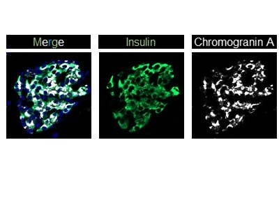
Immunocytochemistry/Immunofluorescence: Chromogranin A Antibody [NB120-15160] - Mouse pancreas cryosections stained with insulin (DAKO, Green) and Chromogranin A (NB120-15160; 1:500; White)![Immunocytochemistry/ Immunofluorescence: Chromogranin A Antibody - BSA Free [NB120-15160]](https://resources.rndsystems.com/images/products/Chromogranin-A-Antibody-Immunocytochemistry-Immunofluorescence-NB120-15160-img0006.jpg "Immunocytochemistry/ Immunofluorescence: Chromogranin A Antibody - BSA Free [NB120-15160]")
Immunocytochemistry/ Immunofluorescence: Chromogranin A Antibody - BSA Free [NB120-15160]
Immunocytochemistry/Immunofluorescence: Chromogranin A Antibody [NB120-15160] - Neuro2a cells were fixed for 10 minutes using 10% formalin and then permeabilized for 5 minutes using 1X PBS + 0.05% Triton-X100. The cells were incubated with anti-Chromogranin A Antibody at 2 ug/ml overnight at 4C and detected with an anti-rabbit Dylight 488 (Green) at a 1:500 dilution. Nuclei were counterstained with DAPI (Blue). Cells were imaged using a 40X objective.
Immunocytochemistry/ Immunofluorescence: Chromogranin A Antibody - BSA Free [NB120-15160] -
Immunocytochemistry/ Immunofluorescence: Chromogranin A Antibody - BSA Free [NB120-15160] - ChromograninA positive hormone-negative (CPHN) cells do not replicate during fetal & infant life. Representative pancreatic sections from fetal (A) & infant (B) donors immunostained for Endocrine cocktail (insulin, glucagon, somatostatin, pancreatic polypeptide, & ghrelin) (white), chromograninA (green), Ki67 (red), & DAPI (blue). Yellow arrows indicate CPHN cells. CPHN cells were rarely positive for Ki67 staining in both fetal & infant groups; no detectable difference was found in the frequency of replicative CPHN cells between fetal & infant pancreatic sections. Scale bars: 100 μm for low power & 50 μm for high magnification images. Image collected & cropped by CiteAb from the following publication (https://pubmed.ncbi.nlm.nih.gov/30687234), licensed under a CC-BY license. Not internally tested by Novus Biologicals.
Immunocytochemistry/ Immunofluorescence: Chromogranin A Antibody - BSA Free [NB120-15160] -
Immunocytochemistry/ Immunofluorescence: Chromogranin A Antibody - BSA Free [NB120-15160] - ChromograninA positive hormone-negative (CPHN) cells express the beta-cell differentiation transcription factor NKX2.2 in both fetuses & infants. Representative pancreatic sections from fetal (A) & infant (B) donors immunostained for Endocrine cocktail (insulin, glucagon, somatostatin, pancreatic polypeptide, & ghrelin) (white), chromograninA (green), the transcription factor NKX2.2 (red) & DAPI (blue). Yellow arrows indicate CPHN cells. Scale bars: 100 μm for low power & 25 μm for high magnification images. Image collected & cropped by CiteAb from the following publication (https://pubmed.ncbi.nlm.nih.gov/30687234), licensed under a CC-BY license. Not internally tested by Novus Biologicals.
Immunocytochemistry/ Immunofluorescence: Chromogranin A Antibody - BSA Free [NB120-15160] -
Immunocytochemistry/ Immunofluorescence: Chromogranin A Antibody - BSA Free [NB120-15160] - ChromograninA positive hormone-negative (CPHN) cells express the endocrine differentiation transcription factor NKX6.1 in both fetal & infant pancreas. Representative pancreatic sections from fetal (A) & infant (B) donors immunostained for Endocrine cocktail (insulin, glucagon, somatostatin, pancreatic polypeptide, & ghrelin) (white), chromograninA (green), the transcription factor NKX6.1 (red) & DAPI (blue). Yellow arrows indicate CPHN cells. Scale bars: 100 μm for low power & 25 μm for high magnification images. Image collected & cropped by CiteAb from the following publication (https://pubmed.ncbi.nlm.nih.gov/30687234), licensed under a CC-BY license. Not internally tested by Novus Biologicals.Applications for Chromogranin A Antibody - BSA Free
Immunocytochemistry/ Immunofluorescence
Immunohistochemistry
Immunohistochemistry-Paraffin
Western Blot
Reviewed Applications
Read 2 reviews rated 5 using NB120-15160 in the following applications:
Formulation, Preparation, and Storage
Purification
Formulation
Format
Preservative
Concentration
Shipping
Stability & Storage
Background: Chromogranin A
Alternate Names
Entrez Gene IDs
Gene Symbol
UniProt
Additional Chromogranin A Products
Product Documents for Chromogranin A Antibody - BSA Free
Certificate of Analysis
To download a Certificate of Analysis, please enter a lot or batch number in the search box below.
Product Specific Notices for Chromogranin A Antibody - BSA Free
This product is for research use only and is not approved for use in humans or in clinical diagnosis. Primary Antibodies are guaranteed for 1 year from date of receipt.
Citations for Chromogranin A Antibody - BSA Free
 Powered by Bioz
Customer Reviews for Chromogranin A Antibody - BSA Free (2)
Have you used Chromogranin A Antibody - BSA Free?
Submit a review and receive an Amazon gift card!
$25/€18/£15/$25CAN/¥2500 Yen for a review with an image
$10/€7/£6/$10CAN/¥1110 Yen for a review without an image
Submit a review
Customer Images
-
Application: Immunohistochemistry-FrozenSample Tested: Pancreas tissue (cryosection)Species: MouseVerified Customer | Posted 11/29/2016Mouse pancreas cryosections stained with insulin (DAKO, Green) and Chromogranin A (NB120-15160; 1:500; White)

-
Application: ImmunofluorescenceSample Tested: Mouse small intestine and colonSpecies: MouseVerified Customer | Posted 05/07/2012
There are no reviews that match your criteria.
Protocols
View specific protocols for Chromogranin A Antibody - BSA Free (NB120-15160):
Antigen Unmasking:
Bring slides to a boil in 10 mM sodium citrate buffer (pH 6.0) then maintain at a sub-boiling temperature for 10 minutes. Cool slides on bench-top for 30 minutes (keep slides in the sodium citrate buffer at all times).
Staining:
1. Wash sections in deionized water three times for 5 minutes each.
2. Wash sections in PBS for 5 minutes.
3. Block each section with 100-400 ul blocking solution (1% BSA in PBS) for 1 hour at room temperature.
4. Remove blocking solution and add 100-400 ul diluted primary antibody. Incubate overnight at 4 C.
5. Remove antibody solution and wash sections in wash buffer three times for 5 minutes each.
6. Add 100-400 ul HRP polymer conjugated secondary antibody. Incubate 30 minutes at room temperature.
7. Wash sections three times in wash buffer for 5 minutes each.
8. Add 100-400 ul DAB substrate to each section and monitor staining closely.
9. As soon as the sections develop, immerse slides in deionized water.
10. Counterstain sections in hematoxylin.
11. Wash sections in deionized water two times for 5 minutes each.
12. Dehydrate sections.
13. Mount coverslips.
Find general support by application which include: protocols, troubleshooting, illustrated assays, videos and webinars.
- Antigen Retrieval Protocol (PIER)
- Antigen Retrieval for Frozen Sections Protocol
- Appropriate Fixation of IHC/ICC Samples
- Cellular Response to Hypoxia Protocols
- Chromogenic IHC Staining of Formalin-Fixed Paraffin-Embedded (FFPE) Tissue Protocol
- Chromogenic Immunohistochemistry Staining of Frozen Tissue
- ClariTSA™ Fluorophore Kits
- Detection & Visualization of Antibody Binding
- Fluorescent IHC Staining of Frozen Tissue Protocol
- Graphic Protocol for Heat-induced Epitope Retrieval
- Graphic Protocol for the Preparation and Fluorescent IHC Staining of Frozen Tissue Sections
- Graphic Protocol for the Preparation and Fluorescent IHC Staining of Paraffin-embedded Tissue Sections
- Graphic Protocol for the Preparation of Gelatin-coated Slides for Histological Tissue Sections
- ICC Cell Smear Protocol for Suspension Cells
- ICC Immunocytochemistry Protocol Videos
- ICC for Adherent Cells
- IHC Sample Preparation (Frozen sections vs Paraffin)
- Immunocytochemistry (ICC) Protocol
- Immunocytochemistry Troubleshooting
- Immunofluorescence of Organoids Embedded in Cultrex Basement Membrane Extract
- Immunofluorescent IHC Staining of Formalin-Fixed Paraffin-Embedded (FFPE) Tissue Protocol
- Immunohistochemistry (IHC) and Immunocytochemistry (ICC) Protocols
- Immunohistochemistry Frozen Troubleshooting
- Immunohistochemistry Paraffin Troubleshooting
- Preparing Samples for IHC/ICC Experiments
- Preventing Non-Specific Staining (Non-Specific Binding)
- Primary Antibody Selection & Optimization
- Protocol for Heat-Induced Epitope Retrieval (HIER)
- Protocol for Making a 4% Formaldehyde Solution in PBS
- Protocol for VisUCyte™ HRP Polymer Detection Reagent
- Protocol for the Fluorescent ICC Staining of Cell Smears - Graphic
- Protocol for the Fluorescent ICC Staining of Cultured Cells on Coverslips - Graphic
- Protocol for the Preparation & Fixation of Cells on Coverslips
- Protocol for the Preparation and Chromogenic IHC Staining of Frozen Tissue Sections
- Protocol for the Preparation and Chromogenic IHC Staining of Frozen Tissue Sections - Graphic
- Protocol for the Preparation and Chromogenic IHC Staining of Paraffin-embedded Tissue Sections
- Protocol for the Preparation and Chromogenic IHC Staining of Paraffin-embedded Tissue Sections - Graphic
- Protocol for the Preparation and Fluorescent ICC Staining of Cells on Coverslips
- Protocol for the Preparation and Fluorescent ICC Staining of Non-adherent Cells
- Protocol for the Preparation and Fluorescent ICC Staining of Stem Cells on Coverslips
- Protocol for the Preparation and Fluorescent IHC Staining of Frozen Tissue Sections
- Protocol for the Preparation and Fluorescent IHC Staining of Paraffin-embedded Tissue Sections
- Protocol for the Preparation of Gelatin-coated Slides for Histological Tissue Sections
- Protocol for the Preparation of a Cell Smear for Non-adherent Cell ICC - Graphic
- R&D Systems Quality Control Western Blot Protocol
- TUNEL and Active Caspase-3 Detection by IHC/ICC Protocol
- The Importance of IHC/ICC Controls
- Troubleshooting Guide: Immunohistochemistry
- Troubleshooting Guide: Western Blot Figures
- Western Blot Conditions
- Western Blot Protocol
- Western Blot Protocol for Cell Lysates
- Western Blot Troubleshooting
- Western Blot Troubleshooting Guide
- View all Protocols, Troubleshooting, Illustrated assays and Webinars
FAQs for Chromogranin A Antibody - BSA Free
-
Q: I have following question about antibodies against Chromogranin A. Your products list the species but canine is not included. Does it mean that the products cannot be used on canine tissues?
A:
When we do not list a species, it means we have not tested an antibody to work with those samples. If we test an antibody and find that it does not work, we will list the species with a minus sign. Unfortunately, we haven't tried these abs on canine tissues and they may work, but we cannot guarantee it. If you would like to purchase one to try, you will be eligible for the Innovators Reward Program.