CITED1 Antibody (5H6) - Azide and BSA Free
Novus Biologicals | Catalog # H00004435-M03
![Western Blot: CITED1 Antibody (5H6) [H00004435-M03]](https://resources.rndsystems.com/images/products/CITED1-Antibody-5H6-Western-Blot-H00004435-M03-img0007.jpg "Western Blot: CITED1 Antibody (5H6) [H00004435-M03]")
Key Product Details
Species Reactivity
Validated:
Cited:
Applications
Validated:
Cited:
Label
Antibody Source
Format
Product Specifications
Immunogen
Clonality
Host
Isotype
Description
Scientific Data Images for CITED1 Antibody (5H6) - Azide and BSA Free
Western Blot: CITED1 Antibody (5H6) [H00004435-M03]
Western Blot: CITED1 Antibody (5H6) [H00004435-M03] - Analysis of CITED1 expression in transfected 293T cell line by CITED1 monoclonal antibody (M03), clone 5H6. Lane 1: CITED1 transfected lysatE (19.9 KDa). Lane 2: Non-transfected lysate.![Immunocytochemistry/ Immunofluorescence: CITED1 Antibody (5H6) [H00004435-M03]](https://resources.rndsystems.com/images/products/CITED1-Antibody-5H6-Immunocytochemistry-Immunofluorescence-H00004435-M03-img0004.jpg "Immunocytochemistry/ Immunofluorescence: CITED1 Antibody (5H6) [H00004435-M03]")
Immunocytochemistry/ Immunofluorescence: CITED1 Antibody (5H6) [H00004435-M03]
Immunocytochemistry/Immunofluorescence: CITED1 Antibody (5H6) [H00004435-M03] - analysis of CITED1 expression in human embryonic kidney cells using anti-CITED1 antibody. Image from verified customer review.![Immunohistochemistry: CITED1 Antibody (5H6) [H00004435-M03]](https://resources.rndsystems.com/images/products/CITED1-Antibody-5H6-Immunohistochemistry-H00004435-M03-img0010.jpg "Immunohistochemistry: CITED1 Antibody (5H6) [H00004435-M03]")
Immunohistochemistry: CITED1 Antibody (5H6) [H00004435-M03]
CITED1-Antibody-5H6-Immunohistochemistry-H00004435-M03-img0010.jpg![Western Blot: CITED1 Antibody (5H6) [H00004435-M03]](https://resources.rndsystems.com/images/products/CITED1-Antibody-5H6-Western-Blot-H00004435-M03-img0006.jpg "Western Blot: CITED1 Antibody (5H6) [H00004435-M03]")
Western Blot: CITED1 Antibody (5H6) [H00004435-M03]
Western Blot: CITED1 Antibody (5H6) [H00004435-M03] - Analysis of CITED1 expression in A-431 (Cat # L015V1).![Immunohistochemistry-Paraffin: CITED1 Antibody (5H6) [H00004435-M03]](https://resources.rndsystems.com/images/products/CITED1-Antibody-5H6-Immunohistochemistry-Paraffin-H00004435-M03-img0005.jpg "Immunohistochemistry-Paraffin: CITED1 Antibody (5H6) [H00004435-M03]")
Immunohistochemistry-Paraffin: CITED1 Antibody (5H6) [H00004435-M03]
Immunohistochemistry-Paraffin: CITED1 Antibody (5H6) [H00004435-M03] - Analysis of monoclonal antibody to CITED1 on formalin-fixed paraffin-embedded human testis. Antibody concentration 1.2 ug/ml![Immunohistochemistry-Paraffin: CITED1 Antibody (5H6) [H00004435-M03]](https://resources.rndsystems.com/images/products/CITED1-Antibody-5H6-Immunohistochemistry-Paraffin-H00004435-M03-img0008.jpg "Immunohistochemistry-Paraffin: CITED1 Antibody (5H6) [H00004435-M03]")
Immunohistochemistry-Paraffin: CITED1 Antibody (5H6) [H00004435-M03]
Immunohistochemistry-Paraffin: CITED1 Antibody (5H6) [H00004435-M03] - Analysis of CITED1 on paraffin embedded human fetal renal tissue. Image from verified customer review.![Immunohistochemistry: CITED1 Antibody (5H6) [H00004435-M03]](https://resources.rndsystems.com/images/products/CITED1-Antibody-5H6-Immunohistochemistry-H00004435-M03-img0009.jpg "Immunohistochemistry: CITED1 Antibody (5H6) [H00004435-M03]")
Immunohistochemistry: CITED1 Antibody (5H6) [H00004435-M03]
CITED1-Antibody-5H6-Immunohistochemistry-H00004435-M03-img0009.jpg![ELISA: CITED1 Antibody (5H6) [H00004435-M03]](https://resources.rndsystems.com/images/products/CITED1-Antibody-5H6-ELISA-H00004435-M03-img0003.jpg "ELISA: CITED1 Antibody (5H6) [H00004435-M03]")
ELISA: CITED1 Antibody (5H6) [H00004435-M03]
ELISA: CITED1 Antibody (5H6) [H00004435-M03] - Detection limit for recombinant GST tagged CITED1 is approximately 0.03ng/ml as a capture antibody. [H00004435-M03] -")
Immunohistochemistry: CITED1 Antibody (5H6) [H00004435-M03] -
Immunohistochemistry: CITED1 Antibody (5H6) [H00004435-M03] - The nephrogenic niche exhibited a complex spatial organization.(A) Pseudotime analysis of the nephrogenic niche (NPC) & the PTA. Two-dimensional DDRTree [18] embedding & the learned graph (shown as a black line) were calculated with Monocle 2 [17]. Labels & colors indicate cell types. (B) Schematic sketch of the CM indicating the distance d from the UB to the edge of the CM (solid arrow) & the relative distance s along the UB (dashed arrow), in which 0 & 1 represent the top & bottom of the CM, respectively. (C) Representative image of SIX2 & CITED1 immunostaining in a w15 human fetal kidney. Dashed lines in the insets indicate the outline of the nuclei, based on DAPI signal. Arrows in the inset point to cells in which CITED1 is concentrated in the nucleus. Scale bar = 50 μm. (D) Quantification of SIX2 & CITED1 immunostaining with respect to the distance d from UB or distance s along the UB; see panel A. Error bars indicate the SEM calculated over all evaluated profiles (n = 24). (E) Representative image of HSPA1A, NR4A1, & CKS2 immunostaining in a w15 human fetal kidney. Scale bar = 20 μm. (F) Representative image of UNCX & CITED1 immunostaining. Arrowheads indicate the presence of immunostaining signal. Scale bar = 100 μm. The numerical data underlying this figure can be found in S1 Data. CM, cap mesenchyme; NPC, nephron progenitor cell; PTA, pretubular aggregate; SEM, standard error of the mean; UB, ureteric bud; w15, week 15. Image collected & cropped by CiteAb from the following publication (https://pubmed.ncbi.nlm.nih.gov/30789893), licensed under a CC-BY license. Not internally tested by Novus Biologicals. [H00004435-M03] -")
Immunocytochemistry/ Immunofluorescence: CITED1 Antibody (5H6) [H00004435-M03] -
Immunocytochemistry/ Immunofluorescence: CITED1 Antibody (5H6) [H00004435-M03] - The nephrogenic niche exhibited a complex spatial organization.(A) Pseudotime analysis of the nephrogenic niche (NPC) & the PTA. Two-dimensional DDRTree [18] embedding & the learned graph (shown as a black line) were calculated with Monocle 2 [17]. Labels & colors indicate cell types. (B) Schematic sketch of the CM indicating the distance d from the UB to the edge of the CM (solid arrow) & the relative distance s along the UB (dashed arrow), in which 0 & 1 represent the top & bottom of the CM, respectively. (C) Representative image of SIX2 & CITED1 immunostaining in a w15 human fetal kidney. Dashed lines in the insets indicate the outline of the nuclei, based on DAPI signal. Arrows in the inset point to cells in which CITED1 is concentrated in the nucleus. Scale bar = 50 μm. (D) Quantification of SIX2 & CITED1 immunostaining with respect to the distance d from UB or distance s along the UB; see panel A. Error bars indicate the SEM calculated over all evaluated profiles (n = 24). (E) Representative image of HSPA1A, NR4A1, & CKS2 immunostaining in a w15 human fetal kidney. Scale bar = 20 μm. (F) Representative image of UNCX & CITED1 immunostaining. Arrowheads indicate the presence of immunostaining signal. Scale bar = 100 μm. The numerical data underlying this figure can be found in S1 Data. CM, cap mesenchyme; NPC, nephron progenitor cell; PTA, pretubular aggregate; SEM, standard error of the mean; UB, ureteric bud; w15, week 15. Image collected & cropped by CiteAb from the following publication (https://pubmed.ncbi.nlm.nih.gov/30789893), licensed under a CC-BY license. Not internally tested by Novus Biologicals. [H00004435-M03] -")
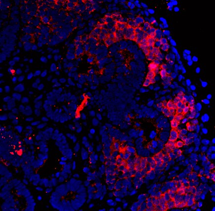
Immunocytochemistry/ Immunofluorescence: CITED1 Antibody (5H6) [H00004435-M03] -
Immunocytochemistry/ Immunofluorescence: CITED1 Antibody (5H6) [H00004435-M03] - The nephrogenic niche exhibited a complex spatial organization.(A) Pseudotime analysis of the nephrogenic niche (NPC) & the PTA. Two-dimensional DDRTree [18] embedding & the learned graph (shown as a black line) were calculated with Monocle 2 [17]. Labels & colors indicate cell types. (B) Schematic sketch of the CM indicating the distance d from the UB to the edge of the CM (solid arrow) & the relative distance s along the UB (dashed arrow), in which 0 & 1 represent the top & bottom of the CM, respectively. (C) Representative image of SIX2 & CITED1 immunostaining in a w15 human fetal kidney. Dashed lines in the insets indicate the outline of the nuclei, based on DAPI signal. Arrows in the inset point to cells in which CITED1 is concentrated in the nucleus. Scale bar = 50 μm. (D) Quantification of SIX2 & CITED1 immunostaining with respect to the distance d from UB or distance s along the UB; see panel A. Error bars indicate the SEM calculated over all evaluated profiles (n = 24). (E) Representative image of HSPA1A, NR4A1, & CKS2 immunostaining in a w15 human fetal kidney. Scale bar = 20 μm. (F) Representative image of UNCX & CITED1 immunostaining. Arrowheads indicate the presence of immunostaining signal. Scale bar = 100 μm. The numerical data underlying this figure can be found in S1 Data. CM, cap mesenchyme; NPC, nephron progenitor cell; PTA, pretubular aggregate; SEM, standard error of the mean; UB, ureteric bud; w15, week 15. Image collected & cropped by CiteAb from the following publication (https://pubmed.ncbi.nlm.nih.gov/30789893), licensed under a CC-BY license. Not internally tested by Novus Biologicals.Applications for CITED1 Antibody (5H6) - Azide and BSA Free
Flow Cytometry
Immunohistochemistry
Immunohistochemistry-Paraffin
Western Blot
Reviewed Applications
Read 2 reviews rated 4 using H00004435-M03 in the following applications:
Flow Cytometry Panel Builder
Bio-Techne Knows Flow Cytometry
Save time and reduce costly mistakes by quickly finding compatible reagents using the Panel Builder Tool.
Advanced Features
- Spectra Viewer - Custom analysis of spectra from multiple fluorochromes
- Spillover Popups - Visualize the spectra of individual fluorochromes
- Antigen Density Selector - Match fluorochrome brightness with antigen density

Formulation, Preparation, and Storage
Purification
Formulation
Format
Preservative
Concentration
Shipping
Stability & Storage
Background: CITED1
Alternate Names
Entrez Gene IDs
Gene Symbol
OMIM
UniProt
Additional CITED1 Products
Product Documents for CITED1 Antibody (5H6) - Azide and BSA Free
Certificate of Analysis
To download a Certificate of Analysis, please enter a lot or batch number in the search box below.
Product Specific Notices for CITED1 Antibody (5H6) - Azide and BSA Free
This product is produced by and distributed for Abnova, a company based in Taiwan.
This product is for research use only and is not approved for use in humans or in clinical diagnosis. Primary Antibodies are guaranteed for 1 year from date of receipt.
Citations for CITED1 Antibody (5H6) - Azide and BSA Free
 Powered by Bioz
Customer Reviews for CITED1 Antibody (5H6) - Azide and BSA Free (2)
Have you used CITED1 Antibody (5H6) - Azide and BSA Free?
Submit a review and receive an Amazon gift card!
$25/€18/£15/$25CAN/¥2500 Yen for a review with an image
$10/€7/£6/$10CAN/¥1110 Yen for a review without an image
Submit a review
Customer Images
-(01-mg)_H00004435-M03_11421.jpg)
-
Application: Immunohistochemistry-ParaffinSample Tested: fetal renal tissueSpecies: HumanVerified Customer | Posted 10/31/2016CITED1 expression in human fetal renal tissue, localized within the cap mesenchymeLow pH antigen retrieval of paraffin embedded tissue

-
Application: ImmunofluorescenceSample Tested: human fetal kidney cellsSpecies: HumanVerified Customer | Posted 10/23/2014cited1 expression in human embryonic kidney cells
There are no reviews that match your criteria.
Protocols
Find general support by application which include: protocols, troubleshooting, illustrated assays, videos and webinars.
- 7-Amino Actinomycin D (7-AAD) Cell Viability Flow Cytometry Protocol
- Antigen Retrieval Protocol (PIER)
- Antigen Retrieval for Frozen Sections Protocol
- Appropriate Fixation of IHC/ICC Samples
- Cellular Response to Hypoxia Protocols
- Chromogenic IHC Staining of Formalin-Fixed Paraffin-Embedded (FFPE) Tissue Protocol
- Chromogenic Immunohistochemistry Staining of Frozen Tissue
- ClariTSA™ Fluorophore Kits
- Detection & Visualization of Antibody Binding
- ELISA Sample Preparation & Collection Guide
- ELISA Troubleshooting Guide
- Extracellular Membrane Flow Cytometry Protocol
- Flow Cytometry Protocol for Cell Surface Markers
- Flow Cytometry Protocol for Staining Membrane Associated Proteins
- Flow Cytometry Staining Protocols
- Flow Cytometry Troubleshooting Guide
- Fluorescent IHC Staining of Frozen Tissue Protocol
- Graphic Protocol for Heat-induced Epitope Retrieval
- Graphic Protocol for the Preparation and Fluorescent IHC Staining of Frozen Tissue Sections
- Graphic Protocol for the Preparation and Fluorescent IHC Staining of Paraffin-embedded Tissue Sections
- Graphic Protocol for the Preparation of Gelatin-coated Slides for Histological Tissue Sections
- How to Run an R&D Systems DuoSet ELISA
- How to Run an R&D Systems Quantikine ELISA
- How to Run an R&D Systems Quantikine™ QuicKit™ ELISA
- ICC Cell Smear Protocol for Suspension Cells
- ICC Immunocytochemistry Protocol Videos
- ICC for Adherent Cells
- IHC Sample Preparation (Frozen sections vs Paraffin)
- Immunocytochemistry (ICC) Protocol
- Immunocytochemistry Troubleshooting
- Immunofluorescence of Organoids Embedded in Cultrex Basement Membrane Extract
- Immunofluorescent IHC Staining of Formalin-Fixed Paraffin-Embedded (FFPE) Tissue Protocol
- Immunohistochemistry (IHC) and Immunocytochemistry (ICC) Protocols
- Immunohistochemistry Frozen Troubleshooting
- Immunohistochemistry Paraffin Troubleshooting
- Intracellular Flow Cytometry Protocol Using Alcohol (Methanol)
- Intracellular Flow Cytometry Protocol Using Detergents
- Intracellular Nuclear Staining Flow Cytometry Protocol Using Detergents
- Intracellular Staining Flow Cytometry Protocol Using Alcohol Permeabilization
- Intracellular Staining Flow Cytometry Protocol Using Detergents to Permeabilize Cells
- Preparing Samples for IHC/ICC Experiments
- Preventing Non-Specific Staining (Non-Specific Binding)
- Primary Antibody Selection & Optimization
- Propidium Iodide Cell Viability Flow Cytometry Protocol
- Protocol for Heat-Induced Epitope Retrieval (HIER)
- Protocol for Liperfluo
- Protocol for Making a 4% Formaldehyde Solution in PBS
- Protocol for VisUCyte™ HRP Polymer Detection Reagent
- Protocol for the Characterization of Human Th22 Cells
- Protocol for the Characterization of Human Th9 Cells
- Protocol for the Fluorescent ICC Staining of Cell Smears - Graphic
- Protocol for the Fluorescent ICC Staining of Cultured Cells on Coverslips - Graphic
- Protocol for the Preparation & Fixation of Cells on Coverslips
- Protocol for the Preparation and Chromogenic IHC Staining of Frozen Tissue Sections
- Protocol for the Preparation and Chromogenic IHC Staining of Frozen Tissue Sections - Graphic
- Protocol for the Preparation and Chromogenic IHC Staining of Paraffin-embedded Tissue Sections
- Protocol for the Preparation and Chromogenic IHC Staining of Paraffin-embedded Tissue Sections - Graphic
- Protocol for the Preparation and Fluorescent ICC Staining of Cells on Coverslips
- Protocol for the Preparation and Fluorescent ICC Staining of Non-adherent Cells
- Protocol for the Preparation and Fluorescent ICC Staining of Stem Cells on Coverslips
- Protocol for the Preparation and Fluorescent IHC Staining of Frozen Tissue Sections
- Protocol for the Preparation and Fluorescent IHC Staining of Paraffin-embedded Tissue Sections
- Protocol for the Preparation of Gelatin-coated Slides for Histological Tissue Sections
- Protocol for the Preparation of a Cell Smear for Non-adherent Cell ICC - Graphic
- Protocol: Annexin V and PI Staining by Flow Cytometry
- Protocol: Annexin V and PI Staining for Apoptosis by Flow Cytometry
- Quantikine HS ELISA Kit Assay Principle, Alkaline Phosphatase
- Quantikine HS ELISA Kit Principle, Streptavidin-HRP Polymer
- R&D Systems Quality Control Western Blot Protocol
- Sandwich ELISA (Colorimetric) – Biotin/Streptavidin Detection Protocol
- Sandwich ELISA (Colorimetric) – Direct Detection Protocol
- TUNEL and Active Caspase-3 Detection by IHC/ICC Protocol
- The Importance of IHC/ICC Controls
- Troubleshooting Guide: ELISA
- Troubleshooting Guide: Fluorokine Flow Cytometry Kits
- Troubleshooting Guide: Immunohistochemistry
- Troubleshooting Guide: Western Blot Figures
- Western Blot Conditions
- Western Blot Protocol
- Western Blot Protocol for Cell Lysates
- Western Blot Troubleshooting
- Western Blot Troubleshooting Guide
- View all Protocols, Troubleshooting, Illustrated assays and Webinars