COLEC10 Antibody (4C1) - Azide and BSA Free
Novus Biologicals | Catalog # H00010584-M01
![Immunocytochemistry/ Immunofluorescence: COLEC10 Antibody (4C1) [H00010584-M01]](https://resources.rndsystems.com/images/products/COLEC10-Antibody-4C1-Immunocytochemistry-Immunofluorescence-H00010584-M01-img0005.jpg "Immunocytochemistry/ Immunofluorescence: COLEC10 Antibody (4C1) [H00010584-M01]")
Loading...
Key Product Details
Species Reactivity
Validated:
Human, Mouse
Cited:
Mouse
Applications
Validated:
Immunohistochemistry, Immunohistochemistry-Paraffin, ELISA, Sandwich ELISA, Immunocytochemistry/ Immunofluorescence
Cited:
IF/IHC
Label
Unconjugated
Antibody Source
Monoclonal Mouse IgG2a Kappa Clone # 4C1
Format
Azide and BSA Free
Loading...
Product Specifications
Immunogen
COLEC10 (NP_006429.1, 121 a.a. ~ 229 a.a) partial recombinant protein with GST tag. MW of the GST tag alone is 26 KDa. CGRYRKFVGQLDISIARLKTSMKFVKNVIAGIRETEEKFYYIVQEEKNYRESLTHCRIRGGMLAMPKDEAANTLIADYVAKSGFFRVFIGVNDLEREGQYMFTDNTPLQ
Reactivity Notes
Use in Mouse reported in scientific literature (PMID:28301481).
Specificity
COLEC10 - collectin sub-family member 10 (C-type lectin) (4C1)
Clonality
Monoclonal
Host
Mouse
Isotype
IgG2a Kappa
Scientific Data Images for COLEC10 Antibody (4C1) - Azide and BSA Free
Immunocytochemistry/ Immunofluorescence: COLEC10 Antibody (4C1) [H00010584-M01]
COLEC10-Antibody-4C1-Immunocytochemistry-Immunofluorescence-H00010584-M01-img0005.jpg![Immunohistochemistry-Paraffin: COLEC10 Antibody (4C1) [H00010584-M01]](https://resources.rndsystems.com/images/products/COLEC10-Antibody-4C1-Immunohistochemistry-Paraffin-H00010584-M01-img0006.jpg "Immunohistochemistry-Paraffin: COLEC10 Antibody (4C1) [H00010584-M01]")
Immunohistochemistry-Paraffin: COLEC10 Antibody (4C1) [H00010584-M01]
COLEC10-Antibody-4C1-Immunohistochemistry-Paraffin-H00010584-M01-img0006.jpg![Immunocytochemistry/ Immunofluorescence: COLEC10 Antibody (4C1) [H00010584-M01]](https://resources.rndsystems.com/images/products/COLEC10-Antibody-4C1-Immunocytochemistry-Immunofluorescence-H00010584-M01-img0003.jpg "Immunocytochemistry/ Immunofluorescence: COLEC10 Antibody (4C1) [H00010584-M01]")
Immunocytochemistry/ Immunofluorescence: COLEC10 Antibody (4C1) [H00010584-M01]
COLEC10-Antibody-4C1-Immunocytochemistry-Immunofluorescence-H00010584-M01-img0003.jpg![Immunocytochemistry/ Immunofluorescence: COLEC10 Antibody (4C1) [H00010584-M01]](https://resources.rndsystems.com/images/products/COLEC10-Antibody-4C1-Immunocytochemistry-Immunofluorescence-H00010584-M01-img0004.jpg "Immunocytochemistry/ Immunofluorescence: COLEC10 Antibody (4C1) [H00010584-M01]")
Immunocytochemistry/ Immunofluorescence: COLEC10 Antibody (4C1) [H00010584-M01]
COLEC10-Antibody-4C1-Immunocytochemistry-Immunofluorescence-H00010584-M01-img0004.jpg![Immunohistochemistry: COLEC10 Antibody (4C1) [H00010584-M01]](https://resources.rndsystems.com/images/products/COLEC10-Antibody-4C1-Immunohistochemistry-H00010584-M01-img0002.jpg "Immunohistochemistry: COLEC10 Antibody (4C1) [H00010584-M01]")
Immunohistochemistry: COLEC10 Antibody (4C1) [H00010584-M01]
COLEC10-Antibody-4C1-Immunohistochemistry-H00010584-M01-img0002.jpg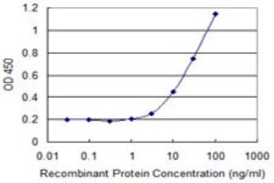
Sandwich ELISA: COLEC10 Antibody (4C1) [H00010584-M01] - Detection limit for recombinant GST tagged COLEC10 is 1 ng/ml as a capture antibody.
 [H00010584-M01] -")
Immunohistochemistry-Paraffin: COLEC10 Antibody (4C1) [H00010584-M01] -
Immunohistochemistry-Paraffin: COLEC10 Antibody (4C1) [H00010584-M01] - Cellular & embryonic localisation of CL-L1.A. Immunostaining of ATDC5 cells with the golgi marker 58K & CL-L1. CL-L1 shows localisation with golgi apparatus (white arrow). B. Laminin & CL-L1 coimmunolocalisation. Laminin shows partial cellular immunolocalisiton with CL-L1 around the golgi area (arrows). Scale bar 50 μm C. Laminin & CL-K1 coimmunolocalisation. CL-K1 staining shows a very strong golgi localisation with partial cytoplasmatic laminin colocalisation. D. CL-L1 immunohistochemistry of a 18.5 days postfertilisation mouse embryo. CL-L1 is expressed in the liver (long arrow) & submucosal patal region (short arrows). E. Immunofluorescence showing co-localisation of CL-L1 & Laminin in E13.5 mouse embryos sections. CL-L1 is expressed in the basal membrane of the ephithelium in the palate shelf of the maxilla (arrows). In contrast Laminin expression is present all around the ephitelium membrane. A faint but clear CL-L1 expression is also observed in the cytoplasm of the epithelium & in the mesenchyme of the palate. PSM; rostral extremity of right palatal shelf of maxilla. Scale bar 100 μm. Image collected & cropped by CiteAb from the following publication (https://pubmed.ncbi.nlm.nih.gov/28301481), licensed under a CC-BY license. Not internally tested by Novus Biologicals. [H00010584-M01] -")
Immunocytochemistry/ Immunofluorescence: COLEC10 Antibody (4C1) [H00010584-M01] -
Immunocytochemistry/ Immunofluorescence: COLEC10 Antibody (4C1) [H00010584-M01] - Cellular & embryonic localisation of CL-L1.A. Immunostaining of ATDC5 cells with the golgi marker 58K & CL-L1. CL-L1 shows localisation with golgi apparatus (white arrow). B. Laminin & CL-L1 coimmunolocalisation. Laminin shows partial cellular immunolocalisiton with CL-L1 around the golgi area (arrows). Scale bar 50 μm C. Laminin & CL-K1 coimmunolocalisation. CL-K1 staining shows a very strong golgi localisation with partial cytoplasmatic laminin colocalisation. D. CL-L1 immunohistochemistry of a 18.5 days postfertilisation mouse embryo. CL-L1 is expressed in the liver (long arrow) & submucosal patal region (short arrows). E. Immunofluorescence showing co-localisation of CL-L1 & Laminin in E13.5 mouse embryos sections. CL-L1 is expressed in the basal membrane of the ephithelium in the palate shelf of the maxilla (arrows). In contrast Laminin expression is present all around the ephitelium membrane. A faint but clear CL-L1 expression is also observed in the cytoplasm of the epithelium & in the mesenchyme of the palate. PSM; rostral extremity of right palatal shelf of maxilla. Scale bar 100 μm. Image collected & cropped by CiteAb from the following publication (https://pubmed.ncbi.nlm.nih.gov/28301481), licensed under a CC-BY license. Not internally tested by Novus Biologicals. [H00010584-M01] -")
Immunocytochemistry/ Immunofluorescence: COLEC10 Antibody (4C1) [H00010584-M01] -
Immunocytochemistry/ Immunofluorescence: COLEC10 Antibody (4C1) [H00010584-M01] - Cellular & embryonic localisation of CL-L1.A. Immunostaining of ATDC5 cells with the golgi marker 58K & CL-L1. CL-L1 shows localisation with golgi apparatus (white arrow). B. Laminin & CL-L1 coimmunolocalisation. Laminin shows partial cellular immunolocalisiton with CL-L1 around the golgi area (arrows). Scale bar 50 μm C. Laminin & CL-K1 coimmunolocalisation. CL-K1 staining shows a very strong golgi localisation with partial cytoplasmatic laminin colocalisation. D. CL-L1 immunohistochemistry of a 18.5 days postfertilisation mouse embryo. CL-L1 is expressed in the liver (long arrow) & submucosal patal region (short arrows). E. Immunofluorescence showing co-localisation of CL-L1 & Laminin in E13.5 mouse embryos sections. CL-L1 is expressed in the basal membrane of the ephithelium in the palate shelf of the maxilla (arrows). In contrast Laminin expression is present all around the ephitelium membrane. A faint but clear CL-L1 expression is also observed in the cytoplasm of the epithelium & in the mesenchyme of the palate. PSM; rostral extremity of right palatal shelf of maxilla. Scale bar 100 μm. Image collected & cropped by CiteAb from the following publication (https://pubmed.ncbi.nlm.nih.gov/28301481), licensed under a CC-BY license. Not internally tested by Novus Biologicals.Applications for COLEC10 Antibody (4C1) - Azide and BSA Free
Application
Recommended Usage
ELISA
Optimal dilutions of this antibody should be experimentally determined.
Immunocytochemistry/ Immunofluorescence
Optimal dilutions of this antibody should be experimentally determined.
Immunohistochemistry
Optimal dilutions of this antibody should be experimentally determined.
Immunohistochemistry-Paraffin
Optimal dilutions of this antibody should be experimentally determined.
Sandwich ELISA
Optimal dilutions of this antibody should be experimentally determined.
Application Notes
This product is useful for ELISA. Use in Immunocytochemistry/immunofluorescence reported in scientific literature (PMID 28301481).
Formulation, Preparation, and Storage
Purification
IgG purified
Formulation
In 1x PBS, pH 7.4
Format
Azide and BSA Free
Preservative
No Preservative
Concentration
Concentrations vary lot to lot. See vial label for concentration. If unlisted please contact technical services.
Shipping
The product is shipped with polar packs. Upon receipt, store it immediately at the temperature recommended below.
Stability & Storage
Aliquot and store at -20C or -80C. Avoid freeze-thaw cycles.
Background: CL-L1/COLEC10
Long Name
Collectin Liver 1
Alternate Names
CL-34, COLEC10, Collectin-10, Collectin-34
Entrez Gene IDs
10584 (Human)
Gene Symbol
COLEC10
UniProt
Additional CL-L1/COLEC10 Products
Product Documents for COLEC10 Antibody (4C1) - Azide and BSA Free
Certificate of Analysis
To download a Certificate of Analysis, please enter a lot or batch number in the search box below.
Product Specific Notices for COLEC10 Antibody (4C1) - Azide and BSA Free
This product is produced by and distributed for Abnova, a company based in Taiwan.
This product is for research use only and is not approved for use in humans or in clinical diagnosis. Primary Antibodies are guaranteed for 1 year from date of receipt.
Related Research Areas
Citations for COLEC10 Antibody (4C1) - Azide and BSA Free
 Powered by Bioz
Customer Reviews for COLEC10 Antibody (4C1) - Azide and BSA Free
There are currently no reviews for this product. Be the first to review COLEC10 Antibody (4C1) - Azide and BSA Free and earn rewards!
Have you used COLEC10 Antibody (4C1) - Azide and BSA Free?
Submit a review and receive an Amazon gift card!
$25/€18/£15/$25CAN/¥2500 Yen for a review with an image
$10/€7/£6/$10CAN/¥1110 Yen for a review without an image
Submit a review
Protocols
Find general support by application which include: protocols, troubleshooting, illustrated assays, videos and webinars.
- Antigen Retrieval Protocol (PIER)
- Antigen Retrieval for Frozen Sections Protocol
- Appropriate Fixation of IHC/ICC Samples
- Cellular Response to Hypoxia Protocols
- Chromogenic IHC Staining of Formalin-Fixed Paraffin-Embedded (FFPE) Tissue Protocol
- Chromogenic Immunohistochemistry Staining of Frozen Tissue
- ClariTSA™ Fluorophore Kits
- Detection & Visualization of Antibody Binding
- ELISA Sample Preparation & Collection Guide
- ELISA Troubleshooting Guide
- Fluorescent IHC Staining of Frozen Tissue Protocol
- Graphic Protocol for Heat-induced Epitope Retrieval
- Graphic Protocol for the Preparation and Fluorescent IHC Staining of Frozen Tissue Sections
- Graphic Protocol for the Preparation and Fluorescent IHC Staining of Paraffin-embedded Tissue Sections
- Graphic Protocol for the Preparation of Gelatin-coated Slides for Histological Tissue Sections
- How to Run an R&D Systems DuoSet ELISA
- How to Run an R&D Systems Quantikine ELISA
- How to Run an R&D Systems Quantikine™ QuicKit™ ELISA
- ICC Cell Smear Protocol for Suspension Cells
- ICC Immunocytochemistry Protocol Videos
- ICC for Adherent Cells
- IHC Sample Preparation (Frozen sections vs Paraffin)
- Immunocytochemistry (ICC) Protocol
- Immunocytochemistry Troubleshooting
- Immunofluorescence of Organoids Embedded in Cultrex Basement Membrane Extract
- Immunofluorescent IHC Staining of Formalin-Fixed Paraffin-Embedded (FFPE) Tissue Protocol
- Immunohistochemistry (IHC) and Immunocytochemistry (ICC) Protocols
- Immunohistochemistry Frozen Troubleshooting
- Immunohistochemistry Paraffin Troubleshooting
- Preparing Samples for IHC/ICC Experiments
- Preventing Non-Specific Staining (Non-Specific Binding)
- Primary Antibody Selection & Optimization
- Protocol for Heat-Induced Epitope Retrieval (HIER)
- Protocol for Making a 4% Formaldehyde Solution in PBS
- Protocol for VisUCyte™ HRP Polymer Detection Reagent
- Protocol for the Fluorescent ICC Staining of Cell Smears - Graphic
- Protocol for the Fluorescent ICC Staining of Cultured Cells on Coverslips - Graphic
- Protocol for the Preparation & Fixation of Cells on Coverslips
- Protocol for the Preparation and Chromogenic IHC Staining of Frozen Tissue Sections
- Protocol for the Preparation and Chromogenic IHC Staining of Frozen Tissue Sections - Graphic
- Protocol for the Preparation and Chromogenic IHC Staining of Paraffin-embedded Tissue Sections
- Protocol for the Preparation and Chromogenic IHC Staining of Paraffin-embedded Tissue Sections - Graphic
- Protocol for the Preparation and Fluorescent ICC Staining of Cells on Coverslips
- Protocol for the Preparation and Fluorescent ICC Staining of Non-adherent Cells
- Protocol for the Preparation and Fluorescent ICC Staining of Stem Cells on Coverslips
- Protocol for the Preparation and Fluorescent IHC Staining of Frozen Tissue Sections
- Protocol for the Preparation and Fluorescent IHC Staining of Paraffin-embedded Tissue Sections
- Protocol for the Preparation of Gelatin-coated Slides for Histological Tissue Sections
- Protocol for the Preparation of a Cell Smear for Non-adherent Cell ICC - Graphic
- Quantikine HS ELISA Kit Assay Principle, Alkaline Phosphatase
- Quantikine HS ELISA Kit Principle, Streptavidin-HRP Polymer
- Sandwich ELISA (Colorimetric) – Biotin/Streptavidin Detection Protocol
- Sandwich ELISA (Colorimetric) – Direct Detection Protocol
- TUNEL and Active Caspase-3 Detection by IHC/ICC Protocol
- The Importance of IHC/ICC Controls
- Troubleshooting Guide: ELISA
- Troubleshooting Guide: Immunohistochemistry
- View all Protocols, Troubleshooting, Illustrated assays and Webinars
Loading...