Common beta Chain Antibody (BION-1) - Chimeric - Azide and BSA Free
Novus Biologicals | Catalog # NBP3-09025
Recombinant Monoclonal Antibody
![Immunocytochemistry/ Immunofluorescence: Common beta Chain Antibody (BION-1) - Chimeric - Azide and BSA Free [NBP3-09025]](https://resources.rndsystems.com/images/products/Common-beta-Chain-Antibody-BION-1-Chimeric-Immunocytochemistry-Immunofluorescence-NBP3-09025-img0003.jpg "Immunocytochemistry/ Immunofluorescence: Common beta Chain Antibody (BION-1) - Chimeric - Azide and BSA Free [NBP3-09025]")
Loading...
Key Product Details
Species Reactivity
Human
Applications
Immunohistochemistry, Western Blot, Block/Neutralize, Flow Cytometry, Immunocytochemistry/ Immunofluorescence, Immunoprecipitation, Functional
Label
Unconjugated
Antibody Source
Recombinant Monoclonal Rabbit IgG Kappa Clone # BION-1
Format
Azide and BSA Free
Loading...
Product Specifications
Immunogen
BALB/c mice were immunized intraperitonally with COS cells transfected DQP (Domain 4 of the common beta-chain) expression constructs. The immunizations were repeated 4 times at 2-week intervals. Four weeks after the final immunization, a mouse was boosted with COS transfectants intravenously. Three days later, splenocytes were harvested and fused with NS-1 myeloma cells. Hybridoma supernatants were screened on CHO DQP cells by flow cytometry, with untransfected CHO cells as a control. All antibodies were from single hybridoma clones as selected by a limiting dilution method. MoAbs were purified from ascites fluid or hybridoma supernatant by a protein A sepharose column.
Specificity
Binds to domain 4 of the common beta-chain and recognises the same binding site used by IL-5, GM-CSF and IL-3, or binds to an epitope in close proximity to it. Residues M363, R364, E366 (B-C loop) and R418 (F-G loop) in the common beta-chain are important for BION-1 binding.
Clonality
Monoclonal
Host
Rabbit
Isotype
IgG Kappa
Scientific Data Images for Common beta Chain Antibody (BION-1) - Chimeric - Azide and BSA Free
Immunocytochemistry/ Immunofluorescence: Common beta Chain Antibody (BION-1) - Chimeric - Azide and BSA Free [NBP3-09025]
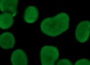
Immunocytochemistry/Immunofluorescence: Common beta Chain Antibody (BION-1) - Chimeric [NBP3-09025] - Human epithelial cells stained with NBP3-09025. ICC/IF image submitted by a verified customer review.![Flow Cytometry: Common beta Chain Antibody (BION-1) - Chimeric - Azide and BSA Free [NBP3-09025]](https://resources.rndsystems.com/images/products/Common-beta-Chain-Antibody-BION-1-Chimeric-Flow-Cytometry-NBP3-09025-img0001.jpg "Flow Cytometry: Common beta Chain Antibody (BION-1) - Chimeric - Azide and BSA Free [NBP3-09025]")
Flow Cytometry: Common beta Chain Antibody (BION-1) - Chimeric - Azide and BSA Free [NBP3-09025]
Flow Cytometry: Common beta Chain Antibody (BION-1) - Chimeric [NBP3-09025] - U937 cells were stained with anti-Fluorescein IgG antibody (4-4-20; isotype control, black line) or the rabbit IgG version of BION-1 ((NBP3-09025, blue line) at a dilution of 1:100 for 1h at RT. After washing, bound antibody was detected using a goat anti-rabbit IgG AlexaFluor(R) 488 antibody at a dilution of 1:1000 and cells analyzed using a FACSCanto flow-cytometer.![Immunocytochemistry/ Immunofluorescence: Common beta Chain Antibody (BION-1) - Chimeric - Azide and BSA Free [NBP3-09025]](https://resources.rndsystems.com/images/products/Common-beta-Chain-Antibody-BION-1-Chimeric-Immunocytochemistry-Immunofluorescence-NBP3-09025-img0002.jpg "Immunocytochemistry/ Immunofluorescence: Common beta Chain Antibody (BION-1) - Chimeric - Azide and BSA Free [NBP3-09025]")
Immunocytochemistry/ Immunofluorescence: Common beta Chain Antibody (BION-1) - Chimeric - Azide and BSA Free [NBP3-09025]
Immunocytochemistry/Immunofluorescence: Common beta Chain Antibody (BION-1) - Chimeric [NBP3-09025] - Immunofluorescence analysis of paraformaldehyde fixed U937 cells immobilized on Shi-fix(TM) cover-slips and stained with the chimeric rabbit IgG version of BION-1 (NBP3-09025) at 10 ug/ml followed by Alexa Fluor(R) 488 secondary antibody (2 ug/ml), showing membrane staining. The nuclear stain is DAPI (blue). Panels show from left-right, top-bottom NBP3-09025, DAPI, merged channels and an isotype control. The isotype control was stained with anti-Fluorescein antibody followed by Alexa Fluor(R) 488 secondary antibody.Applications for Common beta Chain Antibody (BION-1) - Chimeric - Azide and BSA Free
Application
Recommended Usage
Block/Neutralize
Optimal dilutions of this antibody should be experimentally determined.
Flow Cytometry
Optimal dilutions of this antibody should be experimentally determined.
Functional
Optimal dilutions of this antibody should be experimentally determined.
Immunocytochemistry/ Immunofluorescence
Optimal dilutions of this antibody should be experimentally determined.
Immunohistochemistry
Optimal dilutions of this antibody should be experimentally determined.
Immunoprecipitation
Optimal dilutions of this antibody should be experimentally determined.
Western Blot
Optimal dilutions of this antibody should be experimentally determined.
Application Notes
This chimeric rabbit antibody was made using the variable domain sequences of the original Mouse IgG format, for improved compatibility with existing reagents, assays and techniques.
BION-1 has been shown the block eosinophil production, survival, and activation stimulated by IL-5 as well as by GM-CSF and IL-3, thereby influencing inflammation (Sun 1999). Further, BION-1 has been used in immunohistochemistry experiments in order to examine the presence of GM-CSF-receptor protein in human 8-cell embryos and blastocysts (Sjoblom 2002). In addition, a fab fragment derived from BION-1 has been used in the crystallisation of the common beta-chain to analyse the structure of the activation domain of the GM-CSF/IL-3/IL-5 receptor (Rossjohn 200).
BION-1 has been shown the block eosinophil production, survival, and activation stimulated by IL-5 as well as by GM-CSF and IL-3, thereby influencing inflammation (Sun 1999). Further, BION-1 has been used in immunohistochemistry experiments in order to examine the presence of GM-CSF-receptor protein in human 8-cell embryos and blastocysts (Sjoblom 2002). In addition, a fab fragment derived from BION-1 has been used in the crystallisation of the common beta-chain to analyse the structure of the activation domain of the GM-CSF/IL-3/IL-5 receptor (Rossjohn 200).
Reviewed Applications
Read 1 review rated 5 using NBP3-09025 in the following applications:
Flow Cytometry Panel Builder
Bio-Techne Knows Flow Cytometry
Save time and reduce costly mistakes by quickly finding compatible reagents using the Panel Builder Tool.
Advanced Features
- Spectra Viewer - Custom analysis of spectra from multiple fluorochromes
- Spillover Popups - Visualize the spectra of individual fluorochromes
- Antigen Density Selector - Match fluorochrome brightness with antigen density

Formulation, Preparation, and Storage
Purification
Protein A purified
Formulation
PBS
Format
Azide and BSA Free
Preservative
0.02% Proclin 300
Concentration
1 mg/ml
Shipping
The product is shipped with polar packs. Upon receipt, store it immediately at the temperature recommended below.
Stability & Storage
Store at 4C short term. Aliquot and store at -20C long term. Avoid freeze-thaw cycles.
Background: Common beta Chain
Alternate Names
CD131, CSF2RB, GM-CSF R beta, IL-3 R beta, IL-5 R beta
Gene Symbol
CSF2RB
Additional Common beta Chain Products
Product Documents for Common beta Chain Antibody (BION-1) - Chimeric - Azide and BSA Free
Certificate of Analysis
To download a Certificate of Analysis, please enter a lot or batch number in the search box below.
Product Specific Notices for Common beta Chain Antibody (BION-1) - Chimeric - Azide and BSA Free
This product is for research use only and is not approved for use in humans or in clinical diagnosis. Primary Antibodies are guaranteed for 1 year from date of receipt.
Customer Reviews for Common beta Chain Antibody (BION-1) - Chimeric - Azide and BSA Free (1)
5 out of 5
1 Customer Rating
Have you used Common beta Chain Antibody (BION-1) - Chimeric - Azide and BSA Free?
Submit a review and receive an Amazon gift card!
$25/€18/£15/$25CAN/¥2500 Yen for a review with an image
$10/€7/£6/$10CAN/¥1110 Yen for a review without an image
Submit a review
Customer Images
Showing
1
-
1 of
1 review
Showing All
Filter By:
-
Application: ImmunocytochemistrySample Tested: Epithelial cellsSpecies: HumanVerified Customer | Posted 12/07/2021Human epithelial cells

There are no reviews that match your criteria.
Protocols
Find general support by application which include: protocols, troubleshooting, illustrated assays, videos and webinars.
- 7-Amino Actinomycin D (7-AAD) Cell Viability Flow Cytometry Protocol
- Antigen Retrieval Protocol (PIER)
- Antigen Retrieval for Frozen Sections Protocol
- Appropriate Fixation of IHC/ICC Samples
- Cellular Response to Hypoxia Protocols
- Chromogenic IHC Staining of Formalin-Fixed Paraffin-Embedded (FFPE) Tissue Protocol
- Chromogenic Immunohistochemistry Staining of Frozen Tissue
- ClariTSA™ Fluorophore Kits
- Detection & Visualization of Antibody Binding
- Extracellular Membrane Flow Cytometry Protocol
- Flow Cytometry Protocol for Cell Surface Markers
- Flow Cytometry Protocol for Staining Membrane Associated Proteins
- Flow Cytometry Staining Protocols
- Flow Cytometry Troubleshooting Guide
- Fluorescent IHC Staining of Frozen Tissue Protocol
- Graphic Protocol for Heat-induced Epitope Retrieval
- Graphic Protocol for the Preparation and Fluorescent IHC Staining of Frozen Tissue Sections
- Graphic Protocol for the Preparation and Fluorescent IHC Staining of Paraffin-embedded Tissue Sections
- Graphic Protocol for the Preparation of Gelatin-coated Slides for Histological Tissue Sections
- ICC Cell Smear Protocol for Suspension Cells
- ICC Immunocytochemistry Protocol Videos
- ICC for Adherent Cells
- IHC Sample Preparation (Frozen sections vs Paraffin)
- Immunocytochemistry (ICC) Protocol
- Immunocytochemistry Troubleshooting
- Immunofluorescence of Organoids Embedded in Cultrex Basement Membrane Extract
- Immunofluorescent IHC Staining of Formalin-Fixed Paraffin-Embedded (FFPE) Tissue Protocol
- Immunohistochemistry (IHC) and Immunocytochemistry (ICC) Protocols
- Immunohistochemistry Frozen Troubleshooting
- Immunohistochemistry Paraffin Troubleshooting
- Immunoprecipitation Protocol
- Intracellular Flow Cytometry Protocol Using Alcohol (Methanol)
- Intracellular Flow Cytometry Protocol Using Detergents
- Intracellular Nuclear Staining Flow Cytometry Protocol Using Detergents
- Intracellular Staining Flow Cytometry Protocol Using Alcohol Permeabilization
- Intracellular Staining Flow Cytometry Protocol Using Detergents to Permeabilize Cells
- Preparing Samples for IHC/ICC Experiments
- Preventing Non-Specific Staining (Non-Specific Binding)
- Primary Antibody Selection & Optimization
- Propidium Iodide Cell Viability Flow Cytometry Protocol
- Protocol for Heat-Induced Epitope Retrieval (HIER)
- Protocol for Liperfluo
- Protocol for Making a 4% Formaldehyde Solution in PBS
- Protocol for VisUCyte™ HRP Polymer Detection Reagent
- Protocol for the Characterization of Human Th22 Cells
- Protocol for the Characterization of Human Th9 Cells
- Protocol for the Fluorescent ICC Staining of Cell Smears - Graphic
- Protocol for the Fluorescent ICC Staining of Cultured Cells on Coverslips - Graphic
- Protocol for the Preparation & Fixation of Cells on Coverslips
- Protocol for the Preparation and Chromogenic IHC Staining of Frozen Tissue Sections
- Protocol for the Preparation and Chromogenic IHC Staining of Frozen Tissue Sections - Graphic
- Protocol for the Preparation and Chromogenic IHC Staining of Paraffin-embedded Tissue Sections
- Protocol for the Preparation and Chromogenic IHC Staining of Paraffin-embedded Tissue Sections - Graphic
- Protocol for the Preparation and Fluorescent ICC Staining of Cells on Coverslips
- Protocol for the Preparation and Fluorescent ICC Staining of Non-adherent Cells
- Protocol for the Preparation and Fluorescent ICC Staining of Stem Cells on Coverslips
- Protocol for the Preparation and Fluorescent IHC Staining of Frozen Tissue Sections
- Protocol for the Preparation and Fluorescent IHC Staining of Paraffin-embedded Tissue Sections
- Protocol for the Preparation of Gelatin-coated Slides for Histological Tissue Sections
- Protocol for the Preparation of a Cell Smear for Non-adherent Cell ICC - Graphic
- Protocol: Annexin V and PI Staining by Flow Cytometry
- Protocol: Annexin V and PI Staining for Apoptosis by Flow Cytometry
- R&D Systems Quality Control Western Blot Protocol
- TUNEL and Active Caspase-3 Detection by IHC/ICC Protocol
- The Importance of IHC/ICC Controls
- Troubleshooting Guide: Fluorokine Flow Cytometry Kits
- Troubleshooting Guide: Immunohistochemistry
- Troubleshooting Guide: Western Blot Figures
- Western Blot Conditions
- Western Blot Protocol
- Western Blot Protocol for Cell Lysates
- Western Blot Troubleshooting
- Western Blot Troubleshooting Guide
- View all Protocols, Troubleshooting, Illustrated assays and Webinars
Loading...
Associated Pathways
