Cyclooxygenase (COX), also known as prostaglandin G/H synthase, is a membrane bound enzyme partly responsible for the catalysis of prostanoid synthesis. COX is expressed as at least three different isoforms. COX-1 is constitutively expressed and thought to regulate a number of 'housekeeping' functions such as vascular hemostasis, renal blood flow, and glomerular function. COX-2 expression is tightly regulated and induced by inflammatory mediators such as growth factors, cytokines, and endotoxin. COX-3 appears to be much more strictly regulated spatially and is observed in greatest abundance in cerebral cortex.
COX-1 Antibody - BSA Free
Novus Biologicals | Catalog # NBP1-85500
![Immunohistochemistry-Paraffin: COX-1 Antibody [NBP1-85500]](https://resources.rndsystems.com/images/products/COX-1-Antibody-Immunohistochemistry-Paraffin-NBP1-85500-img0012.jpg "Immunohistochemistry-Paraffin: COX-1 Antibody [NBP1-85500]")
Loading...
Key Product Details
Validated by
Orthogonal Validation
Species Reactivity
Validated:
Human
Cited:
Human, Mouse
Predicted:
Rat (90%). Backed by our 100% Guarantee.
Applications
Validated:
Immunohistochemistry, Immunohistochemistry-Paraffin, Western Blot, Immunocytochemistry/ Immunofluorescence, Simple Western
Cited:
Immunohistochemistry-Paraffin, Western Blot
Label
Unconjugated
Antibody Source
Polyclonal Rabbit IgG
Format
BSA Free
Loading...
Product Specifications
Immunogen
This antibody was developed against Recombinant Protein corresponding to amino acids: MPDSFKVGSQEYSYEQFLFNTSMLVDYGVEALVDAFSRQIAGRIGGGRNMDHHILHVAVDVIRESREMRLQPFNEYRKRFGMKPYTSFQELVGEKEMAAELEELYGDIDALEFYPGLLLEKCHPNSIFGESMIEIGAPFSLKGLLGN
Reactivity Notes
Mouse reactivity reported from a verified customer review.
Clonality
Polyclonal
Host
Rabbit
Isotype
IgG
Scientific Data Images for COX-1 Antibody - BSA Free
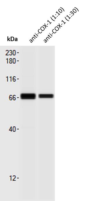
![Western Blot: COX-1 Antibody [NBP1-85500]](https://resources.rndsystems.com/images/products/COX-1-Antibody-Western-Blot-NBP1-85500-img0005.jpg "Western Blot: COX-1 Antibody [NBP1-85500]")
Western Blot: COX-1 Antibody [NBP1-85500]
Western Blot: COX-1 Antibody [NBP1-85500] - Analysis in human cell line HELA.![Immunocytochemistry/ Immunofluorescence: COX-1 Antibody [NBP1-85500]](https://resources.rndsystems.com/images/products/COX-1-Antibody-Immunocytochemistry-Immunofluorescence-NBP1-85500-img0014.jpg "Immunocytochemistry/ Immunofluorescence: COX-1 Antibody [NBP1-85500]")
Immunocytochemistry/ Immunofluorescence: COX-1 Antibody [NBP1-85500]
COX-1-Antibody-Immunocytochemistry-Immunofluorescence-NBP1-85500-img0014.jpg![Immunocytochemistry/ Immunofluorescence: COX-1 Antibody [NBP1-85500]](https://resources.rndsystems.com/images/products/COX-1-Antibody-Immunocytochemistry-Immunofluorescence-NBP1-85500-img0009.jpg "Immunocytochemistry/ Immunofluorescence: COX-1 Antibody [NBP1-85500]")
Immunocytochemistry/ Immunofluorescence: COX-1 Antibody [NBP1-85500]
Immunocytochemistry/Immunofluorescence: COX-1 Antibody [NBP1-85500] - Staining of human cell line BJ shows localization to the Golgi apparatus. Antibody staining is shown in green.![Immunohistochemistry-Paraffin: COX-1 Antibody [NBP1-85500]](https://resources.rndsystems.com/images/products/COX-1-Antibody-Immunohistochemistry-Paraffin-NBP1-85500-img0010.jpg "Immunohistochemistry-Paraffin: COX-1 Antibody [NBP1-85500]")
Immunohistochemistry-Paraffin: COX-1 Antibody [NBP1-85500]
Immunohistochemistry-Paraffin: COX-1 Antibody [NBP1-85500] - Staining of human skeletal muscle shows low expression as expected.![Immunohistochemistry-Paraffin: COX-1 Antibody [NBP1-85500]](https://resources.rndsystems.com/images/products/COX-1-Antibody-Immunohistochemistry-Paraffin-NBP1-85500-img0011.jpg "Immunohistochemistry-Paraffin: COX-1 Antibody [NBP1-85500]")
Immunohistochemistry-Paraffin: COX-1 Antibody [NBP1-85500]
Immunohistochemistry-Paraffin: COX-1 Antibody [NBP1-85500] - Staining of human skin shows high expression.![Simple Western: COX-1 Antibody [NBP1-85500]](https://resources.rndsystems.com/images/products/COX-1-Antibody-Simple-Western-NBP1-85500-img0013.jpg "Simple Western: COX-1 Antibody [NBP1-85500]")
Simple Western: COX-1 Antibody [NBP1-85500]
Simple Western: COX-1 Antibody [NBP1-85500] - Lane view shows one specific signal (~70kDa) in lysates prepared from the mouse tail vein. Image from a verified customer review.
Immunocytochemistry/ Immunofluorescence: COX-1 Antibody - BSA Free [NBP1-85500] -
Validation of gene expression changes from RNA-Seq experiments. a, c Expression of Cpt2 and Krt5 in the urothelium of Ppargfl/fl controls (a) and ShhCre;Ppargfl/fl mutants (c). b, d Cox1 and Krt5 expression in a control (b) and in the ShhCre;Ppargfl/fl mutant urothelium (d). e, g Sod1 and Krt5 expression in a Ppargfl/fl control urothelium (e) and in the urothelium of an ShhCre;Ppargfl/fl mutant (g). f, h Sod2 and Krt5 expression in the urothelium of a Ppargfl/fl control (f) and in a ShhCre;Ppargfl/fl mutant urothelium (h). i, l Uchl1 and Krt5 expression in the urothelium of a wild-type adult tongue (i) and in the urothelium of a ShhCre;Ppargfl/fl mutant (l). j, m Expression of Sprr1a and Krt5 in the urothelium of a control (j) and in a urothelium of a ShhCre;Ppargfl/fl mutant (m). k, n Cldn8 and Krt5 expression in a control urothelium (k) and in a ShhCre;Ppargfl/fl mutant (n). o, q)Krt6 and Krt5 expression in a control urothelium (o) and in a ShhCre;Ppargfl/fl mutant (q). p, r Uchl1 and Krt5 expression in a control urothelium (p) and in a ShhCre;Ppargfl/fl mutant (r) urothelium. Scale bars: 50 μm. Adult wild type, n = 6; adult mutant, n = 5 Image collected and cropped by CiteAb from the following open publication (https://pubmed.ncbi.nlm.nih.gov/31597917), licensed under a CC-BY license. Not internally tested by Novus Biologicals.
Immunocytochemistry/ Immunofluorescence: COX-1 Antibody - BSA Free [NBP1-85500] -
Validation of gene expression changes from RNA-Seq experiments. a, c Expression of Cpt2 and Krt5 in the urothelium of Ppargfl/fl controls (a) and ShhCre;Ppargfl/fl mutants (c). b, d Cox1 and Krt5 expression in a control (b) and in the ShhCre;Ppargfl/fl mutant urothelium (d). e, g Sod1 and Krt5 expression in a Ppargfl/fl control urothelium (e) and in the urothelium of an ShhCre;Ppargfl/fl mutant (g). f, h Sod2 and Krt5 expression in the urothelium of a Ppargfl/fl control (f) and in a ShhCre;Ppargfl/fl mutant urothelium (h). i, l Uchl1 and Krt5 expression in the urothelium of a wild-type adult tongue (i) and in the urothelium of a ShhCre;Ppargfl/fl mutant (l). j, m Expression of Sprr1a and Krt5 in the urothelium of a control (j) and in a urothelium of a ShhCre;Ppargfl/fl mutant (m). k, n Cldn8 and Krt5 expression in a control urothelium (k) and in a ShhCre;Ppargfl/fl mutant (n). o, q)Krt6 and Krt5 expression in a control urothelium (o) and in a ShhCre;Ppargfl/fl mutant (q). p, r Uchl1 and Krt5 expression in a control urothelium (p) and in a ShhCre;Ppargfl/fl mutant (r) urothelium. Scale bars: 50 μm. Adult wild type, n = 6; adult mutant, n = 5 Image collected and cropped by CiteAb from the following open publication (https://pubmed.ncbi.nlm.nih.gov/31597917), licensed under a CC-BY license. Not internally tested by Novus Biologicals.Applications for COX-1 Antibody - BSA Free
Application
Recommended Usage
Immunocytochemistry/ Immunofluorescence
0.25-2 ug/ml
Immunohistochemistry
1:50-1:200
Immunohistochemistry-Paraffin
1:50-1:200
Western Blot
0.04-0.4 ug/ml
Application Notes
IHC-Paraffin, HIER pH 6 retrieval is recommended. ICC/IF, Fixation Permeabilization, Use PFA/Triton X-100. COX-1 antibody validated for Simple Western from a verified customer review.
See Simple Western Antibody Database for Simple Western validation: Tested in Mouse tail vein lysates, separated by Size, apparent MW was 70 kDa
See Simple Western Antibody Database for Simple Western validation: Tested in Mouse tail vein lysates, separated by Size, apparent MW was 70 kDa
Reviewed Applications
Read 1 review rated 5 using NBP1-85500 in the following applications:
Formulation, Preparation, and Storage
Purification
Affinity purified
Formulation
PBS (pH 7.2) and 40% Glycerol
Format
BSA Free
Preservative
0.02% Sodium Azide
Concentration
Concentrations vary lot to lot. See vial label for concentration. If unlisted please contact technical services.
Shipping
The product is shipped with polar packs. Upon receipt, store it immediately at the temperature recommended below.
Stability & Storage
Store at 4C short term. Aliquot and store at -20C long term. Avoid freeze-thaw cycles.
Background: COX-1
Long Name
Cyclooxygenase 1
Alternate Names
COX1, PTGS1
Gene Symbol
PTGS1
Additional COX-1 Products
Product Documents for COX-1 Antibody - BSA Free
Certificate of Analysis
To download a Certificate of Analysis, please enter a lot or batch number in the search box below.
Product Specific Notices for COX-1 Antibody - BSA Free
This product is for research use only and is not approved for use in humans or in clinical diagnosis. Primary Antibodies are guaranteed for 1 year from date of receipt.
Related Research Areas
Citations for COX-1 Antibody - BSA Free
 Powered by Bioz
Customer Reviews for COX-1 Antibody - BSA Free (1)
5 out of 5
1 Customer Rating
Have you used COX-1 Antibody - BSA Free?
Submit a review and receive an Amazon gift card!
$25/€18/£15/$25CAN/¥2500 Yen for a review with an image
$10/€7/£6/$10CAN/¥1110 Yen for a review without an image
Submit a review
Customer Images
Showing
1
-
1 of
1 review
Showing All
Filter By:
-
Application: Simple WesternSample Tested: tail vein lysatesSpecies: MouseVerified Customer | Posted 11/02/2017Simple Western lane view shows one specific signal (~70kDa) in lysates prepared from the mouse tail vein.This run was performed under reducing conditions using the 12-230 kDa separation system in combination with a rabbit detection system. Lysis buffer: RIPA, prot. conc.: 0.7 µg/µl.

There are no reviews that match your criteria.
Protocols
Find general support by application which include: protocols, troubleshooting, illustrated assays, videos and webinars.
- Antigen Retrieval Protocol (PIER)
- Antigen Retrieval for Frozen Sections Protocol
- Appropriate Fixation of IHC/ICC Samples
- Cellular Response to Hypoxia Protocols
- Chromogenic IHC Staining of Formalin-Fixed Paraffin-Embedded (FFPE) Tissue Protocol
- Chromogenic Immunohistochemistry Staining of Frozen Tissue
- ClariTSA™ Fluorophore Kits
- Detection & Visualization of Antibody Binding
- Fluorescent IHC Staining of Frozen Tissue Protocol
- Graphic Protocol for Heat-induced Epitope Retrieval
- Graphic Protocol for the Preparation and Fluorescent IHC Staining of Frozen Tissue Sections
- Graphic Protocol for the Preparation and Fluorescent IHC Staining of Paraffin-embedded Tissue Sections
- Graphic Protocol for the Preparation of Gelatin-coated Slides for Histological Tissue Sections
- ICC Cell Smear Protocol for Suspension Cells
- ICC Immunocytochemistry Protocol Videos
- ICC for Adherent Cells
- IHC Sample Preparation (Frozen sections vs Paraffin)
- Immunocytochemistry (ICC) Protocol
- Immunocytochemistry Troubleshooting
- Immunofluorescence of Organoids Embedded in Cultrex Basement Membrane Extract
- Immunofluorescent IHC Staining of Formalin-Fixed Paraffin-Embedded (FFPE) Tissue Protocol
- Immunohistochemistry (IHC) and Immunocytochemistry (ICC) Protocols
- Immunohistochemistry Frozen Troubleshooting
- Immunohistochemistry Paraffin Troubleshooting
- Preparing Samples for IHC/ICC Experiments
- Preventing Non-Specific Staining (Non-Specific Binding)
- Primary Antibody Selection & Optimization
- Protocol for Heat-Induced Epitope Retrieval (HIER)
- Protocol for Making a 4% Formaldehyde Solution in PBS
- Protocol for VisUCyte™ HRP Polymer Detection Reagent
- Protocol for the Fluorescent ICC Staining of Cell Smears - Graphic
- Protocol for the Fluorescent ICC Staining of Cultured Cells on Coverslips - Graphic
- Protocol for the Preparation & Fixation of Cells on Coverslips
- Protocol for the Preparation and Chromogenic IHC Staining of Frozen Tissue Sections
- Protocol for the Preparation and Chromogenic IHC Staining of Frozen Tissue Sections - Graphic
- Protocol for the Preparation and Chromogenic IHC Staining of Paraffin-embedded Tissue Sections
- Protocol for the Preparation and Chromogenic IHC Staining of Paraffin-embedded Tissue Sections - Graphic
- Protocol for the Preparation and Fluorescent ICC Staining of Cells on Coverslips
- Protocol for the Preparation and Fluorescent ICC Staining of Non-adherent Cells
- Protocol for the Preparation and Fluorescent ICC Staining of Stem Cells on Coverslips
- Protocol for the Preparation and Fluorescent IHC Staining of Frozen Tissue Sections
- Protocol for the Preparation and Fluorescent IHC Staining of Paraffin-embedded Tissue Sections
- Protocol for the Preparation of Gelatin-coated Slides for Histological Tissue Sections
- Protocol for the Preparation of a Cell Smear for Non-adherent Cell ICC - Graphic
- R&D Systems Quality Control Western Blot Protocol
- TUNEL and Active Caspase-3 Detection by IHC/ICC Protocol
- The Importance of IHC/ICC Controls
- Troubleshooting Guide: Immunohistochemistry
- Troubleshooting Guide: Western Blot Figures
- Western Blot Conditions
- Western Blot Protocol
- Western Blot Protocol for Cell Lysates
- Western Blot Troubleshooting
- Western Blot Troubleshooting Guide
- View all Protocols, Troubleshooting, Illustrated assays and Webinars
FAQs for COX-1 Antibody - BSA Free
Showing
1
-
1 of
1 FAQ
Showing All
-
Q: Please could you tell me if Novus Biologicals has an active COX1 Recombinant Protein?
A: Unfortunately we do not currently have any COX1 recombinant proteins that are expected to be active.
Loading...