![Immunohistochemistry-Paraffin: COX-2 Antibody [NB100-689]](https://resources.rndsystems.com/images/products/COX-2-Antibody-Immunohistochemistry-Paraffin-NB100-689-img0010.jpg "Immunohistochemistry-Paraffin: COX-2 Antibody [NB100-689]")
Loading...
Key Product Details
Species Reactivity
Validated:
Human, Rat
Cited:
Human, Mouse, Rat
Applications
Validated:
Immunohistochemistry, Immunohistochemistry-Paraffin, Immunohistochemistry-Frozen, Western Blot, Simple Western
Cited:
Immunohistochemistry-Paraffin, Immunohistochemistry-Frozen, Western Blot, IF/IHC
Label
Unconjugated
Antibody Source
Polyclonal Rabbit IgG
Loading...
Product Specifications
Immunogen
Recombinant protein corresponding to the C-terminus of rat COX2 (the exact amino acid sequence is considered to be proprietary information).
Localization
Microsomal membrane, cytoplasmic
Specificity
This reacts with the 70 kDa protein.
Clonality
Polyclonal
Host
Rabbit
Isotype
IgG
Scientific Data Images for COX-2 Antibody
Immunohistochemistry-Paraffin: COX-2 Antibody [NB100-689]
Immunohistochemistry-Paraffin: COX-2 Antibody [NB100-689] - Formalin fixed paraffin embedded colon carcinoma stained with COX-2 antibody (NB100-689).![Simple Western: COX-2 Antibody [NB100-689]](https://resources.rndsystems.com/images/products/COX-2-Antibody-Simple-Western-NB100-689-img0003.jpg "Simple Western: COX-2 Antibody [NB100-689]")
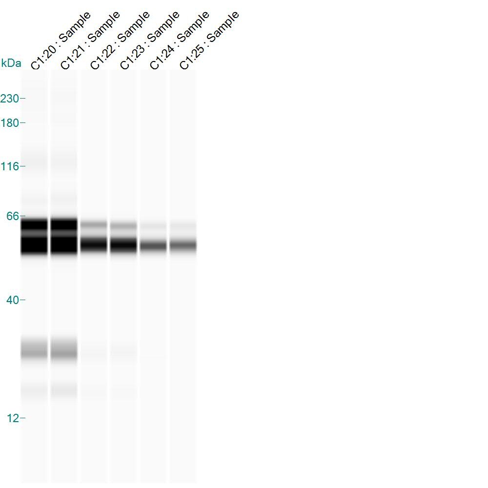
Simple Western: COX-2 Antibody [NB100-689]
Simple Western: COX-2 Antibody [NB100-689] - Simple Western lane view shows a specific band for COX2 in 1.0 mg/ml of Rat Brain at an antibody concentration of 1:100. This experiment was performed under reducing conditions using the 12-230 kDa separation system. Image provided through customer review.
Western Blot: COX-2 Antibody [NB100-689] -
(A) The COX-2 level in U-118 MG, BJ, and HaCaT cells after 24 h treatment with CXB, FL, or CXB+FL mixture at non-toxic concentrations and IC50. Results are presented as medians expressed as percent of non-treated control. The whiskers are lower (25%) and upper (75%) quartile ranges. Symbol * shows statistically significant difference against control, p < 0.05, Kruskal–Wallis test. (B) Image of immunoblots. Image collected and cropped by CiteAb from the following open publication (https://www.mdpi.com/1422-0067/25/6/3226), licensed under a CC-BY license. Not internally tested by Novus Biologicals.
Western Blot: COX-2 Antibody [NB100-689] -
Effects of Rut on the expression of cyclooxygenase-2 (COX-2), interleukin-1 beta (IL-1 beta ), and inducible nitric oxide synthase (iNOS) in LTA-stimulated RAW cells. (A–D) Cells were pretreated with Rut (10 and 20 μM) for 20 min and then stimulated by LTA (10 μg/mL) for 24 h. The levels of (B) COX-2 (C) IL-1 beta and (D) iNOS protein expression were evaluated as described in the Methods section. Data are presented as the means +/- SEM (n = 4); * p < 0.05, ** p < 0.01, and *** p < 0.001, compared with the control group; # p < 0.05 and ## p < 0.01, compared with the LTA group. Image collected and cropped by CiteAb from the following open publication (https://pubmed.ncbi.nlm.nih.gov/35682568), licensed under a CC-BY license. Not internally tested by Novus Biologicals.Applications for COX-2 Antibody
Application
Recommended Usage
Immunohistochemistry
1:50-1:100
Immunohistochemistry-Paraffin
1:50-1:100
Simple Western
1:100
Western Blot
1:100-1:2000
Application Notes
IHC-P: recommended pretreatment of citrate buffer, pH 6.0. Recommended incubation time of 30 min at RT. Use in Immunohistochemistry-Frozen reported in scientific literature (PMID: 31592103).
See Simple Western Antibody Database for Simple Western validation: Tested in Rat Brain, separated by Size, antibody dilution of 1:100, apparent MW was ~55-66 kDa
See Simple Western Antibody Database for Simple Western validation: Tested in Rat Brain, separated by Size, antibody dilution of 1:100, apparent MW was ~55-66 kDa
Reviewed Applications
Read 1 review rated 2 using NB100-689 in the following applications:
Formulation, Preparation, and Storage
Purification
Affinity purified
Formulation
PBS (pH 7.4), 0.2% BSA, Tween-20
Preservative
0.05% Sodium Azide
Concentration
Please see the vial label for concentration. If unlisted please contact technical services.
Shipping
The product is shipped with polar packs. Upon receipt, store it immediately at the temperature recommended below.
Stability & Storage
Store at 4C. Do not freeze.
Background: COX-2
Long Name
Cyclooxygenase 2
Alternate Names
COX2, PGHS-2, PHS-II, PTGS2
Gene Symbol
PTGS2
UniProt
Additional COX-2 Products
Product Documents for COX-2 Antibody
Certificate of Analysis
To download a Certificate of Analysis, please enter a lot or batch number in the search box below.
Product Specific Notices for COX-2 Antibody
This product is for research use only and is not approved for use in humans or in clinical diagnosis. Primary Antibodies are guaranteed for 1 year from date of receipt.
Citations for COX-2 Antibody
 Powered by Bioz
Customer Reviews for COX-2 Antibody (1)
2 out of 5
1 Customer Rating
Have you used COX-2 Antibody?
Submit a review and receive an Amazon gift card!
$25/€18/£15/$25CAN/¥2500 Yen for a review with an image
$10/€7/£6/$10CAN/¥1110 Yen for a review without an image
Submit a review
Customer Images
Showing
1
-
1 of
1 review
Showing All
Filter By:
-
Application: Simple WesternSample Tested: Non membrane associated protein lysate from rat brainsSpecies: RatVerified Customer | Posted 02/19/2015

There are no reviews that match your criteria.
Protocols
Find general support by application which include: protocols, troubleshooting, illustrated assays, videos and webinars.
- Antigen Retrieval Protocol (PIER)
- Antigen Retrieval for Frozen Sections Protocol
- Appropriate Fixation of IHC/ICC Samples
- Cellular Response to Hypoxia Protocols
- Chromogenic IHC Staining of Formalin-Fixed Paraffin-Embedded (FFPE) Tissue Protocol
- Chromogenic Immunohistochemistry Staining of Frozen Tissue
- ClariTSA™ Fluorophore Kits
- Detection & Visualization of Antibody Binding
- Fluorescent IHC Staining of Frozen Tissue Protocol
- Graphic Protocol for Heat-induced Epitope Retrieval
- Graphic Protocol for the Preparation and Fluorescent IHC Staining of Frozen Tissue Sections
- Graphic Protocol for the Preparation and Fluorescent IHC Staining of Paraffin-embedded Tissue Sections
- Graphic Protocol for the Preparation of Gelatin-coated Slides for Histological Tissue Sections
- IHC Sample Preparation (Frozen sections vs Paraffin)
- Immunofluorescent IHC Staining of Formalin-Fixed Paraffin-Embedded (FFPE) Tissue Protocol
- Immunohistochemistry (IHC) and Immunocytochemistry (ICC) Protocols
- Immunohistochemistry Frozen Troubleshooting
- Immunohistochemistry Paraffin Troubleshooting
- Preparing Samples for IHC/ICC Experiments
- Preventing Non-Specific Staining (Non-Specific Binding)
- Primary Antibody Selection & Optimization
- Protocol for Heat-Induced Epitope Retrieval (HIER)
- Protocol for Making a 4% Formaldehyde Solution in PBS
- Protocol for VisUCyte™ HRP Polymer Detection Reagent
- Protocol for the Preparation & Fixation of Cells on Coverslips
- Protocol for the Preparation and Chromogenic IHC Staining of Frozen Tissue Sections
- Protocol for the Preparation and Chromogenic IHC Staining of Frozen Tissue Sections - Graphic
- Protocol for the Preparation and Chromogenic IHC Staining of Paraffin-embedded Tissue Sections
- Protocol for the Preparation and Chromogenic IHC Staining of Paraffin-embedded Tissue Sections - Graphic
- Protocol for the Preparation and Fluorescent IHC Staining of Frozen Tissue Sections
- Protocol for the Preparation and Fluorescent IHC Staining of Paraffin-embedded Tissue Sections
- Protocol for the Preparation of Gelatin-coated Slides for Histological Tissue Sections
- R&D Systems Quality Control Western Blot Protocol
- TUNEL and Active Caspase-3 Detection by IHC/ICC Protocol
- The Importance of IHC/ICC Controls
- Troubleshooting Guide: Immunohistochemistry
- Troubleshooting Guide: Western Blot Figures
- Western Blot Conditions
- Western Blot Protocol
- Western Blot Protocol for Cell Lysates
- Western Blot Troubleshooting
- Western Blot Troubleshooting Guide
- View all Protocols, Troubleshooting, Illustrated assays and Webinars
FAQs for COX-2 Antibody
Showing
1
-
1 of
1 FAQ
Showing All
-
Q: Can you please tell me the concentration for product NB100-689 (Lot #Y295)?
A: The concentration for the lot# Y295 of NB100-689 (COX-2 Antibody) is 0.08 mg/ml.
Loading...