CRISPR-Cas9 Antibody (6G12) - C-terminus - Azide and BSA Free
Novus Biologicals | Catalog # NBP2-80680
Key Product Details
Validated by
Biological Validation
Species Reactivity
Bacteria
Applications
Western Blot, Immunocytochemistry/ Immunofluorescence, Simple Western, Immunoprecipitation, Chromatin Immunoprecipitation (ChIP)
Label
Unconjugated
Antibody Source
Monoclonal Mouse IgG1 kappa Clone # 6G12
Format
Azide and BSA Free
Loading...
Product Specifications
Immunogen
This CRISPR-Cas9 antibody (6G12) - C-terminus - Azide and BSA Free was raised against recombinant C-terminal fragment of S.pyogenes CRISPR/Cas9. [UniProt# Q99ZW2]
Clonality
Monoclonal
Host
Mouse
Isotype
IgG1 kappa
Theoretical MW
158.4 kDa.
Disclaimer note: The observed molecular weight of the protein may vary from the listed predicted molecular weight due to post translational modifications, post translation cleavages, relative charges, and other experimental factors.
Disclaimer note: The observed molecular weight of the protein may vary from the listed predicted molecular weight due to post translational modifications, post translation cleavages, relative charges, and other experimental factors.
Scientific Data Images for CRISPR-Cas9 Antibody (6G12) - C-terminus - Azide and BSA Free
![Western Blot: CRISPR-Cas9 Antibody (6G12)C-terminusAzide and BSA Free [NBP2-80680]](https://resources.rndsystems.com/images/products/CRISPR-Cas9-Antibody-6G12-C-terminus-Azide-and-BSA-Free-Western-Blot-NBP2-80680-img0002.jpg "Western Blot: CRISPR-Cas9 Antibody (6G12)C-terminusAzide and BSA Free [NBP2-80680]")
Western Blot: CRISPR-Cas9 Antibody (6G12)C-terminusAzide and BSA Free [NBP2-80680]
Western Blot: CRISPR-Cas9 Antibody (6G12) - C-terminus - Azide and BSA Free [NBP2-80680] - Whole cell protein from 293T cells transfected with Cas9-Flag (~150 kDa) was separated on a 7.5% gel by SDS-PAGE, transferred to PVDF membrane and blocked in 5% non-fat milk in TBST. The membrane was probed with 2 ug/mL anti-Cas9 (6G12) in 1% milk, and detected with an anti-mouse HRP secondary antibody using chemiluminescence. Image from the standard format of this antibody.![Immunocytochemistry/ Immunofluorescence: CRISPR-Cas9 Antibody (6G12) - C-terminus - Azide and BSA Free [NBP2-80680]](https://resources.rndsystems.com/images/products/CRISPR-Cas9-Antibody-6G12-C-terminus-Azide-and-BSA-Free-Immunocytochemistry-Immunofluorescence-NBP2-80680-img0003.jpg "Immunocytochemistry/ Immunofluorescence: CRISPR-Cas9 Antibody (6G12) - C-terminus - Azide and BSA Free [NBP2-80680]")
Immunocytochemistry/ Immunofluorescence: CRISPR-Cas9 Antibody (6G12) - C-terminus - Azide and BSA Free [NBP2-80680]
Immunocytochemistry/Immunofluorescence: CRISPR-Cas9 Antibody (6G12) - C-terminus - Azide and BSA Free [NBP2-80680] - HeLa cells or HeLa cells expressing Flag-tagged SpCas9 under the control of the PTight (Tet-ON) promoter were treated for 24h with 1ug/uL Doxycyclin, fixed and permeabilized with Methanol/Acetone and blocked in 2% BSA in PBS for 2 hours at RT. Cells were stained with 6G12 hybridoma supernatant at 1:10 at 4C O/N, followed by incubation with anti mouse-Alexa Fluor 488 coupled secondary antibody for 1h at RT. Nuclei were counter-stained with Hoechst 33342. Image from the standard format of this antibody.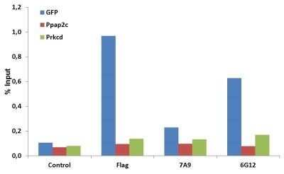
Chromatin Immunoprecipitation: CRISPR-Cas9 Antibody (6G12) - C-terminus - Azide and BSA Free [NBP2-80680] - NIH3T3 cells stably expressing GFP-H2B, nuclease dead Cas9, and a GFP-targeting gRNA were fixed with formaldehyde, harvested and sonicated to get 200-500bp DNA fragments. 50ug chromatin was incubated over night at 4C with the indicated antibodies (200ul hybridoma SN, 5ug anti-Flag [M2, Sigma]) followed by incubation with protein G beads for 3h at 4C. After washing chromatin was eluted from the beads and crosslinking was reversed over night at 65C. After a proteinase K digestion step, DNA was separated using phenol/chloroform/isoamyl alcohol, precipitated with ethanol/sodium acetate and dissolved in water. For qPCR, primers either targeting the GFP gene or as negative control non-targeted regions (Ppap2c +7122 and Prkcd +24069 from transcription start) were used. Image from the standard format of this antibody.
![Western Blot: CRISPR-Cas9 Antibody (6G12)C-terminusAzide and BSA Free [NBP2-80680]](https://resources.rndsystems.com/images/products/CRISPR-Cas9-Antibody-6G12-C-terminus-Azide-and-BSA-Free-Western-Blot-NBP2-80680-img0001.jpg "Western Blot: CRISPR-Cas9 Antibody (6G12)C-terminusAzide and BSA Free [NBP2-80680]")
Western Blot: CRISPR-Cas9 Antibody (6G12)C-terminusAzide and BSA Free [NBP2-80680]
Western Blot: CRISPR-Cas9 Antibody (6G12) - C-terminus - Azide and BSA Free [NBP2-80680] - Control HeLa cells (un-transfected) and HeLa cells expressing Flag-tagged S. pyogenes's CRISPR-Cas9 under the control of PTight (Tet-ON) promoter. Samples were treated for 24 hours with 1ug/uL of Doxycyclin and lysed under native conditions. 30 ug of the whole cell lysate from each sample type per lane was separated by 7.5% SDS-PAGE. Nitrocellulose membrane was incubated with CRISPR-Cas9 antibody clone 6G12 (hybridoma supernatant diluted 1:100 at 4C O/N). After washing, the membranes were incubated with secondary HRP-coupled antibody and bands were visualized by ECL and exposure of X-ray films. Prestained marker bands were visualized with Blue Marker Antibody (NBP2-33376). The image shown is from 1 minute exposure time. Observed molecular weight is ~158 kDa. Image from the standard format of this antibody.![Simple Western: CRISPR-Cas9 Antibody (6G12)C-terminusAzide and BSA Free [NBP2-80680]](https://resources.rndsystems.com/images/products/CRISPR-Cas9-Antibody-6G12-C-terminus-Azide-and-BSA-Free-Simple-Western-NBP2-80680-img0006.jpg "Simple Western: CRISPR-Cas9 Antibody (6G12)C-terminusAzide and BSA Free [NBP2-80680]")
Simple Western: CRISPR-Cas9 Antibody (6G12)C-terminusAzide and BSA Free [NBP2-80680]
Simple Western: CRISPR-Cas9 Antibody (6G12) - C-terminus - Azide and BSA Free [NBP2-80680] - Image shows a specific band for Cas9 in 0.2 mg/mL of HeLa Cas9 lysate but not in Hela WT lysate. This experiment was performed under reducing conditions using the 12-230 kDa separation system. Observed molecular weight is ~158 kDa. Image from the standard format of this antibody.![Immunoprecipitation: CRISPR-Cas9 Antibody (6G12) - C-terminus - Azide and BSA Free [NBP2-80680]](https://resources.rndsystems.com/images/products/CRISPR-Cas9-Antibody-6G12-C-terminus-Azide-and-BSA-Free-Immunoprecipitation-NBP2-80680-img0005.jpg "Immunoprecipitation: CRISPR-Cas9 Antibody (6G12) - C-terminus - Azide and BSA Free [NBP2-80680]")
Immunoprecipitation: CRISPR-Cas9 Antibody (6G12) - C-terminus - Azide and BSA Free [NBP2-80680]
Immunoprecipitation: CRISPR-Cas9 Antibody (6G12) - C-terminus - Azide and BSA Free [NBP2-80680] - HEK293 cells expressing Flag-SpCas9 were lysed under native conditions. SpCas9 was immunoprecipitated at 4C from 300 ug of whole cell lysate with the 6G12 antibody and a 1:1 mixture of protein A/G sepharose. After 4x washing, the bound proteins were boiled off the beads, separated by 7.5% SDS-PAGE and transfered to nitrocellulose membranes, and SpCas9 was detected with a rabbit polyclonal Cas9 antibody. After washing, the membranes were incubated with secondary HRP-coupled antibody and bands were visualized by ECL and exposure of X-ray films. Image from the standard format of this antibody.Applications for CRISPR-Cas9 Antibody (6G12) - C-terminus - Azide and BSA Free
Application
Recommended Usage
Immunocytochemistry/ Immunofluorescence
1:500
Simple Western
10-20 ug/ml
Western Blot
1:1000
Formulation, Preparation, and Storage
Purification
Protein G purified
Formulation
PBS
Format
Azide and BSA Free
Preservative
No Preservative
Concentration
1 mg/ml
Shipping
The product is shipped with polar packs. Upon receipt, store it immediately at the temperature recommended below.
Stability & Storage
Store at 4C short term. Aliquot and store at -20C long term. Avoid freeze-thaw cycles.
Background: CRISPR-Cas9
Using CRISPR-Cas9 technology, double-stranded DNA breaks may be induced within specific targeted genome sequences (target DNA; protospacer) for insertion or removal of DNA sequences for gene editing applications. To target a specific loci, a gRNA that will bind to a specific target sequence of DNA within a genome is created. The gRNA will recognize the DNA sequence, and the Cas9 enzyme will cleave the DNA at the targeted location. Once the targeted DNA is removed by Cas9, the cell's own DNA repair mechanism is used to insert or remove a DNA sequence for genomic editing.
Cas9 detection is used to confirm and evaluate CRISPR Cas9 gRNA transfection efficiency. Western blot analysis of CRISPR-Cas9 gRNA transfected cell lysates with Cas9 antibodies identifies the protein having a theoretical molecular weight of 160kDa. Broad areas of research are benefiting from CRISPR-Cas9 based gene editing tools including studies of basic immunity functions, genetic screening and disease treatment (2). Ethical concerns have led to many countries making it illegal to manipulate human germline cells or perform embryo genome editing.
References
1. Oakes, B. L., Fellmann, C., Rishi, H., Taylor, K. L., Ren, S. M., Nadler, D. C.,... Savage, D. F. (2019). CRISPR-Cas9 Circular Permutants as Programmable Scaffolds for Genome Modification. Cell, 176(1-2), 254-267.e216. doi:10.1016/j.cell.2018.11.052
2. Chiou, S. H., Winters, I. P., Wang, J., Naranjo, S., Dudgeon, C., Tamburini, F. B.,... Winslow, M. M. (2015). Pancreatic cancer modeling using retrograde viral vector delivery and in vivo CRISPR/Cas9-mediated somatic genome editing. Genes Dev, 29(14), 1576-1585. doi:10.1101/gad.264861.115
Long Name
CRISPR-associated Protein 9
Alternate Names
Cas9, CRISPR-associated endonuclease Cas9/Csn1, CRISPR-Cas9/Csn1, CRISPR/Cas9, csn1, SPy_1046, SPy1046, SpyCas9
Additional CRISPR-Cas9 Products
Product Documents for CRISPR-Cas9 Antibody (6G12) - C-terminus - Azide and BSA Free
Certificate of Analysis
To download a Certificate of Analysis, please enter a lot or batch number in the search box below.
Product Specific Notices for CRISPR-Cas9 Antibody (6G12) - C-terminus - Azide and BSA Free
This product is for research use only and is not approved for use in humans or in clinical diagnosis. Primary Antibodies are guaranteed for 1 year from date of receipt.
Customer Reviews for CRISPR-Cas9 Antibody (6G12) - C-terminus - Azide and BSA Free
There are currently no reviews for this product. Be the first to review CRISPR-Cas9 Antibody (6G12) - C-terminus - Azide and BSA Free and earn rewards!
Have you used CRISPR-Cas9 Antibody (6G12) - C-terminus - Azide and BSA Free?
Submit a review and receive an Amazon gift card!
$25/€18/£15/$25CAN/¥2500 Yen for a review with an image
$10/€7/£6/$10CAN/¥1110 Yen for a review without an image
Submit a review
Protocols
Find general support by application which include: protocols, troubleshooting, illustrated assays, videos and webinars.
- Appropriate Fixation of IHC/ICC Samples
- Cellular Response to Hypoxia Protocols
- ChIP Protocol Video
- Chromatin Immunoprecipitation (ChIP) Protocol
- Chromatin Immunoprecipitation Protocol
- ClariTSA™ Fluorophore Kits
- Detection & Visualization of Antibody Binding
- ICC Cell Smear Protocol for Suspension Cells
- ICC Immunocytochemistry Protocol Videos
- ICC for Adherent Cells
- Immunocytochemistry (ICC) Protocol
- Immunocytochemistry Troubleshooting
- Immunofluorescence of Organoids Embedded in Cultrex Basement Membrane Extract
- Immunohistochemistry (IHC) and Immunocytochemistry (ICC) Protocols
- Immunoprecipitation Protocol
- Preparing Samples for IHC/ICC Experiments
- Preventing Non-Specific Staining (Non-Specific Binding)
- Primary Antibody Selection & Optimization
- Protocol for VisUCyte™ HRP Polymer Detection Reagent
- Protocol for the Fluorescent ICC Staining of Cell Smears - Graphic
- Protocol for the Fluorescent ICC Staining of Cultured Cells on Coverslips - Graphic
- Protocol for the Preparation and Fluorescent ICC Staining of Cells on Coverslips
- Protocol for the Preparation and Fluorescent ICC Staining of Non-adherent Cells
- Protocol for the Preparation and Fluorescent ICC Staining of Stem Cells on Coverslips
- Protocol for the Preparation of a Cell Smear for Non-adherent Cell ICC - Graphic
- R&D Systems Quality Control Western Blot Protocol
- TUNEL and Active Caspase-3 Detection by IHC/ICC Protocol
- The Importance of IHC/ICC Controls
- Troubleshooting Guide: Western Blot Figures
- Western Blot Conditions
- Western Blot Protocol
- Western Blot Protocol for Cell Lysates
- Western Blot Troubleshooting
- Western Blot Troubleshooting Guide
- View all Protocols, Troubleshooting, Illustrated assays and Webinars
Loading...