CRISPR-Cas9 Antibody (7A9-3A3) - N-Terminus - BSA Free
Novus Biologicals | Catalog # NBP2-36440
Key Product Details
Species Reactivity
Validated:
Bacteria
Cited:
Human, Mouse, Bacteria, Bacterial, Fungi
Applications
Validated:
Knockout Validated, Immunohistochemistry, Immunohistochemistry-Frozen, Immunohistochemistry Whole-Mount, Western Blot, Immunoblotting, Immunocytochemistry/ Immunofluorescence, Simple Western, Immunoprecipitation
Cited:
Knockout Validated, Immunohistochemistry-Frozen, Western Blot, Immunocytochemistry/ Immunofluorescence, Simple Western, IF/IHC
Label
Unconjugated
Antibody Source
Monoclonal Mouse IgG1 kappa Clone # 7A9-3A3
Format
BSA Free
Loading...
Product Specifications
Immunogen
This CRISPR-Cas9 antibody (7A9-3A3) - N-Terminus was raised against Recombinant Cas9 within the N-terminal region of Streptococcus pyogene. [Uniprot: Q99ZW2].
Specificity
This CRISPR-Cas9 antibody (7A9-3A3) - N-Terminus is specific toCas9 protein from Streptococcus pyogene serotype M1.
Clonality
Monoclonal
Host
Mouse
Isotype
IgG1 kappa
Theoretical MW
158.4 kDa.
Disclaimer note: The observed molecular weight of the protein may vary from the listed predicted molecular weight due to post translational modifications, post translation cleavages, relative charges, and other experimental factors.
Disclaimer note: The observed molecular weight of the protein may vary from the listed predicted molecular weight due to post translational modifications, post translation cleavages, relative charges, and other experimental factors.
Scientific Data Images for CRISPR-Cas9 Antibody (7A9-3A3) - N-Terminus - BSA Free
![Simple Western: CRISPR-Cas9 Antibody (7A9-3A3)N-TerminusBSA Free [NBP2-36440]](https://resources.rndsystems.com/images/products/CRISPR-Cas9-Antibody-7A9-3A3-N-Terminus-Simple-Western-NBP2-36440-img0013.jpg "Simple Western: CRISPR-Cas9 Antibody (7A9-3A3)N-TerminusBSA Free [NBP2-36440]")
Simple Western: CRISPR-Cas9 Antibody (7A9-3A3)N-TerminusBSA Free [NBP2-36440]
Simple Western: CRISPR-Cas9 Antibody (7A9-3A3) - N-Terminus [NBP2-36440] - Image shows a specific band for Cas9 (observed molecular weight ~158 kDa) in HeLa Cas9 lysate but not in Hela WT lysate. This experiment was performed under reducing conditions using the 12-230 kDa separation system.![Immunocytochemistry/ Immunofluorescence: CRISPR-Cas9 Antibody (7A9-3A3) - N-Terminus - BSA Free [NBP2-36440]](https://resources.rndsystems.com/images/products/CRISPR-Cas9-Antibody-7A9-3A3-N-Terminus-Immunocytochemistry-Immunofluorescence-NBP2-36440-img0002.jpg "Immunocytochemistry/ Immunofluorescence: CRISPR-Cas9 Antibody (7A9-3A3) - N-Terminus - BSA Free [NBP2-36440]")
Immunocytochemistry/ Immunofluorescence: CRISPR-Cas9 Antibody (7A9-3A3) - N-Terminus - BSA Free [NBP2-36440]
Immunocytochemistry/Immunofluorescence: CRISPR-Cas9 Antibody (7A9-3A3) - N-Terminus [NBP2-36440] - Hela cells were transiently transfected with an N-terminally Flag-tagged S. pyogenes Cas9 expression vector. The cells were stained with the Cas9 antibody followed by anti mouse-AF488 coupled secondary antibody. Nuclei were counter-stained with Hoechst 33342.![Immunoprecipitation: CRISPR-Cas9 Antibody (7A9-3A3) - N-Terminus - BSA Free [NBP2-36440]](https://resources.rndsystems.com/images/products/CRISPR-Cas9-Antibody-7A9-3A3-N-Terminus-Immunoprecipitation-NBP2-36440-img0011.jpg "Immunoprecipitation: CRISPR-Cas9 Antibody (7A9-3A3) - N-Terminus - BSA Free [NBP2-36440]")
Immunoprecipitation: CRISPR-Cas9 Antibody (7A9-3A3) - N-Terminus - BSA Free [NBP2-36440]
Immunoprecipitation: CRISPR-Cas9 Antibody (7A9-3A3) - N-Terminus [NBP2-36440] - HEK293T expressing N-terminally Flag-tagged S.pyogenes Cas9 were lysed 72h post transfection by resuspending the cells in Hunt buffer and subjecting to 3 freeze-thaw cycles in liquid nitrogen/ice. Proteins were immunoprecipitated from 100ug of whole cell lysate for 1h at 4C with Cas9 supernatant followed by incubation for 1h at 4C with a 1:1 mixture of protein A/G sepharose beads, or for 2h at 4C with Cas9 ab crosslinked to a 1:1 mixture of protein A/G sepharose beads. Beads were washed 2x with Hunt buffer and 1x with TBS. Bound proteins were eluted by boiling in Laemmli, separated by SDS-PAGE and transferred to nitrocellulose. Membrane was blocked, incubated with Cas9 ab, incubated with HRP anti-mouse secondary. *IgG heavy chain![Western Blot: CRISPR-Cas9 Antibody (7A9-3A3)N-TerminusBSA Free [NBP2-36440]](https://resources.rndsystems.com/images/products/CRISPR-Cas9-Antibody-7A9-3A3-N-Terminus-Western-Blot-NBP2-36440-img0008.jpg "Western Blot: CRISPR-Cas9 Antibody (7A9-3A3)N-TerminusBSA Free [NBP2-36440]")
Western Blot: CRISPR-Cas9 Antibody (7A9-3A3)N-TerminusBSA Free [NBP2-36440]
Western Blot: CRISPR-Cas9 Antibody (7A9-3A3) - N-Terminus [NBP2-36440] - Analysis of lysate from Cas9 transfected HEK-293T cells using Cas9 antibody clone 7A9-3A3 at 2ug/ml concentration. The signal was developed using HRP-labelled anti-mouse secondary antibody and ECL based detection. Observed molecular weight is ~158 kDa.![Western Blot: CRISPR-Cas9 Antibody (7A9-3A3)N-TerminusBSA Free [NBP2-36440]](https://resources.rndsystems.com/images/products/CRISPR-Cas9-Antibody-7A9-3A3-N-Terminus-Western-Blot-NBP2-36440-img0016.jpg "Western Blot: CRISPR-Cas9 Antibody (7A9-3A3)N-TerminusBSA Free [NBP2-36440]")
Western Blot: CRISPR-Cas9 Antibody (7A9-3A3)N-TerminusBSA Free [NBP2-36440]
CRISPR-Cas9-Antibody-7A9-3A3-N-Terminus-Western-Blot-NBP2-36440-img0016.jpg![Immunocytochemistry/ Immunofluorescence: CRISPR-Cas9 Antibody (7A9-3A3) - N-Terminus - BSA Free [NBP2-36440]](https://resources.rndsystems.com/images/products/CRISPR-Cas9-Antibody-7A9-3A3-N-Terminus-Immunocytochemistry-Immunofluorescence-NBP2-36440-img0014.jpg "Immunocytochemistry/ Immunofluorescence: CRISPR-Cas9 Antibody (7A9-3A3) - N-Terminus - BSA Free [NBP2-36440]")
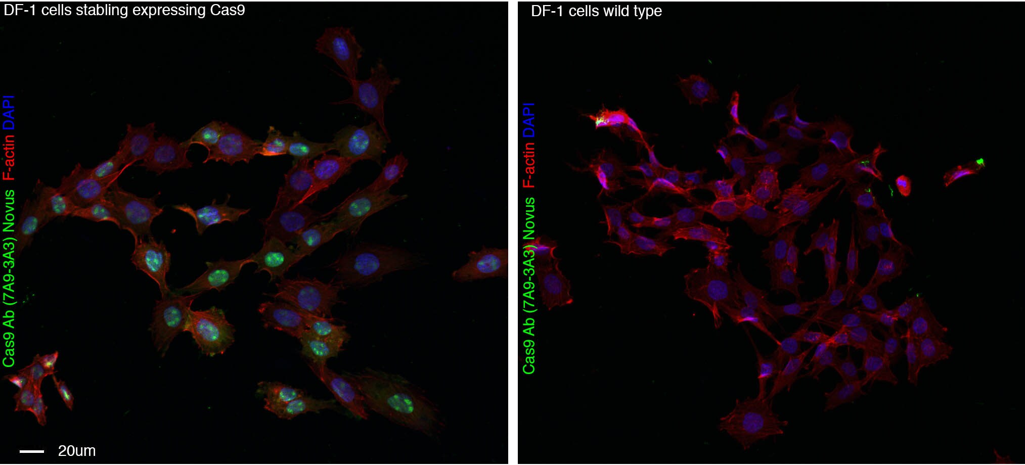
Immunocytochemistry/ Immunofluorescence: CRISPR-Cas9 Antibody (7A9-3A3) - N-Terminus - BSA Free [NBP2-36440]
Immunocytochemistry/Immunofluorescence: CRISPR-Cas9 Antibody (7A9-3A3) - N-Terminus [NBP2-36440] - DF-1 stable cell line (chicken fibroblast) expressing Cas9 (left )or wildtype DF-1 cells (right) stained with Cas9 antibody NBP2-36440 and phalloidin and DAPI to visualise F-actin and DNA respectively. Cells fixed with 4% PFA. Antibody at 1:500 overnight at 4C. ICC/IF image submitted by a verified customer review.![Immunohistochemistry-Frozen: CRISPR-Cas9 Antibody (7A9-3A3) - N-Terminus - BSA Free [NBP2-36440]](https://resources.rndsystems.com/images/products/CRISPR-Cas9-Antibody-7A9-3A3-N-Terminus-Immunohistochemistry-Frozen-NBP2-36440-img0010.jpg "Immunohistochemistry-Frozen: CRISPR-Cas9 Antibody (7A9-3A3) - N-Terminus - BSA Free [NBP2-36440]")
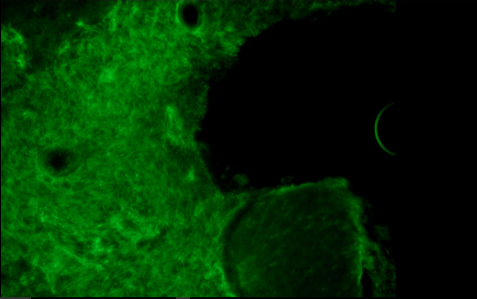
Immunohistochemistry-Frozen: CRISPR-Cas9 Antibody (7A9-3A3) - N-Terminus - BSA Free [NBP2-36440]
Immunohistochemistry-Frozen: CRISPR-Cas9 Antibody (7A9-3A3) - N-Terminus [NBP2-36440] - Analysis of a formalin fixed 20um thick frozen section of mouse brain with GBM xenograft tumor areas (GBM cells over expressing SpyCas9 through lentivirus infection). CRISPR-Cas9 antibody (clone 7A9-3A3) was used at 1:50 dilution. The signal was detected using immunofluoresence labeled secondary antibody via Confocal microscopy. Image submitted via verified customer review.![Western Blot: CRISPR-Cas9 Antibody (7A9-3A3)N-TerminusBSA Free [NBP2-36440]](https://resources.rndsystems.com/images/products/CRISPR-Cas9-Antibody-7A9-3A3-N-Terminus-Western-Blot-NBP2-36440-img0012.jpg "Western Blot: CRISPR-Cas9 Antibody (7A9-3A3)N-TerminusBSA Free [NBP2-36440]")
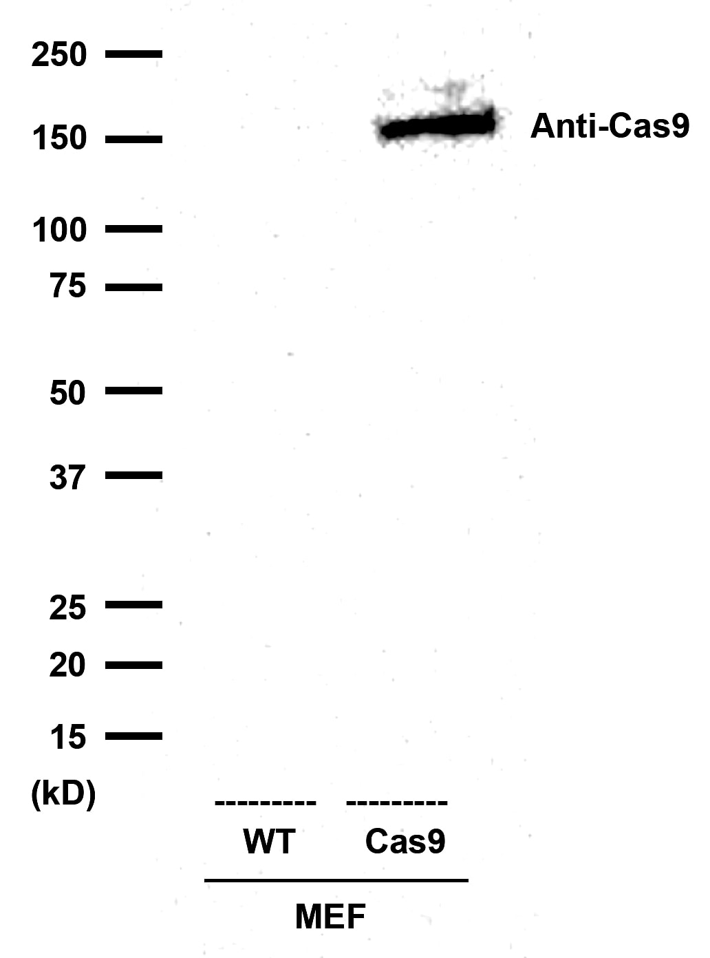
Western Blot: CRISPR-Cas9 Antibody (7A9-3A3)N-TerminusBSA Free [NBP2-36440]
Western Blot: CRISPR-Cas9 Antibody (7A9-3A3) - N-Terminus [NBP2-36440] - 20 ug whole cell lysates from control MEF (MEF-WT) and MEF-Cas9 stable cell line. CRISPR-Cas9 antibody (clone 7A9-3A3) was used at 1:1000 dilution. Observed molecular weight is ~158 kDa. Image submitted via verified customer review.![Western Blot: CRISPR-Cas9 Antibody (7A9-3A3)N-TerminusBSA Free [NBP2-36440]](https://resources.rndsystems.com/images/products/CRISPR-Cas9-Antibody-7A9-3A3-N-Terminus-Western-Blot-NBP2-36440-img0015.jpg "Western Blot: CRISPR-Cas9 Antibody (7A9-3A3)N-TerminusBSA Free [NBP2-36440]")
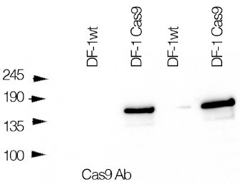
Western Blot: CRISPR-Cas9 Antibody (7A9-3A3)N-TerminusBSA Free [NBP2-36440]
Western Blot: CRISPR-Cas9 Antibody (7A9-3A3) - N-Terminus [NBP2-36440] - Chicken fibroblasts DF-1 cells stably expressing Cas9 in lanes 2 and 4, wild type fibroblasts in lanes 1 and 3 blot, probed with Cas9 antibody. WB image submitted by a verified customer review.![Immunocytochemistry/ Immunofluorescence: CRISPR-Cas9 Antibody (7A9-3A3) - N-Terminus - BSA Free [NBP2-36440]](https://resources.rndsystems.com/images/products/CRISPR-Cas9-Antibody-7A9-3A3-N-Terminus-Immunocytochemistry-Immunofluorescence-NBP2-36440-img0007.jpg "Immunocytochemistry/ Immunofluorescence: CRISPR-Cas9 Antibody (7A9-3A3) - N-Terminus - BSA Free [NBP2-36440]")
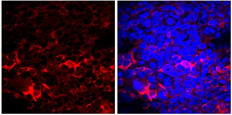
Immunocytochemistry/ Immunofluorescence: CRISPR-Cas9 Antibody (7A9-3A3) - N-Terminus - BSA Free [NBP2-36440]
Immunocytochemistry/Immunofluorescence: CRISPR-Cas9 Antibody (7A9-3A3) - N-Terminus [NBP2-36440] - Analysis of Crispr-Cas9 transfected HEK293 cells using CRISPR-Cas9 antibody (clone 7A9-3A3). Red staining represents CRISPR-Cas9 positivity while DAPI stained nuclei are visible in blue color. Image submitted via verified customer review. - N-Terminus [NBP2-36440]")
Immunoprecipitation: Mouse Monoclonal CRISPR-Cas9 Antibody (7A9-3A3) - N-Terminus [NBP2-36440]
Immunoprecipitation: Mouse Monoclonal CRISPR-Cas9 Antibody (7A9-3A3) - N-Terminus [NBP2-36440] - LC-MS/MS signals of a surrogate peptide from Cas9 and a Cas9 variant after immunoprecipitation using CRISPR-Cas9 Antibody (7A9-3A3) [Biotin] (NBP2-36440B). Image from a verified customer review. - N-Terminus - BSA Free [NBP2-36440] -")
Western Blot: CRISPR-Cas9 Antibody (7A9-3A3) - N-Terminus - BSA Free [NBP2-36440] -
Western Blot: CRISPR-Cas9 Antibody (7A9-3A3) - N-Terminus - BSA Free [NBP2-36440] - Identification of “HDR-enhancer” (HE) domain of CtIP. a Schematic diagram of CtIP protein showing different truncated CtIP proteins have been fused to Cas9 & tested for their ability to stimulate HDR. Various sequence features of CtIP, including tetramerization & dimerization domains, & CDK phosphorylation sites S233, T245, & S276, are indicated. b Identification of a domain of CtIP, called HE, spanning aa 1 to 296, which is able to stimulate HDR when fused to Cas9. Human RG37 fibroblasts transfected w/ indicated plasmids expressing Cas9 or Cas9–CtIP derivatives, T2 guide RNA plasmid, & GFP transgene donor w/ homology arms to targeted AAVS1 locus. Expression of fusion proteins examined by WB (Supplementary Fig. 1). Data are from four independent experiments. Error bars indicate standard deviation. c Functional analysis of HE domain. HEK293 cells transfected w/ indicated Cas9 plasmids, T2 guide RNA, & GFP transgene donor w/ homology arms to AAVS1 targeted locus. HDR-mediated transgene integration measured by FACS analysis of GFP-positive cells, resulting from targeted GFP transgene integration. Indels at cleavage site measured by T7E1 assay. Results are expressed as mean of relative HDR or indel frequencies calculated by normalizing every HDR or indel frequency by induced by Cas9, respectively. Asterisks indicate difference is statistically significant when comparing Cas9–CtIP or Cas9–HE derivatives to Cas9 in nonparametric t-test (*P<0.05, **P<0.005, or ***P<0.0005). Data are from four independent experiments. Error bars indicate standard deviation. The relative expression levels of Cas9 & Cas9–HE derivatives analyzed by WB using anti-Cas9 & control anti-tubulin antibodies Image collected & cropped by CiteAb from following publication (https://pubmed.ncbi.nlm.nih.gov/29556040), licensed under a CC-BY license. Not internally tested by Novus Biologicals. - N-Terminus - BSA Free [NBP2-36440] -")
Western Blot: CRISPR-Cas9 Antibody (7A9-3A3) - N-Terminus - BSA Free [NBP2-36440] -
Western Blot: CRISPR-Cas9 Antibody (7A9-3A3) - N-Terminus - BSA Free [NBP2-36440] - Stimulation of transgene integration by Cas9–HE & Cas9–Geminin. Relative frequencies of HDR & indels induced by Cas9 or fusion of Cas9 to HE domain, Geminin degron, or both. Human HEK293 cells were transfected with the indicated Cas9 plasmids, T2 guide RNA, & GFP transgene donor with homology arms to the AAVS1 targeted locus. HDR-mediated transgene integration was measured by FACS analysis of GFP-positive cells, resulting from targeted GFP transgene integration. Indels at the cleavage site were measured by the T7E1 assay. The results are expressed as the mean of relative HDR or indel frequency calculated by normalizing every HDR or indel frequency by that induced by Cas9. Asterisks indicate that the difference is statistically significant when comparing Cas9–HE, Cas9–HE–Geminin, & Cas9–Geminin to Cas9 in t-test (*P<0.05 or **P<0.005). Data are from three independent experiments. Error bars indicate standard deviation. Relative expression levels of Cas9 & other fusions were analyzed by western blot with anti-Cas9 & control anti-tubulin antibodies. Protein extracts were obtained with lysis buffer containing 150 mM NaCl, which resulted in inefficient solubilization of Cas9 fusions with the HE domain compared to those of Cas9 & Cas9–Geminin Image collected & cropped by CiteAb from the following publication (https://pubmed.ncbi.nlm.nih.gov/29556040), licensed under a CC-BY license. Not internally tested by Novus Biologicals.Applications for CRISPR-Cas9 Antibody (7A9-3A3) - N-Terminus - BSA Free
Application
Recommended Usage
Immunoblotting
reported in scientific literature (PMID 31959836)
Immunocytochemistry/ Immunofluorescence
1:500
Immunohistochemistry-Frozen
reported in scientific literature (PMID 28153089)
Knockout Validated
reported in scientific literature (PMID 31959836)
Western Blot
1:1000
Application Notes
IF and IHC use of CRISPR-Cas9 antibody (clone 7A9-3A3) on 4% formaldehyde fixed and 20um thick frozen-/cryo-sections reported in scientific literature
Reviewed Applications
Read 8 reviews rated 4.6 using NBP2-36440 in the following applications:
Formulation, Preparation, and Storage
Purification
Protein G purified
Formulation
PBS
Format
BSA Free
Preservative
0.02% Sodium Azide
Concentration
1.0 mg/ml
Shipping
The product is shipped with polar packs. Upon receipt, store it immediately at the temperature recommended below.
Stability & Storage
Store at 4C short term. Aliquot and store at -20C long term. Avoid freeze-thaw cycles.
Background: CRISPR-Cas9
Using CRISPR-Cas9 technology, double-stranded DNA breaks may be induced within specific targeted genome sequences (target DNA; protospacer) for insertion or removal of DNA sequences for gene editing applications. To target a specific loci, a gRNA that will bind to a specific target sequence of DNA within a genome is created. The gRNA will recognize the DNA sequence, and the Cas9 enzyme will cleave the DNA at the targeted location. Once the targeted DNA is removed by Cas9, the cell's own DNA repair mechanism is used to insert or remove a DNA sequence for genomic editing.
Cas9 detection is used to confirm and evaluate CRISPR Cas9 gRNA transfection efficiency. Western blot analysis of CRISPR-Cas9 gRNA transfected cell lysates with Cas9 antibodies identifies the protein having a theoretical molecular weight of 160kDa. Broad areas of research are benefiting from CRISPR-Cas9 based gene editing tools including studies of basic immunity functions, genetic screening and disease treatment (2). Ethical concerns have led to many countries making it illegal to manipulate human germline cells or perform embryo genome editing.
References
1. Oakes, B. L., Fellmann, C., Rishi, H., Taylor, K. L., Ren, S. M., Nadler, D. C.,... Savage, D. F. (2019). CRISPR-Cas9 Circular Permutants as Programmable Scaffolds for Genome Modification. Cell, 176(1-2), 254-267.e216. doi:10.1016/j.cell.2018.11.052
2. Chiou, S. H., Winters, I. P., Wang, J., Naranjo, S., Dudgeon, C., Tamburini, F. B.,... Winslow, M. M. (2015). Pancreatic cancer modeling using retrograde viral vector delivery and in vivo CRISPR/Cas9-mediated somatic genome editing. Genes Dev, 29(14), 1576-1585. doi:10.1101/gad.264861.115
Long Name
CRISPR-associated Protein 9
Alternate Names
Cas9, CRISPR-associated endonuclease Cas9/Csn1, CRISPR-Cas9/Csn1, CRISPR/Cas9, csn1, SPy_1046, SPy1046, SpyCas9
Additional CRISPR-Cas9 Products
Product Documents for CRISPR-Cas9 Antibody (7A9-3A3) - N-Terminus - BSA Free
Certificate of Analysis
To download a Certificate of Analysis, please enter a lot or batch number in the search box below.
Product Specific Notices for CRISPR-Cas9 Antibody (7A9-3A3) - N-Terminus - BSA Free
This product is for research use only and is not approved for use in humans or in clinical diagnosis. Primary Antibodies are guaranteed for 1 year from date of receipt.
Citations for CRISPR-Cas9 Antibody (7A9-3A3) - N-Terminus - BSA Free
 Powered by Bioz
Customer Reviews for CRISPR-Cas9 Antibody (7A9-3A3) - N-Terminus - BSA Free (8)
4.6 out of 5
8 Customer Ratings
Have you used CRISPR-Cas9 Antibody (7A9-3A3) - N-Terminus - BSA Free?
Submit a review and receive an Amazon gift card!
$25/€18/£15/$25CAN/¥2500 Yen for a review with an image
$10/€7/£6/$10CAN/¥1110 Yen for a review without an image
Submit a review
Customer Images
Showing
1
-
5 of
8 reviews
Showing All
Filter By:
-
Application: Western BlotSample Tested: DF-1 fibroblastsSpecies: ChickenVerified Customer | Posted 09/06/2021western blot of chicken fibroblasts DF-1 cells stably expressing Cas9 lanes 2 and 4, wild type fibroblasts ! and 3 blot probed with Cas9 antibodymembrane blocked in 5% milk TBST antibody used 1:1000 antibody incubated for 20h at 4 degrees

-
Application: ImmunocytochemistrySample Tested: fixed DF-1 chicken fibroblast cells and DF-1 chicken fibroblastsSpecies: AvianVerified Customer | Posted 08/25/2021DF-1 stable cell line expressing Cas9 (left )or wildtype DF-1 cells (right) stained with Cas9 antibody NBP2-36440 and phalloidin and DAPI to visualise F-actin and DNA respectively.cells fixed with 4%PFA, perm with 0.3% tx100 for 5 mins antibody incubated 1:500 overnight at 4 degrees
Bio-Techne ResponseThis review was submitted through the legacy Novus Innovators Program, reflecting a new species or application tested on a primary antibody.
-
Application: Western BlotSample Tested: MEF (MEF-WT) and MEF-Cas9 stable cell lineSpecies: BacteriaVerified Customer | Posted 03/20/2017Western blots using 20 ug whole cell lysates from our control MEF (MEF-WT) and MEF-Cas9 stable cell linesSample: Whole cell lysates from our control MEF (MEF-WT) and MEF-Cas9 stable cell lines Loading amount: 20ug Primary antibody dilution: 1:1,000 Detection: ECL method Comments on Performance: Good specificity and sensitivity – attached image for a whole membrane
-
Application: Western BlotSample Tested: CRISPR-Cas9 expressing Ewing Sarcoma cell lines (SKES1 and TC71)Species: BacteriaVerified Customer | Posted 03/16/2017First 4 columns: SKES1-wild type without Dox / SKES1-wild type plus Dox / SKES1-Cas9 without Dox / SKES1-Cas9 plus Dox Second 4 columns:TC71-wild type without Dox / TC71-wild type plus Dox / TC71-Cas9 without Dox /TC71-Cas9 plus DoxHuman Cell Lines tested: Ewing Sarcoma cell lines (SKES1 and TC71) Application tested: Western Blot Dilution tested: 1:1000 Detection: ECL method
-
Application: Immunohistochemistry-FrozenSample Tested: Xenograft tissueSpecies: BacteriaVerified Customer | Posted 03/02/2017Immunohistochemistry of brain sections exhibits specific Cas9 staining using CRISPR-Cas9 antibody (7A9-3A3) in GBM the tumor areas after over expression of spCas9 by lentivirus infection.Testing: IHC-Frozen Tissues: 1.LN229-formed glioblastoma xenografts, uninfected or infected with sgRNA G1. 2.GBM8 formed glioblastoma xenograft, treated or not with the mutated nickase version of the virus-encoded Cas9n D10A enzyme and guided by the pair of G1 and G3 sgRNAs. Fixative: 4% formaldehyde Section thickness: 20um Antibody Dilution: 1-50 Detection: Immunofluoresence, Confocal microscopy Data Images: El Fatimy et al. 2017. Mol Ther. 2017 Feb 1;25(2):368-378 [PMID 28153089] Performance; Antibody is more specific than other Cas9 antibodies tested.
-
Application: ImmunocytochemistrySample Tested: LN229 cells (human glioblastoma cell line)Species: BacteriaVerified Customer | Posted 03/02/2017Immunohistochemistry of brain sections exhibits specific Cas9 staining using CRISPR-Cas9 antibody (7A9-3A3) in GBM the tumor areas after over expression of spCas9 by lentivirus infection.Antibody used at 1:100 dilution in immunocytochemistry with Immunofluorescence based detection to determine the lentivirus functional titer (through serial dilutions) in LN229 cells.
-
Application: Western BlotSample Tested: LN229 cells (human glioblastoma cell line), Human primary astrocytes, Mouse primary astrocytes, LN229 human glioblastoma cells transfected with Crispr-Cas9, Primary mouse astrocytes transfected with Crispr-Cas9, Human primary astrocytes transfected with Crispr-Cas9, Mouse primary astrocytes transfected with Crispr-Cas9, LN229 cells (human glioblastoma cell line) transfected with bacterial Crispr-Cas9 and Human or mouse primary astrocytes and neurons transfected with bacterial Crispr-Cas9Species: BacteriaVerified Customer | Posted 03/02/2017Lysates used: 1.Human or mouse primary astrocytes and neurons transduced with miR-10b-editing lentivirus at the MOI levels that led to similar levels of Cas9 expression. 2.Established orthotopic LN229 glioblastoma/GBM tumors that were subjected to intratumoral Injections of 105 TU of Lentiviral miR-10b-Editing Vectors. Dilutionused: 1-2000 Detection Method: ECL
-
Application: ImmunofluorescenceSample Tested: HEK293 cells transfected with Cas9Species: OtherVerified Customer | Posted 07/27/2016Detection of Cas9 (red) using anti-Cas9 antibody
There are no reviews that match your criteria.
Protocols
Find general support by application which include: protocols, troubleshooting, illustrated assays, videos and webinars.
- Antigen Retrieval Protocol (PIER)
- Antigen Retrieval for Frozen Sections Protocol
- Appropriate Fixation of IHC/ICC Samples
- Cellular Response to Hypoxia Protocols
- Chromogenic IHC Staining of Formalin-Fixed Paraffin-Embedded (FFPE) Tissue Protocol
- Chromogenic Immunohistochemistry Staining of Frozen Tissue
- ClariTSA™ Fluorophore Kits
- Detection & Visualization of Antibody Binding
- Fluorescent IHC Staining of Frozen Tissue Protocol
- Graphic Protocol for Heat-induced Epitope Retrieval
- Graphic Protocol for the Preparation and Fluorescent IHC Staining of Frozen Tissue Sections
- Graphic Protocol for the Preparation and Fluorescent IHC Staining of Paraffin-embedded Tissue Sections
- Graphic Protocol for the Preparation of Gelatin-coated Slides for Histological Tissue Sections
- ICC Cell Smear Protocol for Suspension Cells
- ICC Immunocytochemistry Protocol Videos
- ICC for Adherent Cells
- IHC Sample Preparation (Frozen sections vs Paraffin)
- Immunocytochemistry (ICC) Protocol
- Immunocytochemistry Troubleshooting
- Immunofluorescence of Organoids Embedded in Cultrex Basement Membrane Extract
- Immunofluorescent IHC Staining of Formalin-Fixed Paraffin-Embedded (FFPE) Tissue Protocol
- Immunohistochemistry (IHC) and Immunocytochemistry (ICC) Protocols
- Immunohistochemistry Frozen Troubleshooting
- Immunohistochemistry Paraffin Troubleshooting
- Immunoprecipitation Protocol
- Preparing Samples for IHC/ICC Experiments
- Preventing Non-Specific Staining (Non-Specific Binding)
- Primary Antibody Selection & Optimization
- Protocol for Heat-Induced Epitope Retrieval (HIER)
- Protocol for Making a 4% Formaldehyde Solution in PBS
- Protocol for VisUCyte™ HRP Polymer Detection Reagent
- Protocol for the Fluorescent ICC Staining of Cell Smears - Graphic
- Protocol for the Fluorescent ICC Staining of Cultured Cells on Coverslips - Graphic
- Protocol for the Preparation & Fixation of Cells on Coverslips
- Protocol for the Preparation and Chromogenic IHC Staining of Frozen Tissue Sections
- Protocol for the Preparation and Chromogenic IHC Staining of Frozen Tissue Sections - Graphic
- Protocol for the Preparation and Chromogenic IHC Staining of Paraffin-embedded Tissue Sections
- Protocol for the Preparation and Chromogenic IHC Staining of Paraffin-embedded Tissue Sections - Graphic
- Protocol for the Preparation and Fluorescent ICC Staining of Cells on Coverslips
- Protocol for the Preparation and Fluorescent ICC Staining of Non-adherent Cells
- Protocol for the Preparation and Fluorescent ICC Staining of Stem Cells on Coverslips
- Protocol for the Preparation and Fluorescent IHC Staining of Frozen Tissue Sections
- Protocol for the Preparation and Fluorescent IHC Staining of Paraffin-embedded Tissue Sections
- Protocol for the Preparation of Gelatin-coated Slides for Histological Tissue Sections
- Protocol for the Preparation of a Cell Smear for Non-adherent Cell ICC - Graphic
- R&D Systems Quality Control Western Blot Protocol
- TUNEL and Active Caspase-3 Detection by IHC/ICC Protocol
- The Importance of IHC/ICC Controls
- Troubleshooting Guide: Immunohistochemistry
- Troubleshooting Guide: Western Blot Figures
- Western Blot Conditions
- Western Blot Protocol
- Western Blot Protocol for Cell Lysates
- Western Blot Troubleshooting
- Western Blot Troubleshooting Guide
- View all Protocols, Troubleshooting, Illustrated assays and Webinars
FAQs for CRISPR-Cas9 Antibody (7A9-3A3) - N-Terminus - BSA Free
Showing
1
-
2 of
2 FAQs
Showing All
-
Q: Does the antibody NBP2-36440 cross react with the inactive form of CAS9 and CAS9 nickase?
A: Unfortunately, we do not have any information regarding the ability of this clone to detect the Cas9 nickase. Nickase activity involves amino acids 10 and 840 of the Cas9 sequence. This particular antibody was raised against the N-terminal region of the Cas9 protein. It is possible that this product may then detect the modification surrounding amino acid 10, but again, we do not have data at this time that proves this interaction.
-
Q: What is the immunogen sequence for your CRISPR-Cas9 antibody NBP2-36440?
A: CRISPR-Cas9 Antibody (7A9-3A3) - N-Terminus [NBP2-36440] is generated against a recombinant N-terminal fragment of Streptococcus pyogene CRISPR/Cas9 [UniProt# Q99ZW2]. Unfortunately, the exact immunogen info is propriatary but we can confirm that the immunogen is outside the catalytic domain of CRISPR-Cas9.
-
Q: Does the antibody NBP2-36440 cross react with the inactive form of CAS9 and CAS9 nickase?
A: Unfortunately, we do not have any information regarding the ability of this clone to detect the Cas9 nickase. Nickase activity involves amino acids 10 and 840 of the Cas9 sequence. This particular antibody was raised against the N-terminal region of the Cas9 protein. It is possible that this product may then detect the modification surrounding amino acid 10, but again, we do not have data at this time that proves this interaction.
-
Q: What is the immunogen sequence for your CRISPR-Cas9 antibody NBP2-36440?
A: CRISPR-Cas9 Antibody (7A9-3A3) - N-Terminus [NBP2-36440] is generated against a recombinant N-terminal fragment of Streptococcus pyogene CRISPR/Cas9 [UniProt# Q99ZW2]. Unfortunately, the exact immunogen info is propriatary but we can confirm that the immunogen is outside the catalytic domain of CRISPR-Cas9.
Loading...