![Immunocytochemistry/ Immunofluorescence: CTGF/CCN2 Antibody [NBP2-66426]](https://resources.rndsystems.com/images/products/CTGF-CCN2-Antibody-Immunocytochemistry-Immunofluorescence-NBP2-66426-img0001.jpg "Immunocytochemistry/ Immunofluorescence: CTGF/CCN2 Antibody [NBP2-66426]")
Loading...
Key Product Details
Species Reactivity
Mouse, Rat
Applications
Immunohistochemistry, Peptide ELISA, Immunocytochemistry/ Immunofluorescence, Control
Label
Unconjugated
Antibody Source
Polyclonal Goat IgG
Loading...
Product Specifications
Immunogen
Peptide with sequence C-SLYYRKMYGDMA, from the C Terminus of the protein sequence according to NP_001892.
Reactivity Notes
Expected from sequence similarity: Human, Rat and Canine
Clonality
Polyclonal
Host
Goat
Isotype
IgG
Scientific Data Images for CTGF/CCN2 Antibody
Immunocytochemistry/ Immunofluorescence: CTGF/CCN2 Antibody [NBP2-66426]
Immunocytochemistry/Immunofluorescence: CTGF/CCN2 Antibody [NBP2-66426] - Paraformaldehyde fixed NIH3T3 cells, permeabilized with 0.15% Triton. Primary incubation 1hr (5 ug/mL) followed by Alexa Fluor 488 secondary antibody (2 ug/mL), showing cytoplasmic staining. The nuclear stain is DAPI (blue). Negative control: Unimmunized goat IgG (5 ug/mL) followed by Alexa Fluor 488 secondary antibody (2 ug/mL).![Immunohistochemistry: CTGF/CCN2 Antibody [NBP2-66426]](https://resources.rndsystems.com/images/products/CTGF-CCN2-Antibody-Immunohistochemistry-NBP2-66426-img0003.jpg "Immunohistochemistry: CTGF/CCN2 Antibody [NBP2-66426]")
Immunohistochemistry: CTGF/CCN2 Antibody [NBP2-66426]
Immunohistochemistry: CTGF/CCN2 Antibody [NBP2-66426] - (6ug/ml) staining of paraffin embedded Rat Liver. Heat induced antigen retrieval with citrate buffer pH 6, HRP-staining.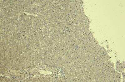
Control: CTGF/CCN2 Antibody [NBP2-66426] - Negative Control showing staining of paraffin embedded Rat Liver, with no primary antibody.
Applications for CTGF/CCN2 Antibody
Application
Recommended Usage
Immunocytochemistry/ Immunofluorescence
5 ug/mL
Immunohistochemistry
6.0 - 8.0 ug/ml
Peptide ELISA
Detection limit 1:10000
Application Notes
Immunofluorescence: Strong expression of the protein seen in the cytoplasm of NIH3T3 cells.
Formulation, Preparation, and Storage
Purification
Antigen Affinity-purified
Formulation
Tris saline (20 mM Tris pH 7.3, 150 mM NaCl), 0.5% BSA
Preservative
0.02% Sodium Azide
Concentration
0.5 mg/ml
Shipping
The product is shipped with polar packs. Upon receipt, store it immediately at the temperature recommended below.
Stability & Storage
Store at 4C short term. Aliquot and store at -20C long term. Avoid freeze-thaw cycles.
Background: CTGF/CCN2
CTGF antibodies are useful tools for angiogenesis and cell structure research, and for studies on certian types of cancer.
Long Name
Connective Tissue Growth Factor
Alternate Names
CCN2, CTGRP, Fisp12, HCS24, IGFBP-8, NOV2
Gene Symbol
CCN2
Additional CTGF/CCN2 Products
Product Documents for CTGF/CCN2 Antibody
Certificate of Analysis
To download a Certificate of Analysis, please enter a lot or batch number in the search box below.
Product Specific Notices for CTGF/CCN2 Antibody
This product is for research use only and is not approved for use in humans or in clinical diagnosis. Primary Antibodies are guaranteed for 1 year from date of receipt.
Customer Reviews for CTGF/CCN2 Antibody
There are currently no reviews for this product. Be the first to review CTGF/CCN2 Antibody and earn rewards!
Have you used CTGF/CCN2 Antibody?
Submit a review and receive an Amazon gift card!
$25/€18/£15/$25CAN/¥2500 Yen for a review with an image
$10/€7/£6/$10CAN/¥1110 Yen for a review without an image
Submit a review
Protocols
Find general support by application which include: protocols, troubleshooting, illustrated assays, videos and webinars.
- Antigen Retrieval Protocol (PIER)
- Antigen Retrieval for Frozen Sections Protocol
- Appropriate Fixation of IHC/ICC Samples
- Cellular Response to Hypoxia Protocols
- Chromogenic IHC Staining of Formalin-Fixed Paraffin-Embedded (FFPE) Tissue Protocol
- Chromogenic Immunohistochemistry Staining of Frozen Tissue
- ClariTSA™ Fluorophore Kits
- Detection & Visualization of Antibody Binding
- ELISA Sample Preparation & Collection Guide
- ELISA Troubleshooting Guide
- Fluorescent IHC Staining of Frozen Tissue Protocol
- Graphic Protocol for Heat-induced Epitope Retrieval
- Graphic Protocol for the Preparation and Fluorescent IHC Staining of Frozen Tissue Sections
- Graphic Protocol for the Preparation and Fluorescent IHC Staining of Paraffin-embedded Tissue Sections
- Graphic Protocol for the Preparation of Gelatin-coated Slides for Histological Tissue Sections
- How to Run an R&D Systems DuoSet ELISA
- How to Run an R&D Systems Quantikine ELISA
- How to Run an R&D Systems Quantikine™ QuicKit™ ELISA
- ICC Cell Smear Protocol for Suspension Cells
- ICC Immunocytochemistry Protocol Videos
- ICC for Adherent Cells
- IHC Sample Preparation (Frozen sections vs Paraffin)
- Immunocytochemistry (ICC) Protocol
- Immunocytochemistry Troubleshooting
- Immunofluorescence of Organoids Embedded in Cultrex Basement Membrane Extract
- Immunofluorescent IHC Staining of Formalin-Fixed Paraffin-Embedded (FFPE) Tissue Protocol
- Immunohistochemistry (IHC) and Immunocytochemistry (ICC) Protocols
- Immunohistochemistry Frozen Troubleshooting
- Immunohistochemistry Paraffin Troubleshooting
- Preparing Samples for IHC/ICC Experiments
- Preventing Non-Specific Staining (Non-Specific Binding)
- Primary Antibody Selection & Optimization
- Protocol for Heat-Induced Epitope Retrieval (HIER)
- Protocol for Making a 4% Formaldehyde Solution in PBS
- Protocol for VisUCyte™ HRP Polymer Detection Reagent
- Protocol for the Fluorescent ICC Staining of Cell Smears - Graphic
- Protocol for the Fluorescent ICC Staining of Cultured Cells on Coverslips - Graphic
- Protocol for the Preparation & Fixation of Cells on Coverslips
- Protocol for the Preparation and Chromogenic IHC Staining of Frozen Tissue Sections
- Protocol for the Preparation and Chromogenic IHC Staining of Frozen Tissue Sections - Graphic
- Protocol for the Preparation and Chromogenic IHC Staining of Paraffin-embedded Tissue Sections
- Protocol for the Preparation and Chromogenic IHC Staining of Paraffin-embedded Tissue Sections - Graphic
- Protocol for the Preparation and Fluorescent ICC Staining of Cells on Coverslips
- Protocol for the Preparation and Fluorescent ICC Staining of Non-adherent Cells
- Protocol for the Preparation and Fluorescent ICC Staining of Stem Cells on Coverslips
- Protocol for the Preparation and Fluorescent IHC Staining of Frozen Tissue Sections
- Protocol for the Preparation and Fluorescent IHC Staining of Paraffin-embedded Tissue Sections
- Protocol for the Preparation of Gelatin-coated Slides for Histological Tissue Sections
- Protocol for the Preparation of a Cell Smear for Non-adherent Cell ICC - Graphic
- Quantikine HS ELISA Kit Assay Principle, Alkaline Phosphatase
- Quantikine HS ELISA Kit Principle, Streptavidin-HRP Polymer
- Sandwich ELISA (Colorimetric) – Biotin/Streptavidin Detection Protocol
- Sandwich ELISA (Colorimetric) – Direct Detection Protocol
- TUNEL and Active Caspase-3 Detection by IHC/ICC Protocol
- The Importance of IHC/ICC Controls
- Troubleshooting Guide: ELISA
- Troubleshooting Guide: Immunohistochemistry
- View all Protocols, Troubleshooting, Illustrated assays and Webinars
FAQs for CTGF/CCN2 Antibody
Showing
1
-
1 of
1 FAQ
Showing All
-
Q: We are looking for a pair of mouse CTGF antibody for ELISA, we prefer no azide and/or glycerol, and in carrier free form (no EDTA, no Tris, no BSA). For the validation, we need approximately 100ug. If it works, we'll order more later.
A:
Unfortunately we do not carry any CTGF antibodies that suit your specifications. We do carry AbSelect antibody purification kits that will remove BSA, Tris and azide from antibody formulations which may be an option for you.
Loading...