![Immunohistochemistry-Paraffin: Cytochrome c Antibody [NBP2-21569]](https://resources.rndsystems.com/images/products/Cytochrome-c-Antibody-Immunohistochemistry-Paraffin-NBP2-21569-img0008.jpg "Immunohistochemistry-Paraffin: Cytochrome c Antibody [NBP2-21569]")
Loading...
Key Product Details
Validated by
Knockout/Knockdown, Orthogonal Validation
Species Reactivity
Validated:
Human, Mouse, Rat, Canine, Drosophila
Cited:
Human
Predicted:
Bovine (90%), Chimpanzee (100%), Porcine (90%). Backed by our 100% Guarantee.
Applications
Validated:
Immunohistochemistry, Immunohistochemistry-Paraffin, Immunohistochemistry-Frozen, Western Blot, Simple Western
Cited:
Immunohistochemistry, Western Blot, IF/IHC
Label
Unconjugated
Antibody Source
Polyclonal Rabbit IgG
Loading...
Product Specifications
Immunogen
Full length human Cytochrome c Recombinant protein.
Reactivity Notes
Chicken (87%), Xenopus laevis (84%).
Localization
Mitochondrion matrix
Marker
Mitochondria Marker
Clonality
Polyclonal
Host
Rabbit
Isotype
IgG
Theoretical MW
12 kDa.
Disclaimer note: The observed molecular weight of the protein may vary from the listed predicted molecular weight due to post translational modifications, post translation cleavages, relative charges, and other experimental factors.
Disclaimer note: The observed molecular weight of the protein may vary from the listed predicted molecular weight due to post translational modifications, post translation cleavages, relative charges, and other experimental factors.
Scientific Data Images for Cytochrome c Antibody
Immunohistochemistry-Paraffin: Cytochrome c Antibody [NBP2-21569]
Immunohistochemistry-Paraffin: Cytochrome c Antibody [NBP2-21569] - Human endometrial carcinoma. Cytochrome C stained by Cytochrome C antibody diluted at 1:500. Antigen Retrieval: Citrate buffer, pH 6.0, 15 min.![Western Blot: Cytochrome c Antibody [NBP2-21569]](https://resources.rndsystems.com/images/products/Cytochrome-C-Antibody-Western-Blot-NBP2-21569-img0002.jpg "Western Blot: Cytochrome c Antibody [NBP2-21569]")
Western Blot: Cytochrome c Antibody [NBP2-21569]
Western Blot: Cytochrome C Antibody [NBP2-21569] - Sample (30 ug of whole cell lysate) A: PC-12 15% SDS PAGE gel, diluted at 1:1000.![Western Blot: Cytochrome c Antibody [NBP2-21569]](https://resources.rndsystems.com/images/products/Cytochrome-C-Antibody-Western-Blot-NBP2-21569-img0003.jpg "Western Blot: Cytochrome c Antibody [NBP2-21569]")
Western Blot: Cytochrome c Antibody [NBP2-21569]
Western Blot: Cytochrome C Antibody [NBP2-21569] - Sample (50 ug of whole cell lysate) A: Mouse Brain, 15% SDS PAGE gel, diluted at 1:1000.![Western Blot: Cytochrome c Antibody [NBP2-21569]](https://resources.rndsystems.com/images/products/Cytochrome-c-Antibody-Western-Blot-NBP2-21569-img0011.jpg "Western Blot: Cytochrome c Antibody [NBP2-21569]")
Western Blot: Cytochrome c Antibody [NBP2-21569]
Western Blot: Cytochrome c Antibody [NBP2-21569] - HepG2 and mitochondria extracts (30 ug) were separated by SDS-PAGE, and the membrane was blotted with Cytochrome C antibody diluted at 1:2000. HRP-conjugated anti-rabbit IgG antibody was used to detect the primary antibody, and the signal was developed with Trident pico Western HRP Substrate.![Immunohistochemistry-Paraffin: Cytochrome c Antibody [NBP2-21569]](https://resources.rndsystems.com/images/products/Cytochrome-c-Antibody-Immunohistochemistry-Paraffin-NBP2-21569-img0007.jpg "Immunohistochemistry-Paraffin: Cytochrome c Antibody [NBP2-21569]")
Immunohistochemistry-Paraffin: Cytochrome c Antibody [NBP2-21569]
Immunohistochemistry-Paraffin: Cytochrome c Antibody [NBP2-21569] - Human cervical carcinoma. Cytochrome C stained by Cytochrome C antibody diluted at 1:500. Antigen Retrieval: Citrate buffer, pH 6.0, 15 min.
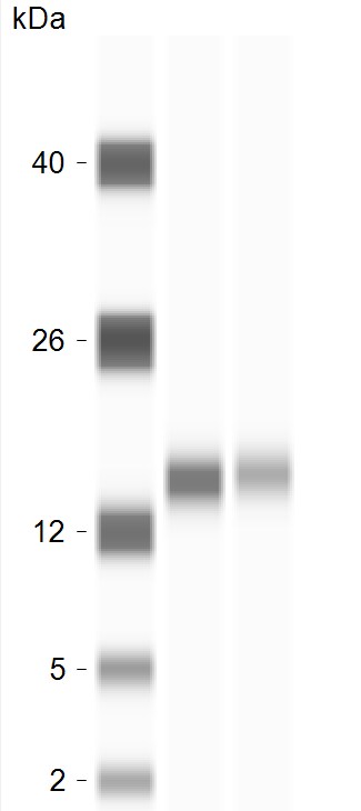
Simple Western: Cytochrome c Antibody [NBP2-21569] -
Simple Western: Cytochrome c Antibody [NBP2-21569] - Cytochrome C Antibodies (NBP2-21569), ProteinSimple Western Blot on Jess Instrument; 1 microgram of human brain tissue lysate was tested with the antibodies diluted 10 and 20 times. Image from verified customer review.
Western Blot: Cytochrome c Antibody [NBP2-21569] -
Western Blot: Cytochrome c Antibody [NBP2-21569] - Whole cell extract (30 ug) was separated by 15% SDS-PAGE, and the membrane was blotted with Cytochrome c antibody (NBP2-21569) diluted at 1:1000. The HRP-conjugated anti-rabbit IgG antibody was used to detect the primary antibody.
Western Blot: Cytochrome c Antibody [NBP2-21569] -
Western Blot: Cytochrome c Antibody [NBP2-21569] - Whole cell extract (30 ug) was separated by 15% SDS-PAGE, and the membrane was blotted with Cytochrome c antibody (NBP2-21569) diluted at 1:1000. The HRP-conjugated anti-rabbit IgG antibody was used to detect the primary antibody, and the signal was developed with Trident ECL plus-Enhanced.
Western Blot: Cytochrome c Antibody [NBP2-21569] -
Western Blot: Cytochrome c Antibody [NBP2-21569] - Various whole cell extracts (30 ug) were separated by 15% SDS-PAGE, and the membrane was blotted with Cytochrome C antibody diluted at 1:1000. The HRP-conjugated anti-rabbit IgG antibody was used to detect the primary antibody, and the signal was developed with Trident ECL plus-Enhanced. Corresponding RNA expression data for the same cell lines are based on Human Protein Atlas program.
Western Blot: Cytochrome c Antibody [NBP2-21569] -
Non-transfected (-) and transfected (+) 293T whole cell extracts (30 ug) were separated by 15% SDS-PAGE, and the membrane was blotted with Cytochrome C antibody (NBP2-21569) diluted at 1:1000. The HRP-conjugated anti-rabbit IgG antibody was used to detect the primary antibody.Applications for Cytochrome c Antibody
Application
Recommended Usage
Immunohistochemistry
1:100-1:1000
Immunohistochemistry-Frozen
Assay dependent
Immunohistochemistry-Paraffin
1:100-1:1000
Western Blot
1:500-1:3000
Application Notes
See Simple Western Antibody Database for Simple Western validation
Reviewed Applications
Read 1 review rated 5 using NBP2-21569 in the following applications:
Formulation, Preparation, and Storage
Purification
Antigen Affinity-purified
Formulation
PBS, 1% BSA, 20% Glycerol
Preservative
0.025% Proclin 300
Concentration
Concentrations vary lot to lot. See vial label for concentration. If unlisted please contact technical services.
Shipping
The product is shipped with polar packs. Upon receipt, store it immediately at the temperature recommended below.
Stability & Storage
Aliquot and store at -20C or -80C. Avoid freeze-thaw cycles.
Background: Cytochrome c
Alternate Names
CYCS
Gene Symbol
CYCS
Additional Cytochrome c Products
Product Documents for Cytochrome c Antibody
Certificate of Analysis
To download a Certificate of Analysis, please enter a lot or batch number in the search box below.
Product Specific Notices for Cytochrome c Antibody
This product is for research use only and is not approved for use in humans or in clinical diagnosis. Primary Antibodies are guaranteed for 1 year from date of receipt.
Related Research Areas
Citations for Cytochrome c Antibody
 Powered by Bioz
Customer Reviews for Cytochrome c Antibody (1)
5 out of 5
1 Customer Rating
Have you used Cytochrome c Antibody?
Submit a review and receive an Amazon gift card!
$25/€18/£15/$25CAN/¥2500 Yen for a review with an image
$10/€7/£6/$10CAN/¥1110 Yen for a review without an image
Submit a review
Customer Images
Showing
1
-
1 of
1 review
Showing All
Filter By:
-
Application: Simple WesternSample Tested: human brain lysateSpecies: HumanVerified Customer | Posted 04/06/2023Cytochrome C Antibodies NBP2-21569, ProteinSimple Western Blot on Jess Instrument; 1 microgram of human brain tissue lysate was tested with the antibodies diluted 10 and 20 times.
 Bio-Techne ResponseThis review was submitted through the legacy Novus Innovators Program, reflecting a new species or application tested on a primary antibody.
There are no reviews that match your criteria.
Protocols
Find general support by application which include: protocols, troubleshooting, illustrated assays, videos and webinars.
- Antigen Retrieval Protocol (PIER)
- Antigen Retrieval for Frozen Sections Protocol
- Appropriate Fixation of IHC/ICC Samples
- Cellular Response to Hypoxia Protocols
- Chromogenic IHC Staining of Formalin-Fixed Paraffin-Embedded (FFPE) Tissue Protocol
- Chromogenic Immunohistochemistry Staining of Frozen Tissue
- ClariTSA™ Fluorophore Kits
- Detection & Visualization of Antibody Binding
- Fluorescent IHC Staining of Frozen Tissue Protocol
- Graphic Protocol for Heat-induced Epitope Retrieval
- Graphic Protocol for the Preparation and Fluorescent IHC Staining of Frozen Tissue Sections
- Graphic Protocol for the Preparation and Fluorescent IHC Staining of Paraffin-embedded Tissue Sections
- Graphic Protocol for the Preparation of Gelatin-coated Slides for Histological Tissue Sections
- IHC Sample Preparation (Frozen sections vs Paraffin)
- Immunofluorescent IHC Staining of Formalin-Fixed Paraffin-Embedded (FFPE) Tissue Protocol
- Immunohistochemistry (IHC) and Immunocytochemistry (ICC) Protocols
- Immunohistochemistry Frozen Troubleshooting
- Immunohistochemistry Paraffin Troubleshooting
- Preparing Samples for IHC/ICC Experiments
- Preventing Non-Specific Staining (Non-Specific Binding)
- Primary Antibody Selection & Optimization
- Protocol for Heat-Induced Epitope Retrieval (HIER)
- Protocol for Making a 4% Formaldehyde Solution in PBS
- Protocol for VisUCyte™ HRP Polymer Detection Reagent
- Protocol for the Preparation & Fixation of Cells on Coverslips
- Protocol for the Preparation and Chromogenic IHC Staining of Frozen Tissue Sections
- Protocol for the Preparation and Chromogenic IHC Staining of Frozen Tissue Sections - Graphic
- Protocol for the Preparation and Chromogenic IHC Staining of Paraffin-embedded Tissue Sections
- Protocol for the Preparation and Chromogenic IHC Staining of Paraffin-embedded Tissue Sections - Graphic
- Protocol for the Preparation and Fluorescent IHC Staining of Frozen Tissue Sections
- Protocol for the Preparation and Fluorescent IHC Staining of Paraffin-embedded Tissue Sections
- Protocol for the Preparation of Gelatin-coated Slides for Histological Tissue Sections
- R&D Systems Quality Control Western Blot Protocol
- TUNEL and Active Caspase-3 Detection by IHC/ICC Protocol
- The Importance of IHC/ICC Controls
- Troubleshooting Guide: Immunohistochemistry
- Troubleshooting Guide: Western Blot Figures
- Western Blot Conditions
- Western Blot Protocol
- Western Blot Protocol for Cell Lysates
- Western Blot Troubleshooting
- Western Blot Troubleshooting Guide
- View all Protocols, Troubleshooting, Illustrated assays and Webinars
Loading...
Associated Pathways

