Cytochrome P450 1A1 Antibody (6G5) - BSA Free
Novus Biologicals | Catalog # NBP2-37526
![Western Blot: Cytochrome P450 1A1 Antibody (6G5)BSA Free [NBP2-37526]](https://resources.rndsystems.com/images/products/Cytochrome-P450-1A1-Antibody-6G5-Western-Blot-NBP2-37526-img0002.jpg "Western Blot: Cytochrome P450 1A1 Antibody (6G5)BSA Free [NBP2-37526]")
Loading...
Key Product Details
Species Reactivity
Validated:
Human
Cited:
Human
Applications
Validated:
Immunohistochemistry, Immunohistochemistry-Paraffin, Western Blot, ELISA, Simple Western
Cited:
Western Blot
Label
Unconjugated
Antibody Source
Monoclonal Mouse IgG1 Clone # 6G5
Format
BSA Free
Loading...
Product Specifications
Immunogen
Purified recombinant fragment of human Cytochrome P450 1A1 expressed in E. Coli.
Clonality
Monoclonal
Host
Mouse
Isotype
IgG1
Theoretical MW
58 kDa.
Disclaimer note: The observed molecular weight of the protein may vary from the listed predicted molecular weight due to post translational modifications, post translation cleavages, relative charges, and other experimental factors.
Disclaimer note: The observed molecular weight of the protein may vary from the listed predicted molecular weight due to post translational modifications, post translation cleavages, relative charges, and other experimental factors.
Scientific Data Images for Cytochrome P450 1A1 Antibody (6G5) - BSA Free
Western Blot: Cytochrome P450 1A1 Antibody (6G5)BSA Free [NBP2-37526]
Western Blot: Cytochrome P450 1A1 Antibody (6G5) [NBP2-37526] - Analysis using CYP1A1 mAb against human CYP1A1 (AA: 203-461) recombinant protein. (Expected MW is 60 kDa)![Immunohistochemistry-Paraffin: Cytochrome P450 1A1 Antibody (6G5) - BSA Free [NBP2-37526]](https://resources.rndsystems.com/images/products/Cytochrome-P450-1A1-Antibody-6G5-Immunohistochemistry-Paraffin-NBP2-37526-img0006.jpg "Immunohistochemistry-Paraffin: Cytochrome P450 1A1 Antibody (6G5) - BSA Free [NBP2-37526]")
Immunohistochemistry-Paraffin: Cytochrome P450 1A1 Antibody (6G5) - BSA Free [NBP2-37526]
Immunohistochemistry-Paraffin: Cytochrome P450 1A1 Antibody (6G5) [NBP2-37526] - Analysis of rectum cancer tissues using CYP1A1 mouse mAb with DAB staining.![Immunohistochemistry-Paraffin: Cytochrome P450 1A1 Antibody (6G5) - BSA Free [NBP2-37526]](https://resources.rndsystems.com/images/products/Cytochrome-P450-1A1-Antibody-6G5-Immunohistochemistry-NBP2-37526-img0003.jpg "Immunohistochemistry-Paraffin: Cytochrome P450 1A1 Antibody (6G5) - BSA Free [NBP2-37526]")
Immunohistochemistry-Paraffin: Cytochrome P450 1A1 Antibody (6G5) - BSA Free [NBP2-37526]
Immunohistochemistry-Paraffin: Cytochrome P450 1A1 Antibody (6G5) [NBP2-37526] - Analysis of ovarian cancer tissues using CYP1A1 mouse mAb with DAB staining.![ELISA: Cytochrome P450 1A1 Antibody (6G5) - BSA Free [NBP2-37526]](https://resources.rndsystems.com/images/products/Cytochrome-P450-1A1-Antibody-6G5-ELISA-NBP2-37526-img0001.jpg "ELISA: Cytochrome P450 1A1 Antibody (6G5) - BSA Free [NBP2-37526]")
ELISA: Cytochrome P450 1A1 Antibody (6G5) - BSA Free [NBP2-37526]
ELISA: Cytochrome P450 1A1 Antibody (6G5) [NBP2-37526] - Red: Control Antigen (100ng); Purple: Antigen (10ng); Green: Antigen (50ng); Blue: Antigen (100ng);![Simple Western: Cytochrome P450 1A1 Antibody (6G5)BSA Free [NBP2-37526]](https://resources.rndsystems.com/images/products/Cytochrome-P450-1A1-Antibody-6G5-Simple-Western-NBP2-37526-img0004.jpg "Simple Western: Cytochrome P450 1A1 Antibody (6G5)BSA Free [NBP2-37526]")
Simple Western: Cytochrome P450 1A1 Antibody (6G5)BSA Free [NBP2-37526]
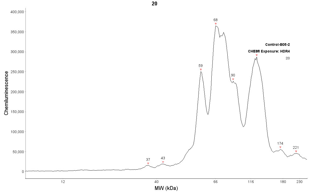
Simple Western: Cytochrome P450 1A1 Antibody (6G5) [NBP2-37526] - Human foreskin cell lysates. Protein concentration is 400 ug/mL. CYP1A1 antibody is diluted 1:20. Detection with chemiluminescence. Image from verified customer review.Applications for Cytochrome P450 1A1 Antibody (6G5) - BSA Free
Application
Recommended Usage
ELISA
1:10000
Immunohistochemistry
1:200 - 1:1000
Simple Western
1:20
Western Blot
1:500 - 1:2000
Application Notes
Use in Simple Western reported by customer review.
See Simple Western Antibody Database for Simple Western validation: Tested in Human foreskin cell lysates, separated by Size, antibody dilution of 1:20
See Simple Western Antibody Database for Simple Western validation: Tested in Human foreskin cell lysates, separated by Size, antibody dilution of 1:20
Reviewed Applications
Read 1 review rated 3 using NBP2-37526 in the following applications:
Formulation, Preparation, and Storage
Purification
Protein G purified
Formulation
PBS
Format
BSA Free
Preservative
0.05% Sodium Azide
Concentration
1 mg/ml
Shipping
The product is shipped with polar packs. Upon receipt, store it immediately at the temperature recommended below.
Stability & Storage
Store at 4C short term. Aliquot and store at -20C long term. Avoid freeze-thaw cycles.
Background: Cytochrome P450 1A1
Long Name
Cytochrome P450 1A1
Alternate Names
CYP1A1, CYPIA1, Cytochrome P450-C, Cytochrome P450-P1
Gene Symbol
CYP1A1
Additional Cytochrome P450 1A1 Products
Product Documents for Cytochrome P450 1A1 Antibody (6G5) - BSA Free
Certificate of Analysis
To download a Certificate of Analysis, please enter a lot or batch number in the search box below.
Product Specific Notices for Cytochrome P450 1A1 Antibody (6G5) - BSA Free
This product is for research use only and is not approved for use in humans or in clinical diagnosis. Primary Antibodies are guaranteed for 1 year from date of receipt.
Citations for Cytochrome P450 1A1 Antibody (6G5) - BSA Free
 Powered by Bioz
Customer Reviews for Cytochrome P450 1A1 Antibody (6G5) - BSA Free (1)
3 out of 5
1 Customer Rating
Have you used Cytochrome P450 1A1 Antibody (6G5) - BSA Free?
Submit a review and receive an Amazon gift card!
$25/€18/£15/$25CAN/¥2500 Yen for a review with an image
$10/€7/£6/$10CAN/¥1110 Yen for a review without an image
Submit a review
Customer Images
Showing
1
-
1 of
1 review
Showing All
Filter By:
-
Application: Simple WesternSample Tested: reconstructed human epidermis and Human foreskin, adult ski, engineered human skin, keratinocytes, HaCaT cellsSpecies: HumanVerified Customer | Posted 05/24/2019Protein concentration is 400 ug/mL. CYP1A1 antibody is diluted 1:20. Detection with chemiluminescence.JESS default run settings are used.

There are no reviews that match your criteria.
Protocols
Find general support by application which include: protocols, troubleshooting, illustrated assays, videos and webinars.
- Antigen Retrieval Protocol (PIER)
- Antigen Retrieval for Frozen Sections Protocol
- Appropriate Fixation of IHC/ICC Samples
- Cellular Response to Hypoxia Protocols
- Chromogenic IHC Staining of Formalin-Fixed Paraffin-Embedded (FFPE) Tissue Protocol
- Chromogenic Immunohistochemistry Staining of Frozen Tissue
- ClariTSA™ Fluorophore Kits
- Detection & Visualization of Antibody Binding
- ELISA Sample Preparation & Collection Guide
- ELISA Troubleshooting Guide
- Fluorescent IHC Staining of Frozen Tissue Protocol
- Graphic Protocol for Heat-induced Epitope Retrieval
- Graphic Protocol for the Preparation and Fluorescent IHC Staining of Frozen Tissue Sections
- Graphic Protocol for the Preparation and Fluorescent IHC Staining of Paraffin-embedded Tissue Sections
- Graphic Protocol for the Preparation of Gelatin-coated Slides for Histological Tissue Sections
- How to Run an R&D Systems DuoSet ELISA
- How to Run an R&D Systems Quantikine ELISA
- How to Run an R&D Systems Quantikine™ QuicKit™ ELISA
- IHC Sample Preparation (Frozen sections vs Paraffin)
- Immunofluorescent IHC Staining of Formalin-Fixed Paraffin-Embedded (FFPE) Tissue Protocol
- Immunohistochemistry (IHC) and Immunocytochemistry (ICC) Protocols
- Immunohistochemistry Frozen Troubleshooting
- Immunohistochemistry Paraffin Troubleshooting
- Preparing Samples for IHC/ICC Experiments
- Preventing Non-Specific Staining (Non-Specific Binding)
- Primary Antibody Selection & Optimization
- Protocol for Heat-Induced Epitope Retrieval (HIER)
- Protocol for Making a 4% Formaldehyde Solution in PBS
- Protocol for VisUCyte™ HRP Polymer Detection Reagent
- Protocol for the Preparation & Fixation of Cells on Coverslips
- Protocol for the Preparation and Chromogenic IHC Staining of Frozen Tissue Sections
- Protocol for the Preparation and Chromogenic IHC Staining of Frozen Tissue Sections - Graphic
- Protocol for the Preparation and Chromogenic IHC Staining of Paraffin-embedded Tissue Sections
- Protocol for the Preparation and Chromogenic IHC Staining of Paraffin-embedded Tissue Sections - Graphic
- Protocol for the Preparation and Fluorescent IHC Staining of Frozen Tissue Sections
- Protocol for the Preparation and Fluorescent IHC Staining of Paraffin-embedded Tissue Sections
- Protocol for the Preparation of Gelatin-coated Slides for Histological Tissue Sections
- Quantikine HS ELISA Kit Assay Principle, Alkaline Phosphatase
- Quantikine HS ELISA Kit Principle, Streptavidin-HRP Polymer
- R&D Systems Quality Control Western Blot Protocol
- Sandwich ELISA (Colorimetric) – Biotin/Streptavidin Detection Protocol
- Sandwich ELISA (Colorimetric) – Direct Detection Protocol
- TUNEL and Active Caspase-3 Detection by IHC/ICC Protocol
- The Importance of IHC/ICC Controls
- Troubleshooting Guide: ELISA
- Troubleshooting Guide: Immunohistochemistry
- Troubleshooting Guide: Western Blot Figures
- Western Blot Conditions
- Western Blot Protocol
- Western Blot Protocol for Cell Lysates
- Western Blot Troubleshooting
- Western Blot Troubleshooting Guide
- View all Protocols, Troubleshooting, Illustrated assays and Webinars
Loading...