Cytochrome P450 2E1 Antibody - BSA Free
Novus Biologicals | Catalog # NBP1-85367
![Immunohistochemistry-Paraffin: Cytochrome P450 2E1 Antibody [NBP1-85367]](https://resources.rndsystems.com/images/products/Cytochrome-P450-2E1-Antibody-Immunohistochemistry-Paraffin-NBP1-85367-img0019.jpg "Immunohistochemistry-Paraffin: Cytochrome P450 2E1 Antibody [NBP1-85367]")
Loading...
Key Product Details
Validated by
Orthogonal Validation, Independent Antibodies
Species Reactivity
Validated:
Human, Mouse, Rat
Cited:
Human, Mouse
Applications
Validated:
Immunohistochemistry, Immunohistochemistry-Paraffin, Western Blot
Cited:
Western Blot, Westen Blot
Label
Unconjugated
Antibody Source
Polyclonal Rabbit IgG
Format
BSA Free
Loading...
Product Specifications
Immunogen
This antibody was developed against Recombinant Protein corresponding to amino acids: EKLHEEIDRVIGPSRIPAIKDRQEMPYMDAVVHEIQRFITLVPSNLPHEATRDTIFRGYLIPKGTVVVPTLDSVLYDNQEFPDPEKFKPEHFLNENGKFKYSDYFKPFSTGKRVCAG
Clonality
Polyclonal
Host
Rabbit
Isotype
IgG
Scientific Data Images for Cytochrome P450 2E1 Antibody - BSA Free
![Immunohistochemistry-Paraffin: Cytochrome P450 2E1 Antibody [NBP1-85367]](https://resources.rndsystems.com/images/products/Cytochrome-P450-2E1-Antibody-Immunohistochemistry-Paraffin-NBP1-85367-img0018.jpg "Immunohistochemistry-Paraffin: Cytochrome P450 2E1 Antibody [NBP1-85367]")
Immunohistochemistry-Paraffin: Cytochrome P450 2E1 Antibody [NBP1-85367]
Immunohistochemistry-Paraffin: Cytochrome P450 2E1 Antibody [NBP1-85367] - Staining of human endometrium, liver, liver cancer and tonsil using Anti-CYP2E1 antibody NBP1-85367 (A) shows similar protein distribution across tissues to independent antibody NBP1-85366 (B).![Western Blot: Cytochrome P450 2E1 Antibody [NBP1-85367]](https://resources.rndsystems.com/images/products/Cytochrome-P450-2E1-Antibody-Western-Blot-NBP1-85367-img0011.jpg "Western Blot: Cytochrome P450 2E1 Antibody [NBP1-85367]")
Western Blot: Cytochrome P450 2E1 Antibody [NBP1-85367]
Western Blot: Cytochrome P450 2E1 Antibody [NBP1-85367] - Western blot analysis in mouse liver tissue and rat liver tissue.![Immunohistochemistry-Paraffin: Cytochrome P450 2E1 Antibody [NBP1-85367]](https://resources.rndsystems.com/images/products/Cytochrome-P450-2E1-Antibody-Immunohistochemistry-Paraffin-NBP1-85367-img0023.jpg "Immunohistochemistry-Paraffin: Cytochrome P450 2E1 Antibody [NBP1-85367]")
Immunohistochemistry-Paraffin: Cytochrome P450 2E1 Antibody [NBP1-85367]
Immunohistochemistry-Paraffin: Cytochrome P450 2E1 Antibody [NBP1-85367] - Staining of human endometrium shows no positivity in glandular or stromal cells as expected.![Western Blot: Cytochrome P450 2E1 Antibody [NBP1-85367]](https://resources.rndsystems.com/images/products/Cytochrome-P450-2E1-Antibody-Western-Blot-NBP1-85367-img0009.jpg "Western Blot: Cytochrome P450 2E1 Antibody [NBP1-85367]")
Western Blot: Cytochrome P450 2E1 Antibody [NBP1-85367]
Western Blot: Cytochrome P450 2E1 Antibody [NBP1-85367] - Analysis in human liver tissue.![Immunohistochemistry-Paraffin: Cytochrome P450 2E1 Antibody [NBP1-85367]](https://resources.rndsystems.com/images/products/Cytochrome-P450-2E1-Antibody-Immunohistochemistry-Paraffin-NBP1-85367-img0020.jpg "Immunohistochemistry-Paraffin: Cytochrome P450 2E1 Antibody [NBP1-85367]")
Immunohistochemistry-Paraffin: Cytochrome P450 2E1 Antibody [NBP1-85367]
Immunohistochemistry-Paraffin: Cytochrome P450 2E1 Antibody [NBP1-85367] - Staining of human liver cancer shows moderate cytoplasmic positivity in tumor cells.![Immunohistochemistry-Paraffin: Cytochrome P450 2E1 Antibody [NBP1-85367]](https://resources.rndsystems.com/images/products/Cytochrome-P450-2E1-Antibody-Immunohistochemistry-Paraffin-NBP1-85367-img0022.jpg "Immunohistochemistry-Paraffin: Cytochrome P450 2E1 Antibody [NBP1-85367]")
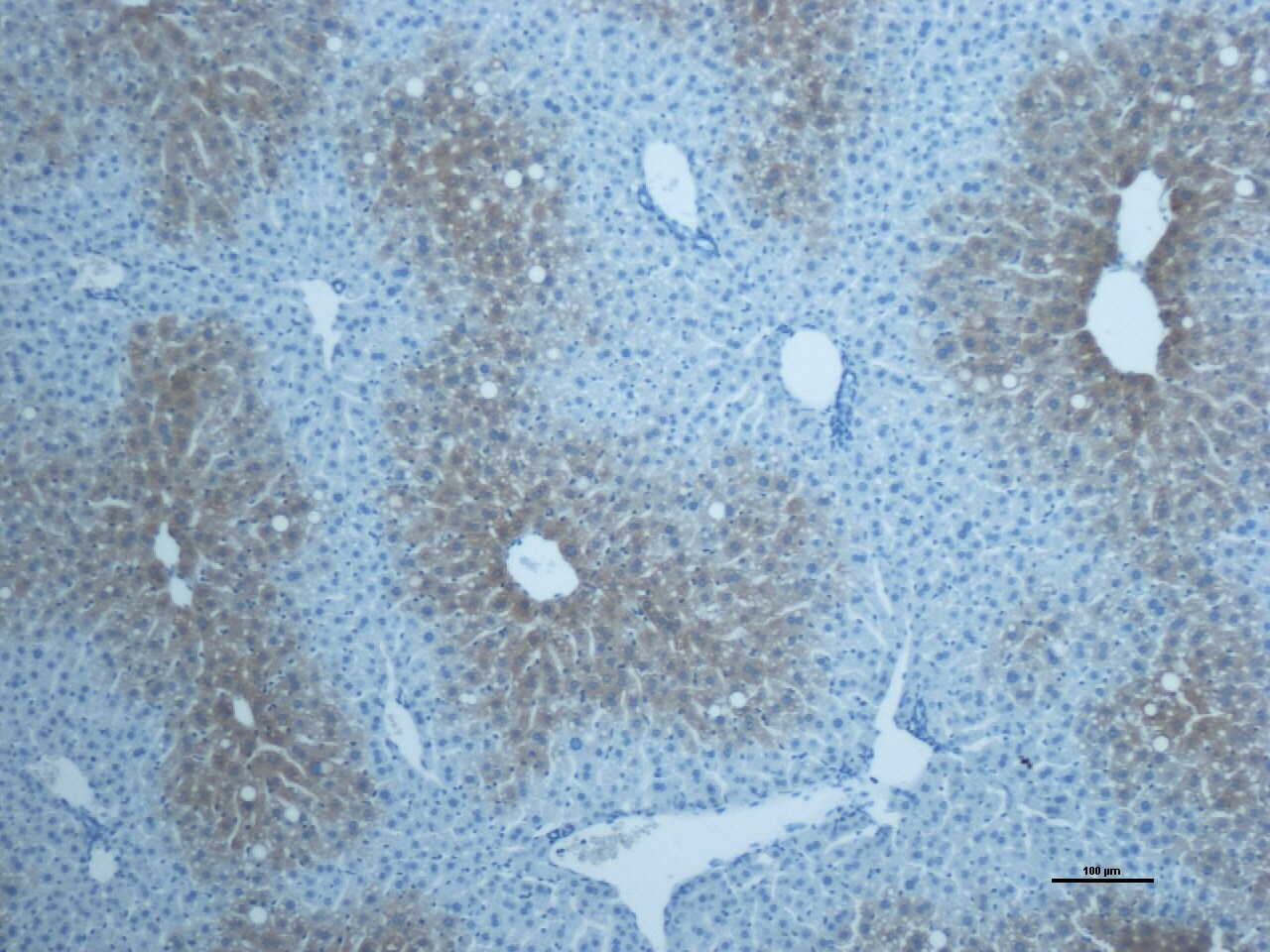
Immunohistochemistry-Paraffin: Cytochrome P450 2E1 Antibody [NBP1-85367]
Immunohistochemistry-Paraffin: Cytochrome P450 2E1 Antibody [NBP1-85367] - Staining of human liver shows strong cytoplasmic positivity in hepatocytes.![Immunohistochemistry-Paraffin: Cytochrome P450 2E1 Antibody [NBP1-85367]](https://resources.rndsystems.com/images/products/Cytochrome-P450-2E1-Antibody-Immunohistochemistry-Paraffin-NBP1-85367-img0021.jpg "Immunohistochemistry-Paraffin: Cytochrome P450 2E1 Antibody [NBP1-85367]")
Immunohistochemistry-Paraffin: Cytochrome P450 2E1 Antibody [NBP1-85367]
Immunohistochemistry-Paraffin: Cytochrome P450 2E1 Antibody [NBP1-85367] - Staining of human tonsil shows no positivity in germinal center cells as expected.
Western Blot: Rabbit Polyclonal Cytochrome P450 2E1 Antibody [NBP1-85367]
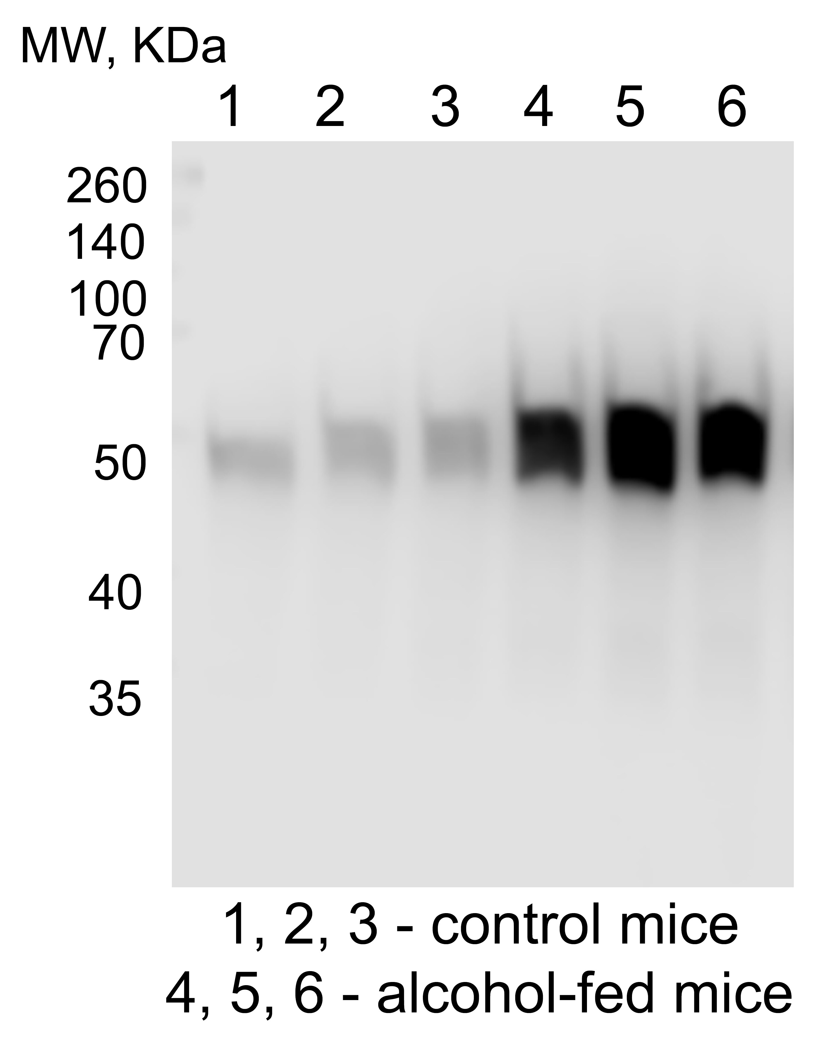
Western Blot: Rabbit Polyclonal Cytochrome P450 2E1 Antibody [NBP1-85367] - Western Blot of the liver homogenates from control and alcohol-fed mice. Image from a verified customer review.
Western Blot: Rabbit Polyclonal Cytochrome P450 2E1 Antibody [NBP1-85367]
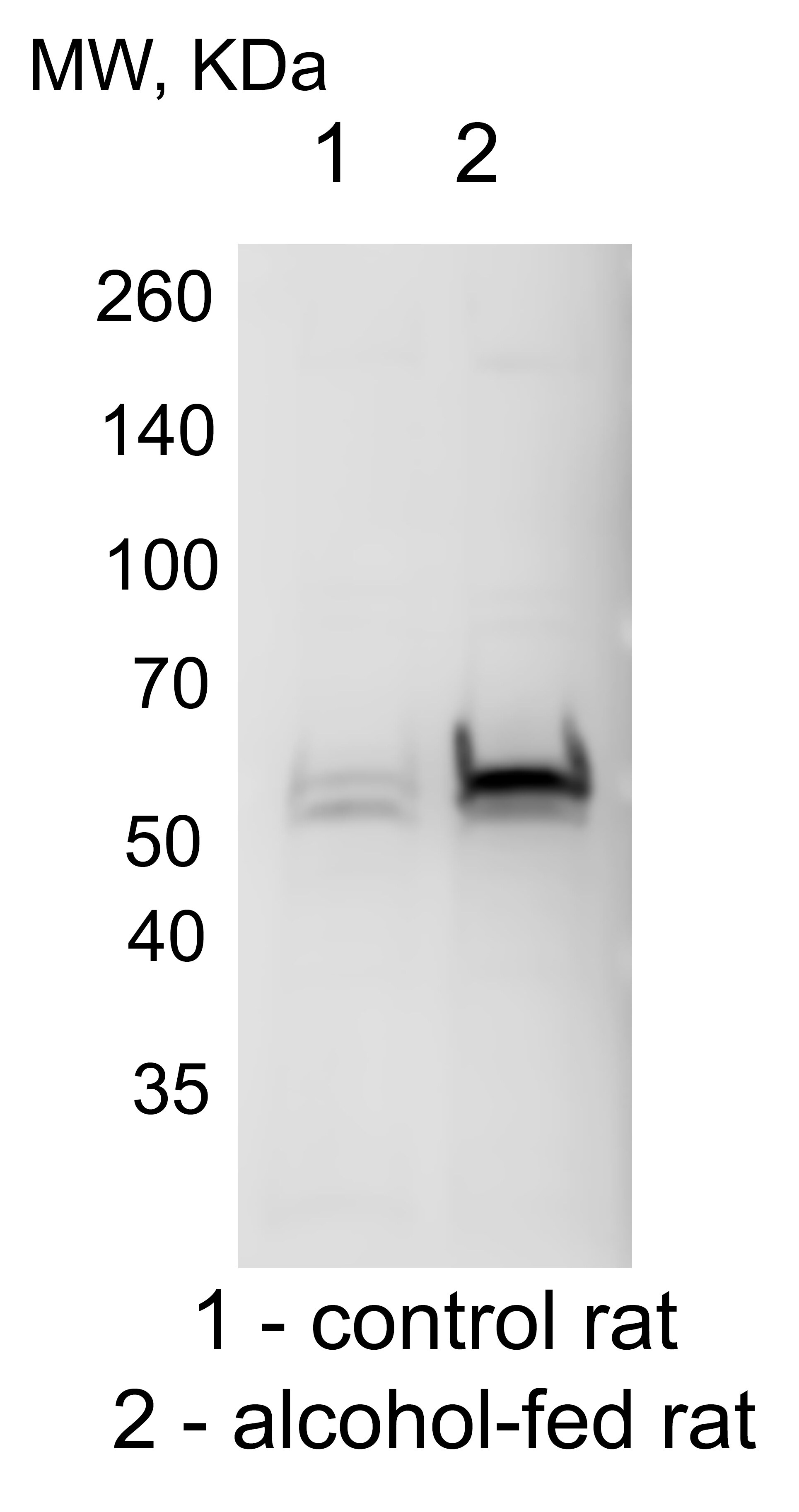
Western Blot: Rabbit Polyclonal Cytochrome P450 2E1 Antibody [NBP1-85367] - Cytochrome P450 2E1 western blot of the homogenates from control and alcohol-fed rats. Image from a verified customer review.
Western Blot: Rabbit Polyclonal Cytochrome P450 2E1 Antibody [NBP1-85367]
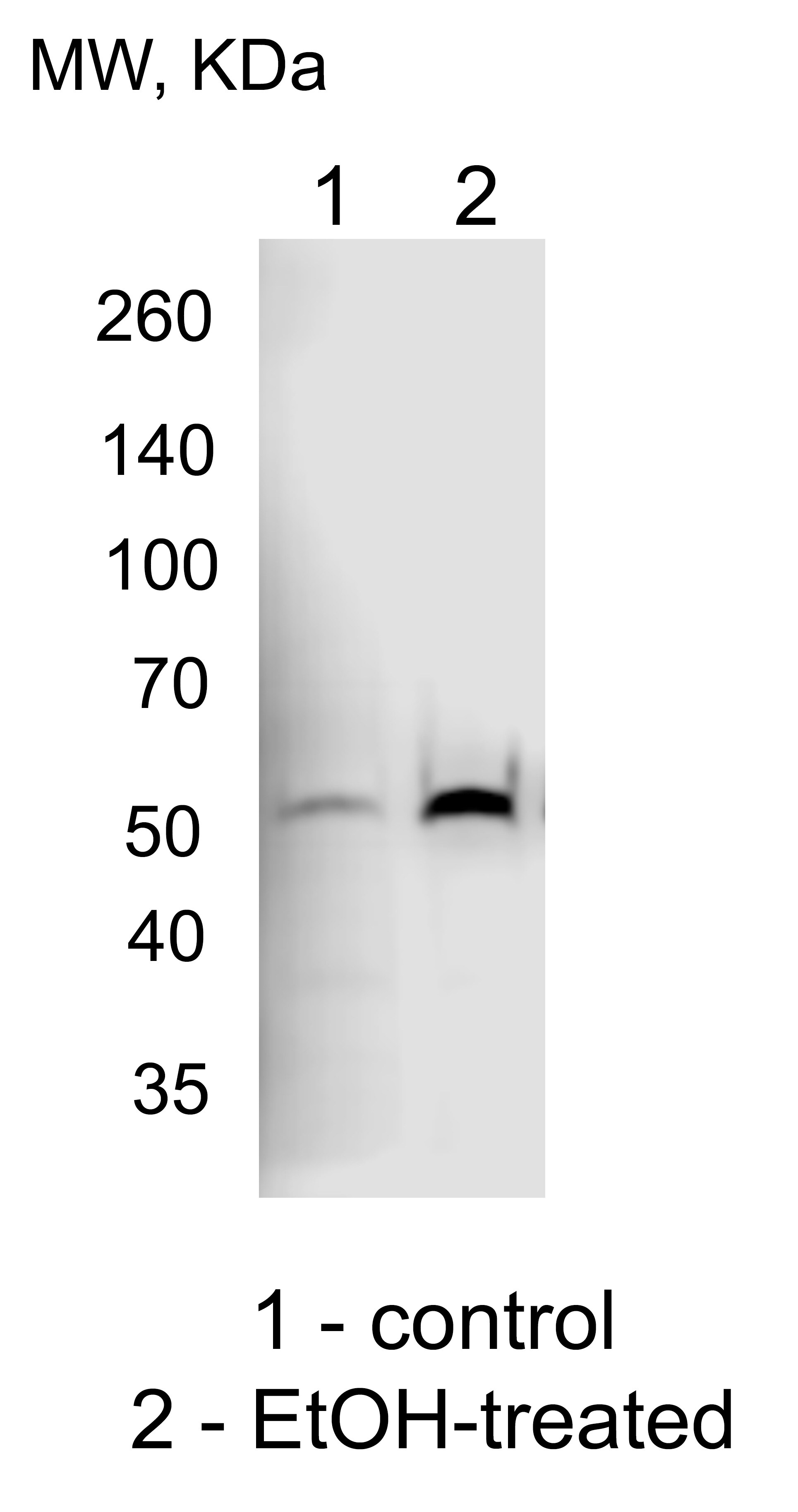
Western Blot: Rabbit Polyclonal Cytochrome P450 2E1 Antibody [NBP1-85367] - Cytochrome P450 2E1 western blot of the lysate of VL-17A cells (Hep G2 cells that stably express both Cytochrome P450 2E1 and ADH): control and treated with EtOH. Image from a verified customer review.Applications for Cytochrome P450 2E1 Antibody - BSA Free
Application
Recommended Usage
Immunohistochemistry
1:200 - 1:500
Immunohistochemistry-Paraffin
1:200 - 1:500
Western Blot
0.04-0.4 ug/ml
Application Notes
For IHC-Paraffin, HIER pH 6 retrieval is recommended.
Reviewed Applications
Read 5 reviews rated 5 using NBP1-85367 in the following applications:
Formulation, Preparation, and Storage
Purification
Affinity purified
Formulation
PBS (pH 7.2) and 40% Glycerol
Format
BSA Free
Preservative
0.02% Sodium Azide
Concentration
Concentrations vary lot to lot. See vial label for concentration. If unlisted please contact technical services.
Shipping
The product is shipped with polar packs. Upon receipt, store it immediately at the temperature recommended below.
Stability & Storage
Store at 4C short term. Aliquot and store at -20C long term. Avoid freeze-thaw cycles.
Background: Cytochrome P450 2E1
Alternate Names
4-nitrophenol 2-hydroxylase, CPE1, CYP2Ecytochrome P450 2E1, CYPIIE1, cytochrome P450, family 2, subfamily E, polypeptide 1, cytochrome P450, subfamily IIE (ethanol-inducible), polypeptide 1, Cytochrome P450-J, EC 1.14.13.-, EC 1.14.13.n7, EC 1.14.14.1, flavoprotein-linked monooxygenase, microsomal monooxygenase, P450C2E, P450-J, xenobiotic monooxygenase
Gene Symbol
CYP2E1
Additional Cytochrome P450 2E1 Products
Product Documents for Cytochrome P450 2E1 Antibody - BSA Free
Certificate of Analysis
To download a Certificate of Analysis, please enter a lot or batch number in the search box below.
Product Specific Notices for Cytochrome P450 2E1 Antibody - BSA Free
This product is for research use only and is not approved for use in humans or in clinical diagnosis. Primary Antibodies are guaranteed for 1 year from date of receipt.
Citations for Cytochrome P450 2E1 Antibody - BSA Free
 Powered by Bioz
Customer Reviews for Cytochrome P450 2E1 Antibody - BSA Free (5)
5 out of 5
5 Customer Ratings
Have you used Cytochrome P450 2E1 Antibody - BSA Free?
Submit a review and receive an Amazon gift card!
$25/€18/£15/$25CAN/¥2500 Yen for a review with an image
$10/€7/£6/$10CAN/¥1110 Yen for a review without an image
Submit a review
Customer Images
Showing
1
-
5 of
5 reviews
Showing All
Filter By:
-
Application: Western BlotSample Tested: HepG2 CELLSSpecies: HumanVerified Customer | Posted 11/10/2024CYP2E1 W-B of the lysate of VL-17A cells (Hep G2 cells that stably express both CYP2E1 and ADH): control and treated with EtOH.

-
Application: Western BlotSample Tested: Rat hepatocytesSpecies: RatVerified Customer | Posted 11/09/2024CYP2E1 W-B of the homogenates from control and alcohol-fed rats
-
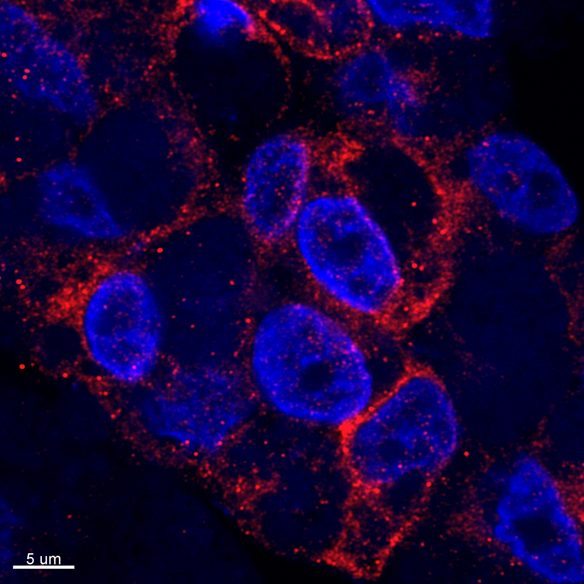
Application: Immunocytochemistry/ImmunofluorescenceSample Tested: HepG2 hepatoma cellsSpecies: HumanVerified Customer | Posted 11/06/2024VA-13 cells (mouse ADH1-transfected HepG2 cells) stained with Cytochrome P450 2E1 Antibody
Bio-Techne ResponseThis review was submitted through the legacy Novus Innovators Program, reflecting a new species or application tested on a primary antibody.
-
Application: Western BlotSample Tested: Liver homogenates sampleSpecies: MouseVerified Customer | Posted 11/06/2024W-B of the liver homogenates from control and alcohol-fed mice
-
Application: Immunohistochemistry-ParaffinSample Tested: mouse liver FFPE section and mouse liver paraffin sectionSpecies: MouseVerified Customer | Posted 12/21/2019Antigen retrieval performed in citrate buffer for 3 minutes; NBP1-85367 antibody dilution was1:100, incubation was 2h at room temperature; DAB developing time was 5 minutes
There are no reviews that match your criteria.
Protocols
Find general support by application which include: protocols, troubleshooting, illustrated assays, videos and webinars.
- Antigen Retrieval Protocol (PIER)
- Antigen Retrieval for Frozen Sections Protocol
- Appropriate Fixation of IHC/ICC Samples
- Cellular Response to Hypoxia Protocols
- Chromogenic IHC Staining of Formalin-Fixed Paraffin-Embedded (FFPE) Tissue Protocol
- Chromogenic Immunohistochemistry Staining of Frozen Tissue
- ClariTSA™ Fluorophore Kits
- Detection & Visualization of Antibody Binding
- Fluorescent IHC Staining of Frozen Tissue Protocol
- Graphic Protocol for Heat-induced Epitope Retrieval
- Graphic Protocol for the Preparation and Fluorescent IHC Staining of Frozen Tissue Sections
- Graphic Protocol for the Preparation and Fluorescent IHC Staining of Paraffin-embedded Tissue Sections
- Graphic Protocol for the Preparation of Gelatin-coated Slides for Histological Tissue Sections
- IHC Sample Preparation (Frozen sections vs Paraffin)
- Immunofluorescent IHC Staining of Formalin-Fixed Paraffin-Embedded (FFPE) Tissue Protocol
- Immunohistochemistry (IHC) and Immunocytochemistry (ICC) Protocols
- Immunohistochemistry Frozen Troubleshooting
- Immunohistochemistry Paraffin Troubleshooting
- Preparing Samples for IHC/ICC Experiments
- Preventing Non-Specific Staining (Non-Specific Binding)
- Primary Antibody Selection & Optimization
- Protocol for Heat-Induced Epitope Retrieval (HIER)
- Protocol for Making a 4% Formaldehyde Solution in PBS
- Protocol for VisUCyte™ HRP Polymer Detection Reagent
- Protocol for the Preparation & Fixation of Cells on Coverslips
- Protocol for the Preparation and Chromogenic IHC Staining of Frozen Tissue Sections
- Protocol for the Preparation and Chromogenic IHC Staining of Frozen Tissue Sections - Graphic
- Protocol for the Preparation and Chromogenic IHC Staining of Paraffin-embedded Tissue Sections
- Protocol for the Preparation and Chromogenic IHC Staining of Paraffin-embedded Tissue Sections - Graphic
- Protocol for the Preparation and Fluorescent IHC Staining of Frozen Tissue Sections
- Protocol for the Preparation and Fluorescent IHC Staining of Paraffin-embedded Tissue Sections
- Protocol for the Preparation of Gelatin-coated Slides for Histological Tissue Sections
- R&D Systems Quality Control Western Blot Protocol
- TUNEL and Active Caspase-3 Detection by IHC/ICC Protocol
- The Importance of IHC/ICC Controls
- Troubleshooting Guide: Immunohistochemistry
- Troubleshooting Guide: Western Blot Figures
- Western Blot Conditions
- Western Blot Protocol
- Western Blot Protocol for Cell Lysates
- Western Blot Troubleshooting
- Western Blot Troubleshooting Guide
- View all Protocols, Troubleshooting, Illustrated assays and Webinars
Loading...