Cytokeratin, pan Antibody (PCK-26)
Novus Biologicals | Catalog # NB120-6401
![Western Blot: Cytokeratin, pan Antibody (PCK-26) [NB120-6401]](https://resources.rndsystems.com/images/products/pan-Cytokeratin-Antibody-PCK-26-Western-Blot-NB120-6401-img0011.jpg "Western Blot: Cytokeratin, pan Antibody (PCK-26) [NB120-6401]")
Loading...
Key Product Details
Species Reactivity
Validated:
Human, Mouse, Rat, Porcine, Bovine, Canine, Chicken, Equine, Feline, Fish, Goat, Guinea Pig, Hamster, Rabbit, Reptile, Sheep
Cited:
Human, Mouse, Rat, Feline, Ovine, Reptile
Applications
Validated:
Immunohistochemistry, Immunohistochemistry-Paraffin, Immunohistochemistry-Frozen, Western Blot, Immunocytochemistry/ Immunofluorescence
Cited:
Immunohistochemistry, Immunohistochemistry-Paraffin, Western Blot, Immunocytochemistry/ Immunofluorescence, IF/IHC
Label
Unconjugated
Antibody Source
Monoclonal Mouse IgG1 Clone # PCK-26
Loading...
Product Specifications
Immunogen
This Cytokeratin, pan Antibody (PCK-26) was developed against the full length native protein (purified) (Human epidermis).
Reactivity Notes
Use in Equine reported in scientific literature (PMID:34372610) Cross reacts with: carp, lizard, snake. Please note that this antibody is reactive to Mouse and derived from the same host, Mouse. Additional Mouse on Mouse blocking steps may be required for IHC and ICC experiments. Please contact Technical Support for more information.
Localization
Cytoplasmic
Marker
Epithelial marker
Specificity
PCK-26 recognizes the 58 kDa cytokeratin 5, the 56 kDa cytokeratin 6 and the 52 kDa cytokeratin 8 band in immunoblotting (Lane EB et al). The antibody recognizes an epitope located on the Type II cytokeratins 1, 5, 6, and 8. PCK-26 is a broad spectrum antibody which reacts specifically with a variety of normal, reactive, and neoplastic epithelial tissues. The antibody reacts with simple, cornifying, and non-cornifying squamous epithelia and pseudostratified epithelia.
Clonality
Monoclonal
Host
Mouse
Isotype
IgG1
Scientific Data Images for Cytokeratin, pan Antibody (PCK-26)
Western Blot: Cytokeratin, pan Antibody (PCK-26) [NB120-6401]
Western Blot: Cytokeratin, pan Antibody (PCK-26) [NB120-6401] - Whole extract of HeLa cells was separated on SDS-PAGE and probed with Monoclonal Anti-Cytokeratin, pan Clone: PCK-26. WB was developed using Goat Anti-Mouse IgG-Peroxidase and a chemiluminescent substrate. Lane 1: Antibody dilution 1:300. Lane 2: Antibody dilution 1:500. Lane 3: Antibody dilution 1:1000.![Immunocytochemistry/ Immunofluorescence: Cytokeratin, pan Antibody (PCK-26) [NB120-6401]](https://resources.rndsystems.com/images/products/Cytokeratin-pan-Antibody-PCK-26-Immunocytochemistry-Immunofluorescence-NB120-6401-img0012.jpg "Immunocytochemistry/ Immunofluorescence: Cytokeratin, pan Antibody (PCK-26) [NB120-6401]")
Immunocytochemistry/ Immunofluorescence: Cytokeratin, pan Antibody (PCK-26) [NB120-6401]
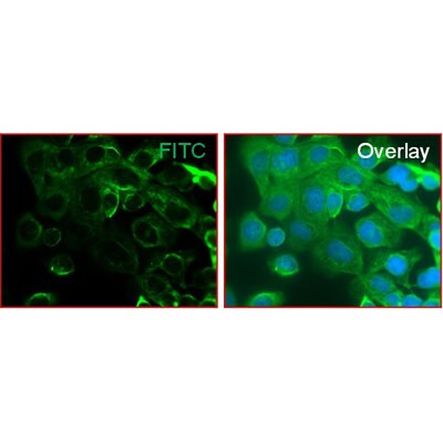
Immunocytochemistry/Immunofluorescence: Cytokeratin, pan Antibody (PCK-26) [NB120-6401] - Human pancreas cancer cells. Antibody at 1:250 dilution. Overnight incubation. No permeabilization. ICC/IF image submitted by a verified customer review.![Immunohistochemistry-Paraffin: Cytokeratin, pan Antibody (PCK-26) [NB120-6401]](https://resources.rndsystems.com/images/products/pan-Cytokeratin-Antibody-PCK-26-Immunohistochemistry-Paraffin-NB120-6401-img0006.jpg "Immunohistochemistry-Paraffin: Cytokeratin, pan Antibody (PCK-26) [NB120-6401]")
Immunohistochemistry-Paraffin: Cytokeratin, pan Antibody (PCK-26) [NB120-6401]
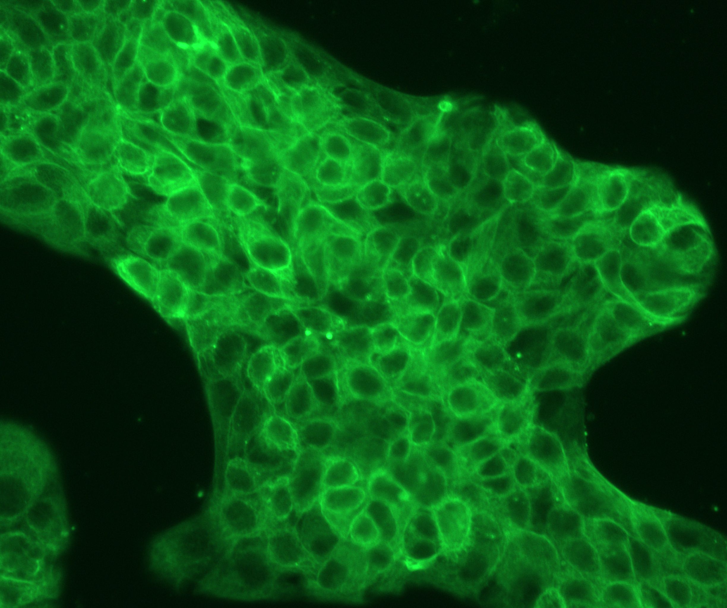
Immunohistochemistry-Paraffin: Cytokeratin, pan Antibody (PCK-26) [NB120-6401] - Staining of FFPE human placenta sections with 1:300 Monoclonal Anti-Cytokeratin, pan Clone: PCK-26, followed by Goat Anti Mouse IgG (Fab)-FITC.![Immunohistochemistry: Cytokeratin, pan Antibody (PCK-26) [NB120-6401]](https://resources.rndsystems.com/images/products/Cytokeratin-pan-Antibody-PCK-26-Immunohistochemistry-NB120-6401-img0014.jpg "Immunohistochemistry: Cytokeratin, pan Antibody (PCK-26) [NB120-6401]")
Immunohistochemistry: Cytokeratin, pan Antibody (PCK-26) [NB120-6401]
Cytokeratin-pan-Antibody-PCK-26-Immunohistochemistry-NB120-6401-img0014.jpg![Immunohistochemistry: Cytokeratin, pan Antibody (PCK-26) [NB120-6401]](https://resources.rndsystems.com/images/products/Cytokeratin-pan-Antibody-PCK-26-Immunohistochemistry-NB120-6401-img0013.jpg "Immunohistochemistry: Cytokeratin, pan Antibody (PCK-26) [NB120-6401]")
Immunohistochemistry: Cytokeratin, pan Antibody (PCK-26) [NB120-6401]
Cytokeratin-pan-Antibody-PCK-26-Immunohistochemistry-NB120-6401-img0013.jpg [NB120-6401] -")
Immunocytochemistry/ Immunofluorescence: Cytokeratin, pan Antibody (PCK-26) [NB120-6401] -
Immunocytochemistry/ Immunofluorescence: Cytokeratin, pan Antibody (PCK-26) [NB120-6401] - Detection of EMT & increased motility in NHRI-HN1 cells. (A) Summary of the most enriched pathways associated with tumorigenesis in syngeneic mice by comparing tumorigenic NHRI-HN1 cells with nontumorigenic cells, including M1-2, M2-3 & NHRI-HN2, using GSEA analysis. Blue indicates a negative normalized enrichment score (NES) & orange indicates a positive NES. (B) Morphology of M1-2 & NHRI-HN1 cells via phase-contrast microscopy at 100× magnification. (C) Relative adhesion activity in M1-2 & NHRI-HN1 cells, determined by normalizing the mean OD 490 nm value of NHRI-HN1 cells to that of M1-2 cells. (D) Cells stained with Alexa Fluor 488 phalloidin, anti-pan-CK & anti-EGFR antibodies at 400× magnification. (E) Immunoblot analysis of epithelial (E-cadherin, alpha -catenin & beta -catenin), mesenchymal (N-cadherin & Vimentin) proteins & EMT-related transcription factors, including Twist, Snail & Slug in M1-2 & NHRI-HN1 cells. Protein levels were normalized to an internal control, alpha -tubulin. Ratios were determined by dividing the normalized protein levels in NHRI-HN1 cells by that in M1-2 cells. (F) Representative images (left) & relative data (right) for migration activity of M1-2 & NHRI-HN1 cells. (G) Representative images (left) & relative data (right) for invasion activity of M1-2 & NHRI-HN1 cells at 200× magnification. The relative migration or invasion activity was determined by normalizing the mean number of cells that have migrated or invaded per field of NHRI-HN1 cells to that of M1-2 cells. Error bars represent SE; ** p < 0.01; *** p < 0.001. Image collected & cropped by CiteAb from the following publication (https://pubmed.ncbi.nlm.nih.gov/31878324), licensed under a CC-BY license. Not internally tested by Novus Biologicals.Applications for Cytokeratin, pan Antibody (PCK-26)
Application
Recommended Usage
Immunocytochemistry/ Immunofluorescence
1:300
Immunohistochemistry-Frozen
1:300 - 1:600
Immunohistochemistry-Paraffin
1:300 - 1:600
Western Blot
1:100 - 1:2000
Reviewed Applications
Read 2 reviews rated 5 using NB120-6401 in the following applications:
Formulation, Preparation, and Storage
Purification
Unpurified
Formulation
Ascites
Preservative
0.09% Sodium Azide
Concentration
This product is unpurified. The exact concentration of antibody is not quantifiable.
Shipping
The product is shipped with polar packs. Upon receipt, store it immediately at the temperature recommended below.
Stability & Storage
Store at 4C short term. Aliquot and store at -20C long term. Avoid freeze-thaw cycles.
Background: Cytokeratin, pan
Epithelial cells express multiple subtypes of cytokeratins which can be used to classify epithelial cell type or differentiation status, as well tumor progression or diagnosis (2). Cytokeratins are important for both stability and integrity of epithelial cells and function in intracellular signaling, from wound healing to apoptosis (1). Cytokeratins are useful immunohistochemistry tumor markers and antibodies to cytokeratins are a common pathological tool (1,3,6). Cytokeratin pan antibody is an antibody cocktail mixture that can detect multiple cytokeratins and reacts to multiple epithelial tissues (1,3,6). For example, AE-1/AE-3 is a commonly used specific pan cytokeratin that detects cytokeratins 1-8, 10, 14-16 and 19 (1,3,6).
Given the role of cytokeratins in the structural integrity of epithelial cells, mutations in cytokeratins have been shown to play a role in a variety of human diseases including epidermolysis bullosa simplex (EBS) (4,5). EBS is an autosomal dominant disorder that is caused by missense mutations in either CK5 or CK14 (5). Other known cytokeratin-related disorders include bullous ichthyosis, a skin disorder characterized by redness, blistering, and hyperkeratosis, and epidermolytic palmoplantar keratoderma (EPPK), which results in hyperkeratosis on the palms and soles of the body (7).
References
1. Awasthi, P., Thahriani, A., Bhattacharya, A., Awasthi, P., & Keratins, B. A. (2016). Keratins or cytokeratins: a review article. Journal of Advanced Medical and Dental Sciences Research. https://10.21276/jamdsr.2016.4.4.30
2. Southgate, J., Harnden, P., & Trejdosiewicz, L. K. (1999). Cytokeratin expression patterns in normal and malignant urothelium: a review of the biological and diagnostic implications. Histology and histopathology. https://doi.org/10.14670/HH-14.657
3. Belaldavar, C., Mane, D. R., Hallikerimath, S., Kale, A. D. (2016). Cytokeratins: Its role and expression profile in oral health and disease. Journal of Oral and Maxillofacial Surgery, Medicine, and Pathology. https://doi.org/10.1016/j.ajoms.2015.08.001.
4. Linder S. (2007). Cytokeratin markers come of age. Tumour biology : the journal of the International Society for Oncodevelopmental Biology and Medicine. https://doi.org/10.1159/000107582
5. Jacob, J. T., Coulombe, P. A., Kwan, R., & Omary, M. B. (2018). Types I and II Keratin Intermediate Filaments. Cold Spring Harbor perspectives in biology. https://doi.org/10.1101/cshperspect.a018275
6. Ordonez N. G. (2013). Broad-spectrum immunohistochemical epithelial markers: a review. Human pathology. https://doi.org/10.1016/j.humpath.2012.11.016
7. McLean, W. H., & Moore, C. B. (2011). Keratin disorders: from gene to therapy. Human molecular genetics. https://doi.org/10.1093/hmg/ddr379
Alternate Names
AEI2, CK1, EHK, EPPK, K1, KRT1A, NEPPK
Gene Symbol
KRT1
UniProt
Additional Cytokeratin, pan Products
Product Documents for Cytokeratin, pan Antibody (PCK-26)
Certificate of Analysis
To download a Certificate of Analysis, please enter a lot or batch number in the search box below.
Product Specific Notices for Cytokeratin, pan Antibody (PCK-26)
This product is for research use only and is not approved for use in humans or in clinical diagnosis. Primary Antibodies are guaranteed for 1 year from date of receipt.
Citations for Cytokeratin, pan Antibody (PCK-26)
 Powered by Bioz
Customer Reviews for Cytokeratin, pan Antibody (PCK-26) (2)
5 out of 5
2 Customer Ratings
Have you used Cytokeratin, pan Antibody (PCK-26)?
Submit a review and receive an Amazon gift card!
$25/€18/£15/$25CAN/¥2500 Yen for a review with an image
$10/€7/£6/$10CAN/¥1110 Yen for a review without an image
Submit a review
Customer Images
Showing
1
-
2 of
2 reviews
Showing All
Filter By:
-
Application: ImmunocytochemistrySample Tested: human pancreas cancer cellsSpecies: HumanVerified Customer | Posted 08/09/2019Dilution 1:250 Incubation time: overnight, 4C No permeabilization

-
Application: ImmunofluorescenceSample Tested: Prostate cancer (Human) cell lineSpecies: HumanVerified Customer | Posted 02/05/2013Pan cytokeratin in PNT2 cells
There are no reviews that match your criteria.
Protocols
Find general support by application which include: protocols, troubleshooting, illustrated assays, videos and webinars.
- Antigen Retrieval Protocol (PIER)
- Antigen Retrieval for Frozen Sections Protocol
- Appropriate Fixation of IHC/ICC Samples
- Cellular Response to Hypoxia Protocols
- Chromogenic IHC Staining of Formalin-Fixed Paraffin-Embedded (FFPE) Tissue Protocol
- Chromogenic Immunohistochemistry Staining of Frozen Tissue
- ClariTSA™ Fluorophore Kits
- Detection & Visualization of Antibody Binding
- Fluorescent IHC Staining of Frozen Tissue Protocol
- Graphic Protocol for Heat-induced Epitope Retrieval
- Graphic Protocol for the Preparation and Fluorescent IHC Staining of Frozen Tissue Sections
- Graphic Protocol for the Preparation and Fluorescent IHC Staining of Paraffin-embedded Tissue Sections
- Graphic Protocol for the Preparation of Gelatin-coated Slides for Histological Tissue Sections
- ICC Cell Smear Protocol for Suspension Cells
- ICC Immunocytochemistry Protocol Videos
- ICC for Adherent Cells
- IHC Sample Preparation (Frozen sections vs Paraffin)
- Immunocytochemistry (ICC) Protocol
- Immunocytochemistry Troubleshooting
- Immunofluorescence of Organoids Embedded in Cultrex Basement Membrane Extract
- Immunofluorescent IHC Staining of Formalin-Fixed Paraffin-Embedded (FFPE) Tissue Protocol
- Immunohistochemistry (IHC) and Immunocytochemistry (ICC) Protocols
- Immunohistochemistry Frozen Troubleshooting
- Immunohistochemistry Paraffin Troubleshooting
- Preparing Samples for IHC/ICC Experiments
- Preventing Non-Specific Staining (Non-Specific Binding)
- Primary Antibody Selection & Optimization
- Protocol for Heat-Induced Epitope Retrieval (HIER)
- Protocol for Making a 4% Formaldehyde Solution in PBS
- Protocol for VisUCyte™ HRP Polymer Detection Reagent
- Protocol for the Fluorescent ICC Staining of Cell Smears - Graphic
- Protocol for the Fluorescent ICC Staining of Cultured Cells on Coverslips - Graphic
- Protocol for the Preparation & Fixation of Cells on Coverslips
- Protocol for the Preparation and Chromogenic IHC Staining of Frozen Tissue Sections
- Protocol for the Preparation and Chromogenic IHC Staining of Frozen Tissue Sections - Graphic
- Protocol for the Preparation and Chromogenic IHC Staining of Paraffin-embedded Tissue Sections
- Protocol for the Preparation and Chromogenic IHC Staining of Paraffin-embedded Tissue Sections - Graphic
- Protocol for the Preparation and Fluorescent ICC Staining of Cells on Coverslips
- Protocol for the Preparation and Fluorescent ICC Staining of Non-adherent Cells
- Protocol for the Preparation and Fluorescent ICC Staining of Stem Cells on Coverslips
- Protocol for the Preparation and Fluorescent IHC Staining of Frozen Tissue Sections
- Protocol for the Preparation and Fluorescent IHC Staining of Paraffin-embedded Tissue Sections
- Protocol for the Preparation of Gelatin-coated Slides for Histological Tissue Sections
- Protocol for the Preparation of a Cell Smear for Non-adherent Cell ICC - Graphic
- R&D Systems Quality Control Western Blot Protocol
- TUNEL and Active Caspase-3 Detection by IHC/ICC Protocol
- The Importance of IHC/ICC Controls
- Troubleshooting Guide: Immunohistochemistry
- Troubleshooting Guide: Western Blot Figures
- Western Blot Conditions
- Western Blot Protocol
- Western Blot Protocol for Cell Lysates
- Western Blot Troubleshooting
- Western Blot Troubleshooting Guide
- View all Protocols, Troubleshooting, Illustrated assays and Webinars
Loading...