DDIT4 Antibody - BSA Free
Novus Biologicals | Catalog # NBP1-22966
![Western Blot: DDIT4 Antibody [NBP1-22966]](https://resources.rndsystems.com/images/products/DDIT4-Antibody-Western-Blot-NBP1-22966-img0005.jpg "Western Blot: DDIT4 Antibody [NBP1-22966]")
Key Product Details
Validated by
Biological Validation
Species Reactivity
Validated:
Human, Mouse
Cited:
Human, Mouse
Predicted:
Bovine (100%), Canine (100%), Chimpanzee (100%), Equine (100%), Monkey (100%), Orangutan (100%), Primate (100%), Rabbit (100%), Rat (100%). Backed by our 100% Guarantee.
Applications
Validated:
Western Blot, Immunocytochemistry/ Immunofluorescence, Immunoprecipitation
Cited:
Western Blot
Label
Unconjugated
Antibody Source
Polyclonal Rabbit IgG
Format
BSA Free
Loading...
Product Specifications
Immunogen
The immunogen recognized by this antibody maps to a region between residue 1 and 50 of human protein regulated in development and DNA damage response 1 (DNA-damage-inducible transcript 4) using the numbering given in entry NP_061931.1 (GeneID 54541).
Reactivity Notes
Mouse reactivity reported in scientific literature (PMID: 25867045)
Clonality
Polyclonal
Host
Rabbit
Isotype
IgG
Scientific Data Images for DDIT4 Antibody - BSA Free
Western Blot: DDIT4 Antibody [NBP1-22966]
DDIT4-Antibody-Western-Blot-NBP1-22966-img0005.jpg![Immunocytochemistry/ Immunofluorescence: DDIT4 Antibody [NBP1-22966]](https://resources.rndsystems.com/images/products/DDIT4-Antibody-Immunocytochemistry-Immunofluorescence-NBP1-22966-img0004.jpg "Immunocytochemistry/ Immunofluorescence: DDIT4 Antibody [NBP1-22966]")
Immunocytochemistry/ Immunofluorescence: DDIT4 Antibody [NBP1-22966]
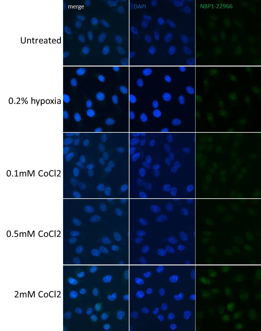
Immunocytochemistry/Immunofluorescence: DDIT4 Antibody [NBP1-22966] - HeLa cells stained with NBP1-22966 after 0.2% hypoxia or CoCl2 overnight. Image from verified customer review.![Western Blot: DDIT4 Antibody [NBP1-22966]](https://resources.rndsystems.com/images/products/DDIT4-Antibody-Western-Blot-NBP1-22966-img0002.jpg "Western Blot: DDIT4 Antibody [NBP1-22966]")
Western Blot: DDIT4 Antibody [NBP1-22966]
Western Blot: DDIT4 Antibody [NBP1-22966] - Whole cell lysate (5, 15 and 50 mcg for WB; 1 mg for IP, 20% of IP loaded) from HeLa cells. Lysate was prepared from cells that had been treated with cobalt chloride (A and B) or mock treated (A). NBP1-22966 used for WB at 0.4 mcg/ml (A) and 1 mcg/ml (B) and used for IP at 10 mcg/mg lysate.![Western Blot: DDIT4 Antibody [NBP1-22966]](https://resources.rndsystems.com/images/products/DDIT4-Antibody-Western-Blot-NBP1-22966-img0006.jpg "Western Blot: DDIT4 Antibody [NBP1-22966]")
Western Blot: DDIT4 Antibody [NBP1-22966]
DDIT4-Antibody-Western-Blot-NBP1-22966-img0006.jpg![Immunocytochemistry/ Immunofluorescence: DDIT4 Antibody [NBP1-22966]](https://resources.rndsystems.com/images/products/DDIT4-Antibody-Immunocytochemistry-Immunofluorescence-NBP1-22966-img0003.jpg "Immunocytochemistry/ Immunofluorescence: DDIT4 Antibody [NBP1-22966]")
Immunocytochemistry/ Immunofluorescence: DDIT4 Antibody [NBP1-22966]
Immunocytochemistry/Immunofluorescence: DDIT4 Antibody [NBP1-22966] - NBF-fixed asynchronous, cobalt-treated HeLa cells. Antibody: Affinity purified rabbit anti-REDD1 used at a dilution of 1:500 (2 ug/ml). Detection: Red-fluorescent goat anti-rabbit IgG H&L crossadsorbed Antibody DyLight®594 used at 1:100.Applications for DDIT4 Antibody - BSA Free
Application
Recommended Usage
Immunocytochemistry/ Immunofluorescence
1:500 to 1:2000
Immunoprecipitation
5-15 ug/mg lysate
Western Blot
1:1000-1:4000
Application Notes
Formaldehyde fixation is recommended. Permeabilization with Triton X-100 is recommended for formaldehydefixed cells.
Reviewed Applications
Read 1 review rated 4 using NBP1-22966 in the following applications:
Formulation, Preparation, and Storage
Purification
Immunogen affinity purified
Formulation
Tris-Citrate/Phosphate (pH 7.0 - 8.0)
Format
BSA Free
Preservative
0.09% Sodium Azide
Concentration
1.0 mg/ml
Shipping
The product is shipped with polar packs. Upon receipt, store it immediately at the temperature recommended below.
Stability & Storage
Store at 4C. Do not freeze.
Background: DDIT4
Alternate Names
Dig2, DNA-damage-inducible transcript 4, FLJ20500, HIF-1 responsive RTP801 (RTP801), Protein regulated in development and DNA damage response 1, REDD-1DNA damage-inducible transcript 4 protein, REDD1HIF-1 responsive protein RTP801, RTP801RP11-442H21.1
Entrez Gene IDs
54541 (Human)
Gene Symbol
DDIT4
UniProt
Additional DDIT4 Products
Product Documents for DDIT4 Antibody - BSA Free
Certificate of Analysis
To download a Certificate of Analysis, please enter a lot or batch number in the search box below.
Product Specific Notices for DDIT4 Antibody - BSA Free
This product is for research use only and is not approved for use in humans or in clinical diagnosis. Primary Antibodies are guaranteed for 1 year from date of receipt.
Citations for DDIT4 Antibody - BSA Free
 Powered by Bioz
Customer Reviews for DDIT4 Antibody - BSA Free (1)
4 out of 5
1 Customer Rating
Have you used DDIT4 Antibody - BSA Free?
Submit a review and receive an Amazon gift card!
$25/€18/£15/$25CAN/¥2500 Yen for a review with an image
$10/€7/£6/$10CAN/¥1110 Yen for a review without an image
Submit a review
Customer Images
Showing
1
-
1 of
1 review
Showing All
Filter By:
-
Application: ImmunocytochemistrySample Tested: hela cellSpecies: HumanVerified Customer | Posted 05/24/2019Immunofluorescence/Immunocytochemistry: HeLa stained with NBP1-22966 after 0.2% hypoxia or CoCl2 overnight.HeLa cells treated with 0.2% hypoxia or different doses CoCl2 overnight; fixed in 4% PFA 10 minutes (directly in hypoxia chamber or quickly after washing off CoCl2/media as half-life DDIT4 is 5-10 minutes); permeabilize 0.5% Triton X-100 5 minutes; dilute primary antibody 1/500 incubate O/N @4oC; wash 3X PBS; Alexa 488 donkey anti-rabbit F(ab”)2 secondary antibody 1/1000 1 hour at rt; wash 5X PBS; mount DAPI/Vectashield; images acquired on Olympus BX63 microscope with Bioview Allegro software.

There are no reviews that match your criteria.
Protocols
Find general support by application which include: protocols, troubleshooting, illustrated assays, videos and webinars.
- Appropriate Fixation of IHC/ICC Samples
- Cellular Response to Hypoxia Protocols
- ClariTSA™ Fluorophore Kits
- Detection & Visualization of Antibody Binding
- ICC Cell Smear Protocol for Suspension Cells
- ICC Immunocytochemistry Protocol Videos
- ICC for Adherent Cells
- Immunocytochemistry (ICC) Protocol
- Immunocytochemistry Troubleshooting
- Immunofluorescence of Organoids Embedded in Cultrex Basement Membrane Extract
- Immunohistochemistry (IHC) and Immunocytochemistry (ICC) Protocols
- Immunoprecipitation Protocol
- Preparing Samples for IHC/ICC Experiments
- Preventing Non-Specific Staining (Non-Specific Binding)
- Primary Antibody Selection & Optimization
- Protocol for VisUCyte™ HRP Polymer Detection Reagent
- Protocol for the Fluorescent ICC Staining of Cell Smears - Graphic
- Protocol for the Fluorescent ICC Staining of Cultured Cells on Coverslips - Graphic
- Protocol for the Preparation and Fluorescent ICC Staining of Cells on Coverslips
- Protocol for the Preparation and Fluorescent ICC Staining of Non-adherent Cells
- Protocol for the Preparation and Fluorescent ICC Staining of Stem Cells on Coverslips
- Protocol for the Preparation of a Cell Smear for Non-adherent Cell ICC - Graphic
- R&D Systems Quality Control Western Blot Protocol
- TUNEL and Active Caspase-3 Detection by IHC/ICC Protocol
- The Importance of IHC/ICC Controls
- Troubleshooting Guide: Western Blot Figures
- Western Blot Conditions
- Western Blot Protocol
- Western Blot Protocol for Cell Lysates
- Western Blot Troubleshooting
- Western Blot Troubleshooting Guide
- View all Protocols, Troubleshooting, Illustrated assays and Webinars
Loading...