![Western Blot: DDX6 Antibody [NB200-191]](https://resources.rndsystems.com/images/products/DDX6-Antibody-Western-Blot-NB200-191-img0014.jpg "Western Blot: DDX6 Antibody [NB200-191]")
Loading...
Key Product Details
Species Reactivity
Validated:
Human, Mouse, Hamster
Cited:
Human, Mouse, Rat
Predicted:
Chicken (100%), Orangutan (100%). Backed by our 100% Guarantee.
Applications
Validated:
Immunohistochemistry, Immunohistochemistry-Paraffin, Western Blot, Immunocytochemistry/ Immunofluorescence, Immunoprecipitation
Cited:
Immunohistochemistry-Paraffin, Western Blot, Immunocytochemistry/ Immunofluorescence
Label
Unconjugated
Antibody Source
Polyclonal Rabbit IgG
Loading...
Product Specifications
Immunogen
The immunogen recognized by NB200-191 maps to a region between residues 1 and 50 of human DEAD (Asp-Glu-Ala-Asp) box polypeptide 6 using the numbering given Swiss-Prot entry P26196 (GeneID 1656).
Reactivity Notes
Hamster reactivity reported from a verified customer reivew.
Clonality
Polyclonal
Host
Rabbit
Isotype
IgG
Scientific Data Images for DDX6 Antibody
Western Blot: DDX6 Antibody [NB200-191]
DDX6-Antibody-Western-Blot-NB200-191-img0014.jpg![Immunocytochemistry/ Immunofluorescence: DDX6 Antibody [NB200-191]](https://resources.rndsystems.com/images/products/DDX6-Antibody-Immunocytochemistry-Immunofluorescence-NB200-191-img0008.jpg "Immunocytochemistry/ Immunofluorescence: DDX6 Antibody [NB200-191]")
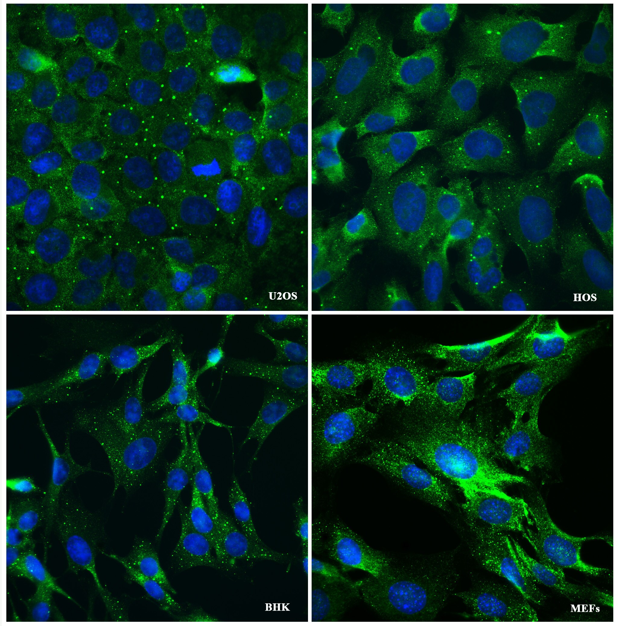
Immunocytochemistry/ Immunofluorescence: DDX6 Antibody [NB200-191]
Immunocytochemistry/Immunofluorescence: DDX6 Antibody [NB200-191] - In normal condition, cells were fixed and stained for DDX6, that usually punctuate in process bodies (PBs). Primary Antibody rabbit-anti-DDX6 diluted 1:1000. Image submitted by a verified customer review.![Immunohistochemistry-Paraffin: DDX6 Antibody [NB200-191]](https://resources.rndsystems.com/images/products/DDX6-Antibody-Immunohistochemistry-Paraffin-NB200-191-img0013.jpg "Immunohistochemistry-Paraffin: DDX6 Antibody [NB200-191]")
Immunohistochemistry-Paraffin: DDX6 Antibody [NB200-191]
Immunohistochemistry-Paraffin: DDX6 Antibody [NB200-191] - Section of human colon carcinoma (left) and mouse renal cell carcinoma(right). Antibody: Affinity purified rabbit anti-DDX6 used at a dilution of 1:200 (1ug/ml). Detection: DAB.![Immunoprecipitation: DDX6 Antibody [NB200-191]](https://resources.rndsystems.com/images/products/DDX6-Antibody-Immunoprecipitation-NB200-191-img0012.jpg "Immunoprecipitation: DDX6 Antibody [NB200-191]")
Immunoprecipitation: DDX6 Antibody [NB200-191]
Immunoprecipitation: DDX6 Antibody [NB200-191] - Detection of human DDX6 by Western Blot and Immunoprecipitation. Samples: Whole cell lysate (15 and 50 ug) from HeLa cells. Antibodies: Affinity purified rabbit anti-DDX6 antibody NB200-191 used at 0.04 ug/ml for WB (A and B) and at 3 ug/mg lysate for IP. DDX6 was also immunoprecipitated using rabbit anti-DDX6 antibody NB200-192, but not with 'DDX6 Ab', which is to another epitope from DDX6. Normal rabbit IgG was used as a negative control. Detection: Chemiluminescence with an exposure time of 1 second (A & B).
Western Blot: DDX6 Antibody [NB200-191] -
Western Blot: DDX6 Antibody [NB200-191] - Staufen1 protein but not mRNA steady-state levels are increased in neurodegenerative disease cells & tissues. Western blot analysis of SCA2- FBs (a) & LBCs (b) show increased STAU1 levels compared with normal controls. DDX6 levels are unchanged. HD & SCA3 patient (polyQ expanded) FBs were used as additional controls. Four normal & five SCA2 FBs, & two normal & three SCA2 LBCs were used. c, d Western blot analyses of ATXN2Q127 (c) & BAC-Q72 (d) mouse cerebellar extracts (24 weeks of age) showing increased Stau1 levels compared with wild-type or BAC-Q22 controls (n = 2–3 animals per group). e Western blot of FB extracts from an ALS patient with the TDP-43G298S mutation show increased STAU1 levels. beta -Actin was used as loading control & representative blots of three independent experiments are shown. f–hSTAU1 RNA levels are unaltered in SCA2 & ALS cells & SCA2 mice. qRT-PCR analyses of STAU1 mRNA in SCA2 FBs & ALS FB with TDP-43G298S mutation (f) or SCA2 LBCs (g). h qRT-PCR analyses of cerebellar RNAs from ATXN2Q127 & BAC-Q72 mice compared to wild-type littermates (24 weeks of age; n = animals per group). Gene expression levels were normalized to Actb Image collected & cropped by CiteAb from the following publication (https://pubmed.ncbi.nlm.nih.gov/30194296), licensed under a CC-BY license. Not internally tested by Novus Biologicals.Applications for DDX6 Antibody
Application
Recommended Usage
Immunohistochemistry
1:100-1:500
Immunohistochemistry-Paraffin
1:100-1:500
Immunoprecipitation
2-5 ug/mg lysate
Western Blot
1:5000-1:15000
Application Notes
ICC/IF was reported in scientific literature (PMID: 25800057) and a verified customer review. Optimal working dilutions should be determined experimentally by the investigator. Prepare working dilution immediately before use. Epitope retrieval with Tris-EDTA pH 9.0 is recommended for FFPE tissue sections.
Reviewed Applications
Read 1 review rated 4 using NB200-191 in the following applications:
Formulation, Preparation, and Storage
Purification
Immunogen affinity purified
Formulation
TBS, 0.1% BSA
Preservative
0.09% Sodium Azide
Concentration
0.2 mg/ml
Shipping
The product is shipped with polar packs. Upon receipt, store it immediately at the temperature recommended below.
Stability & Storage
Store at 4C. Do not freeze.
Background: DDX6
Alternate Names
ATP-dependent RNA helicase p54, DEAD (Asp-Glu-Ala-Asp) box polypeptide 6, DEAD box protein 6, DEAD/H (Asp-Glu-Ala-Asp/His) box polypeptide 6 (RNA helicase, 54kD), EC 3.6.1, EC 3.6.4.13, FLJ36338, HLR2DEAD box-6, Oncogene RCK, probable ATP-dependent RNA helicase DDX6, RCKP54
Entrez Gene IDs
1656 (Human)
Gene Symbol
DDX6
UniProt
Additional DDX6 Products
Product Documents for DDX6 Antibody
Certificate of Analysis
To download a Certificate of Analysis, please enter a lot or batch number in the search box below.
Product Specific Notices for DDX6 Antibody
This product is for research use only and is not approved for use in humans or in clinical diagnosis. Primary Antibodies are guaranteed for 1 year from date of receipt.
Citations for DDX6 Antibody
 Powered by Bioz
Customer Reviews for DDX6 Antibody (1)
4 out of 5
1 Customer Rating
Have you used DDX6 Antibody?
Submit a review and receive an Amazon gift card!
$25/€18/£15/$25CAN/¥2500 Yen for a review with an image
$10/€7/£6/$10CAN/¥1110 Yen for a review without an image
Submit a review
Customer Images
Showing
1
-
1 of
1 review
Showing All
Filter By:
-
Application: Immunofluorescence and microscopySample Tested: MEFs, BHK (Baby Hamster Kidney fibroblasts), U2OS cells and HOS human osteosarcoma cell lineSpecies: Human, Mouse and HamsterVerified Customer | Posted 08/22/2017In normal condition, cells were fixed and stained for DDX6, that usually punctuate in process bodies (PBs).Cells were seeded on coverslips in 12-well plates overnight. Then, cells were fixed with 3.7% formaldehyde in PBS and blocked and permeabilized in PBS containing 5% horse serum (Sigma) and 0.3% Triton X-100 for 16 h at 4° C, followed by incubation with primary antibodies: rabbit-anti-DDX6 (1:1000) (NB200-191) for 1 h at room temperature. Coverslips were then washed with PBS and incubated with secondary antibodies (Alexa 488-conjugated donkey-anti-rabbit (1:200) and Hoechst 33258, followed by one more washing step and mounting on glass slides.

There are no reviews that match your criteria.
Protocols
Find general support by application which include: protocols, troubleshooting, illustrated assays, videos and webinars.
- Antigen Retrieval Protocol (PIER)
- Antigen Retrieval for Frozen Sections Protocol
- Appropriate Fixation of IHC/ICC Samples
- Cellular Response to Hypoxia Protocols
- Chromogenic IHC Staining of Formalin-Fixed Paraffin-Embedded (FFPE) Tissue Protocol
- Chromogenic Immunohistochemistry Staining of Frozen Tissue
- ClariTSA™ Fluorophore Kits
- Detection & Visualization of Antibody Binding
- Fluorescent IHC Staining of Frozen Tissue Protocol
- Graphic Protocol for Heat-induced Epitope Retrieval
- Graphic Protocol for the Preparation and Fluorescent IHC Staining of Frozen Tissue Sections
- Graphic Protocol for the Preparation and Fluorescent IHC Staining of Paraffin-embedded Tissue Sections
- Graphic Protocol for the Preparation of Gelatin-coated Slides for Histological Tissue Sections
- ICC Cell Smear Protocol for Suspension Cells
- ICC Immunocytochemistry Protocol Videos
- ICC for Adherent Cells
- IHC Sample Preparation (Frozen sections vs Paraffin)
- Immunocytochemistry (ICC) Protocol
- Immunocytochemistry Troubleshooting
- Immunofluorescence of Organoids Embedded in Cultrex Basement Membrane Extract
- Immunofluorescent IHC Staining of Formalin-Fixed Paraffin-Embedded (FFPE) Tissue Protocol
- Immunohistochemistry (IHC) and Immunocytochemistry (ICC) Protocols
- Immunohistochemistry Frozen Troubleshooting
- Immunohistochemistry Paraffin Troubleshooting
- Immunoprecipitation Protocol
- Preparing Samples for IHC/ICC Experiments
- Preventing Non-Specific Staining (Non-Specific Binding)
- Primary Antibody Selection & Optimization
- Protocol for Heat-Induced Epitope Retrieval (HIER)
- Protocol for Making a 4% Formaldehyde Solution in PBS
- Protocol for VisUCyte™ HRP Polymer Detection Reagent
- Protocol for the Fluorescent ICC Staining of Cell Smears - Graphic
- Protocol for the Fluorescent ICC Staining of Cultured Cells on Coverslips - Graphic
- Protocol for the Preparation & Fixation of Cells on Coverslips
- Protocol for the Preparation and Chromogenic IHC Staining of Frozen Tissue Sections
- Protocol for the Preparation and Chromogenic IHC Staining of Frozen Tissue Sections - Graphic
- Protocol for the Preparation and Chromogenic IHC Staining of Paraffin-embedded Tissue Sections
- Protocol for the Preparation and Chromogenic IHC Staining of Paraffin-embedded Tissue Sections - Graphic
- Protocol for the Preparation and Fluorescent ICC Staining of Cells on Coverslips
- Protocol for the Preparation and Fluorescent ICC Staining of Non-adherent Cells
- Protocol for the Preparation and Fluorescent ICC Staining of Stem Cells on Coverslips
- Protocol for the Preparation and Fluorescent IHC Staining of Frozen Tissue Sections
- Protocol for the Preparation and Fluorescent IHC Staining of Paraffin-embedded Tissue Sections
- Protocol for the Preparation of Gelatin-coated Slides for Histological Tissue Sections
- Protocol for the Preparation of a Cell Smear for Non-adherent Cell ICC - Graphic
- R&D Systems Quality Control Western Blot Protocol
- TUNEL and Active Caspase-3 Detection by IHC/ICC Protocol
- The Importance of IHC/ICC Controls
- Troubleshooting Guide: Immunohistochemistry
- Troubleshooting Guide: Western Blot Figures
- Western Blot Conditions
- Western Blot Protocol
- Western Blot Protocol for Cell Lysates
- Western Blot Troubleshooting
- Western Blot Troubleshooting Guide
- View all Protocols, Troubleshooting, Illustrated assays and Webinars
Loading...