Desmoglein-3 Antibody (5H10) - BSA Free
Novus Biologicals | Catalog # NBP1-78984
Key Product Details
Species Reactivity
Validated:
Human, Mouse
Cited:
Human
Applications
Validated:
Immunohistochemistry, Immunohistochemistry-Paraffin, Western Blot, Immunocytochemistry/ Immunofluorescence, Immunoprecipitation, Dot Blot
Cited:
Western Blot, Immunocytochemistry/ Immunofluorescence, Immunoprecipitation, Cytometric Bead Assay Standard
Label
Unconjugated
Antibody Source
Monoclonal Mouse IgG1 Clone # 5H10
Format
BSA Free
Loading...
Product Specifications
Immunogen
The extracellular domain of human Desmoglein 3 [Swiss-Prot# P32926]
Localization
Cell membrane; Single-pass type I membrane protein. Cell junction > desmosome
Clonality
Monoclonal
Host
Mouse
Isotype
IgG1
Theoretical MW
130 kDa.
Disclaimer note: The observed molecular weight of the protein may vary from the listed predicted molecular weight due to post translational modifications, post translation cleavages, relative charges, and other experimental factors.
Disclaimer note: The observed molecular weight of the protein may vary from the listed predicted molecular weight due to post translational modifications, post translation cleavages, relative charges, and other experimental factors.
Scientific Data Images for Desmoglein-3 Antibody (5H10) - BSA Free
![Western Blot: Desmoglein-3 Antibody (5H10) [NBP1-78984]](https://resources.rndsystems.com/images/products/Desmoglein-3-Antibody-5H10-Western-Blot-NBP1-78984-img0008.jpg "Western Blot: Desmoglein-3 Antibody (5H10) [NBP1-78984]")
Western Blot: Desmoglein-3 Antibody (5H10) [NBP1-78984]
Desmoglein-3-Antibody-5H10-Western-Blot-NBP1-78984-img0008.jpg![Immunocytochemistry/ Immunofluorescence: Desmoglein-3 Antibody (5H10) [NBP1-78984]](https://resources.rndsystems.com/images/products/Desmoglein-3-Antibody-5H10-Immunocytochemistry-NBP1-78984-img0007.jpg "Immunocytochemistry/ Immunofluorescence: Desmoglein-3 Antibody (5H10) [NBP1-78984]")
Immunocytochemistry/ Immunofluorescence: Desmoglein-3 Antibody (5H10) [NBP1-78984]
Immunocytochemistry/Immunofluorescence: Desmoglein-3 Antibody (5H10) [NBP1-78984] - The cultured primary mouse skin keratinocytes were stained at 1:150. The image shows the cytosolic staining. This image was submitted via customer review.![Immunohistochemistry: Desmoglein-3 Antibody (5H10) [NBP1-78984]](https://resources.rndsystems.com/images/products/Desmoglein-3-Antibody-5H10-Immunohistochemistry-NBP1-78984-img0005.jpg "Immunohistochemistry: Desmoglein-3 Antibody (5H10) [NBP1-78984]")
Immunohistochemistry: Desmoglein-3 Antibody (5H10) [NBP1-78984]
Immunohistochemistry: Desmoglein-3 Antibody (5H10) [NBP1-78984] - Analysis of Desmoglein 3 on human skin cancer using NBP1-78984.![Western Blot: Desmoglein-3 Antibody (5H10) [NBP1-78984]](https://resources.rndsystems.com/images/products/Desmoglein-3-Antibody-5H10-Western-Blot-NBP1-78984-img0001.jpg "Western Blot: Desmoglein-3 Antibody (5H10) [NBP1-78984]")
Western Blot: Desmoglein-3 Antibody (5H10) [NBP1-78984]
Western Blot: Desmoglein-3 Antibody (5H10) [NBP1-78984] - Analysis of Desmoglein 3 expression in HaCat cell lysate using NBP1-78984.![Immunocytochemistry/ Immunofluorescence: Desmoglein-3 Antibody (5H10) [NBP1-78984]](https://resources.rndsystems.com/images/products/Desmoglein-3-Antibody-5H10-Immunocytochemistry-Immunofluorescence-NBP1-78984-img0004.jpg "Immunocytochemistry/ Immunofluorescence: Desmoglein-3 Antibody (5H10) [NBP1-78984]")
Immunocytochemistry/ Immunofluorescence: Desmoglein-3 Antibody (5H10) [NBP1-78984]
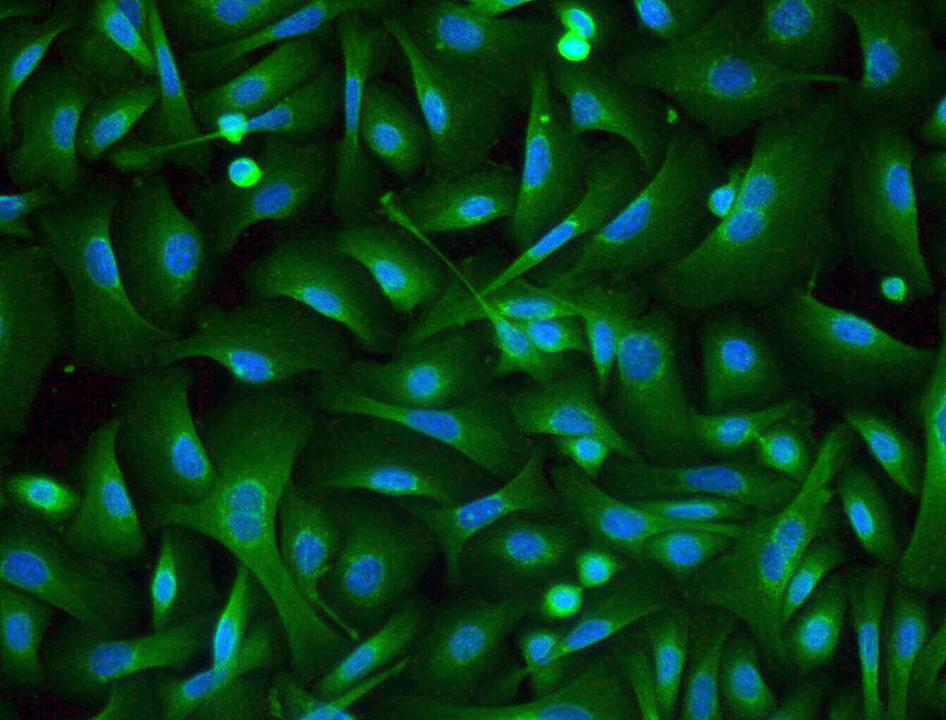
Immunocytochemistry/Immunofluorescence: Desmoglein-3 Antibody (5H10) [NBP1-78984] - Desmoglein 3 (5H10) antibody was tested in A431 cells with FITC (green). Nuclei were counterstained with Dapi (blue).![Immunocytochemistry/ Immunofluorescence: Desmoglein-3 Antibody (5H10) [NBP1-78984]](https://resources.rndsystems.com/images/products/Desmoglein-3-Antibody-5H10-Immunocytochemistry-Immunofluorescence-NBP1-78984-img0006.jpg "Immunocytochemistry/ Immunofluorescence: Desmoglein-3 Antibody (5H10) [NBP1-78984]")
Immunocytochemistry/ Immunofluorescence: Desmoglein-3 Antibody (5H10) [NBP1-78984]
Immunocytochemistry/Immunofluorescence: Desmoglein-3 Antibody (5H10) [NBP1-78984] - Desmoglein 3 (5H10) antibody was tested in oral HNSCC cells fixed with 1% formaldehyde permeabilized in 0.1% Triton X-100. in A431 Human Cell Line.")
Desmoglein-3 Antibody (5H10) in A431 Human Cell Line.
Desmoglein-3 Antibody (5H10) was detected in immersion fixed A431 human skin carcinoma cell line using Mouse anti-Desmoglein-3 Antibody (5H10) Protein G Purified Monoclonal Antibody conjugated to Alexa Fluor® 488 (Catalog # NBP1-78984AF488) (green) at 10 µg/mL overnight at 4C. Cells were counterstained with DAPI (blue). Cells were imaged using a 100X objective and digitally deconvolved.Applications for Desmoglein-3 Antibody (5H10) - BSA Free
Application
Recommended Usage
Dot Blot
reported in scientific literature (PMID 15588331)
Immunocytochemistry/ Immunofluorescence
1:100
Immunohistochemistry
1:200
Immunohistochemistry-Paraffin
1:200
Immunoprecipitation
1:10-1:500
Western Blot
1:1000
Application Notes
In Western blot, a band can be seen at approximately 130 kDa.
Reviewed Applications
Read 1 review rated 4 using NBP1-78984 in the following applications:
Formulation, Preparation, and Storage
Purification
Protein G purified
Formulation
Tris-Glycine and 0.15M NaCl
Format
BSA Free
Preservative
0.05% Sodium Azide
Concentration
1 mg/ml
Shipping
The product is shipped with polar packs. Upon receipt, store it immediately at the temperature recommended below.
Stability & Storage
Store at 4C short term. Aliquot and store at -20C long term. Avoid freeze-thaw cycles.
Background: Desmoglein-3
Additional Desmoglein-3 Products
Product Documents for Desmoglein-3 Antibody (5H10) - BSA Free
Certificate of Analysis
To download a Certificate of Analysis, please enter a lot or batch number in the search box below.
Product Specific Notices for Desmoglein-3 Antibody (5H10) - BSA Free
This product is for research use only and is not approved for use in humans or in clinical diagnosis. Primary Antibodies are guaranteed for 1 year from date of receipt.
Related Research Areas
Citations for Desmoglein-3 Antibody (5H10) - BSA Free
 Powered by Bioz
Customer Reviews for Desmoglein-3 Antibody (5H10) - BSA Free (1)
4 out of 5
1 Customer Rating
Have you used Desmoglein-3 Antibody (5H10) - BSA Free?
Submit a review and receive an Amazon gift card!
$25/€18/£15/$25CAN/¥2500 Yen for a review with an image
$10/€7/£6/$10CAN/¥1110 Yen for a review without an image
Submit a review
Customer Images
Showing
1
-
1 of
1 review
Showing All
Filter By:
-
Application: ImmunocytochemistrySample Tested: Primary skin keratinocytesSpecies: MouseVerified Customer | Posted 06/22/2017The cultured primary mouse skin keratinocytes were stained at 1:150. The image shows the cytosolic staining.

There are no reviews that match your criteria.
Protocols
View specific protocols for Desmoglein-3 Antibody (5H10) - BSA Free (NBP1-78984):
Immunocytochemistry Protocol
Culture cells to appropriate density in 35 mm culture dishes or 6-well plates.
1. Remove culture medium and add 10% formalin to the dish. Fix at room temperature for 30 minutes.
2. Remove the formalin and add ice cold methanol. Incubate for 5-10 minutes.
3. Remove methanol and add washing solution (i.e. PBS). Be sure to not let the specimen dry out. Wash three times for 10 minutes.
4. To block nonspecific antibody binding incubate in 10% normal goat serum from 1 hour to overnight at room temperature.
5. Add primary antibody at appropriate dilution and incubate at room temperature from 2 hours to overnight at room temperature.
6. Remove primary antibody and replace with washing solution. Wash three times for 10 minutes.
7. Add secondary antibody at appropriate dilution. Incubate for 1 hour at room temperature.
8. Remove antibody and replace with wash solution, then wash for 10 minutes. Add Hoechst 33258 to wash solution at 1:25,0000 and incubate for 10 minutes. Wash a third time for 10 minutes.
9. Cells can be viewed directly after washing. The plates can also be stored in PBS containing Azide covered in Parafilm (TM). Cells can also be cover-slipped using Fluoromount, with appropriate sealing.
*The above information is only intended as a guide. The researcher should determine what protocol best meets their needs. Please follow safe laboratory procedures."
Immunohistochemistry-Paraffin Embedded Sections
Antigen Unmasking:
Bring slides to a boil in 10 mM sodium citrate buffer (pH 6.0) then maintain at a sub-boiling temperature for 10 minutes. Cool slides on bench-top for 30 minutes.
Staining:
1. Wash sections in deionized water three times for 5 minutes each.
2. Wash sections in wash buffer for 5 minutes.
3. Block each section with 100-400 ul blocking solution for 1 hour at room temperature.
4. Remove blocking solution and add 100-400 ul diluted primary antibody. Incubate overnight at 4C.
5. Remove antibody solution and wash sections in wash buffer three times for 5 minutes each.
6. Add 100-400 ul biotinylated diluted secondary antibody. Incubate 30 minutes at room temperature.
7. Remove secondary antibody solution and wash sections three times with wash buffer for 5 minutes each.
8. Add 100-400 ul Streptavidin-HRP reagent to each section and incubate for 30 minutes at room temperature.
9. Wash sections three times in wash buffer for 5 minutes each.
10. Add 100-400 ul DAB substrate to each section and monitor staining closely.
11. As soon as the sections develop, immerse slides in deionized water.
12. Counterstain sections in hematoxylin.
13. Wash sections in deionized water two times for 5 minutes each.
14. Dehydrate sections.
15. Mount coverslips.
*The above information is only intended as a guide. The researcher should determine what protocol best meets their needs. Please follow safe laboratory procedures."
Find general support by application which include: protocols, troubleshooting, illustrated assays, videos and webinars.
- Antigen Retrieval Protocol (PIER)
- Antigen Retrieval for Frozen Sections Protocol
- Appropriate Fixation of IHC/ICC Samples
- Cellular Response to Hypoxia Protocols
- Chromogenic IHC Staining of Formalin-Fixed Paraffin-Embedded (FFPE) Tissue Protocol
- Chromogenic Immunohistochemistry Staining of Frozen Tissue
- ClariTSA™ Fluorophore Kits
- Detection & Visualization of Antibody Binding
- Fluorescent IHC Staining of Frozen Tissue Protocol
- Graphic Protocol for Heat-induced Epitope Retrieval
- Graphic Protocol for the Preparation and Fluorescent IHC Staining of Frozen Tissue Sections
- Graphic Protocol for the Preparation and Fluorescent IHC Staining of Paraffin-embedded Tissue Sections
- Graphic Protocol for the Preparation of Gelatin-coated Slides for Histological Tissue Sections
- ICC Cell Smear Protocol for Suspension Cells
- ICC Immunocytochemistry Protocol Videos
- ICC for Adherent Cells
- IHC Sample Preparation (Frozen sections vs Paraffin)
- Immunocytochemistry (ICC) Protocol
- Immunocytochemistry Troubleshooting
- Immunofluorescence of Organoids Embedded in Cultrex Basement Membrane Extract
- Immunofluorescent IHC Staining of Formalin-Fixed Paraffin-Embedded (FFPE) Tissue Protocol
- Immunohistochemistry (IHC) and Immunocytochemistry (ICC) Protocols
- Immunohistochemistry Frozen Troubleshooting
- Immunohistochemistry Paraffin Troubleshooting
- Immunoprecipitation Protocol
- Preparing Samples for IHC/ICC Experiments
- Preventing Non-Specific Staining (Non-Specific Binding)
- Primary Antibody Selection & Optimization
- Protocol for Heat-Induced Epitope Retrieval (HIER)
- Protocol for Making a 4% Formaldehyde Solution in PBS
- Protocol for VisUCyte™ HRP Polymer Detection Reagent
- Protocol for the Fluorescent ICC Staining of Cell Smears - Graphic
- Protocol for the Fluorescent ICC Staining of Cultured Cells on Coverslips - Graphic
- Protocol for the Preparation & Fixation of Cells on Coverslips
- Protocol for the Preparation and Chromogenic IHC Staining of Frozen Tissue Sections
- Protocol for the Preparation and Chromogenic IHC Staining of Frozen Tissue Sections - Graphic
- Protocol for the Preparation and Chromogenic IHC Staining of Paraffin-embedded Tissue Sections
- Protocol for the Preparation and Chromogenic IHC Staining of Paraffin-embedded Tissue Sections - Graphic
- Protocol for the Preparation and Fluorescent ICC Staining of Cells on Coverslips
- Protocol for the Preparation and Fluorescent ICC Staining of Non-adherent Cells
- Protocol for the Preparation and Fluorescent ICC Staining of Stem Cells on Coverslips
- Protocol for the Preparation and Fluorescent IHC Staining of Frozen Tissue Sections
- Protocol for the Preparation and Fluorescent IHC Staining of Paraffin-embedded Tissue Sections
- Protocol for the Preparation of Gelatin-coated Slides for Histological Tissue Sections
- Protocol for the Preparation of a Cell Smear for Non-adherent Cell ICC - Graphic
- R&D Systems Quality Control Western Blot Protocol
- TUNEL and Active Caspase-3 Detection by IHC/ICC Protocol
- The Importance of IHC/ICC Controls
- Troubleshooting Guide: Immunohistochemistry
- Troubleshooting Guide: Western Blot Figures
- Western Blot Conditions
- Western Blot Protocol
- Western Blot Protocol for Cell Lysates
- Western Blot Troubleshooting
- Western Blot Troubleshooting Guide
- View all Protocols, Troubleshooting, Illustrated assays and Webinars
Loading...