DNMT3A Antibody (64B1446) - BSA Free
Novus Biologicals | Catalog # NB120-13888
Key Product Details
Validated by
Knockout/Knockdown, Biological Validation
Species Reactivity
Validated:
Human, Mouse, Rat
Cited:
Human, Mouse, Rat
Applications
Validated:
Knockout Validated, Immunohistochemistry, Immunohistochemistry-Paraffin, Immunohistochemistry-Frozen, Immunohistochemistry Whole-Mount, Western Blot, Flow Cytometry, Immunocytochemistry/ Immunofluorescence, Chromatin Immunoprecipitation, Chromatin Immunoprecipitation (ChIP), Knockdown Validated, CyTOF-ready
Cited:
Knockout Validated, Immunohistochemistry, Immunohistochemistry-Paraffin, Immunohistochemistry-Frozen, Immunohistochemistry Whole-Mount, Western Blot, Immunocytochemistry/ Immunofluorescence, Chromatin Immunoprecipitation (ChIP), Chemotaxis, IF/IHC
Label
Unconjugated
Antibody Source
Monoclonal Mouse IgG1 kappa Clone # 64B1446
Format
BSA Free
Loading...
Product Specifications
Immunogen
This DNMT3A Antibody (64B1446) was raised against bacteria expressed recombinant mouse Dnmt3a. The epitope was found to lie near the C-terminus (a.a. 705-908), see Chen et (2002) for details.
Reactivity Notes
Use in Rat reported in scientific literature (PMID:34874218).
Localization
Nuclear
Clonality
Monoclonal
Host
Mouse
Isotype
IgG1 kappa
Scientific Data Images for DNMT3A Antibody (64B1446) - BSA Free
![Western Blot: DNMT3A Antibody (64B1446)BSA Free [NB120-13888]](https://resources.rndsystems.com/images/products/DNMT3A-Antibody-64B1446-Western-Blot-NB120-13888-img0020.jpg "Western Blot: DNMT3A Antibody (64B1446)BSA Free [NB120-13888]")
Western Blot: DNMT3A Antibody (64B1446)BSA Free [NB120-13888]
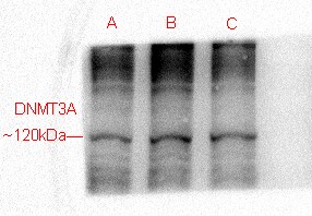
Western Blot: DNMT3A Antibody (64B1446) [NB120-13888] - Analysis of (A) Dnmt3a transfected 293 cell lysate and (B) untransfected 293 cell lysate using Dnmt3a antibody at 1 ug/mL.![Immunocytochemistry/ Immunofluorescence: DNMT3A Antibody (64B1446) - BSA Free [NB120-13888]](https://resources.rndsystems.com/images/products/DNMT3A-Antibody-64B1446-Immunocytochemistry-Immunofluorescence-NB120-13888-img0016.jpg "Immunocytochemistry/ Immunofluorescence: DNMT3A Antibody (64B1446) - BSA Free [NB120-13888]")
Immunocytochemistry/ Immunofluorescence: DNMT3A Antibody (64B1446) - BSA Free [NB120-13888]
Immunocytochemistry/Immunofluorescence: DNMT3A Antibody (64B1446) [NB120-13888] - Ntera2 cells were fixed for 10 minutes using 10% formalin and then permeabilized for 5 minutes using 1X TBS + 0.5% Triton-X100. The cells were incubated with anti-DNMT3A (68B1446) [NB120-13888] at a 1:200 dilution overnight at 4C and detected with an anti-mouse Dylight 488 (Green) at a 1:500 dilution. Actin was detected with Phalloidin 568 (Red) at a 1:200 dilution. Nuclei were counterstained with DAPI (Blue). Cells were imaged using a 40X objective.![Immunohistochemistry-Paraffin: DNMT3A Antibody (64B1446) - BSA Free [NB120-13888]](https://resources.rndsystems.com/images/products/DNMT3A-Antibody-64B1446-Immunohistochemistry-Paraffin-NB120-13888-img0012.jpg "Immunohistochemistry-Paraffin: DNMT3A Antibody (64B1446) - BSA Free [NB120-13888]")
Immunohistochemistry-Paraffin: DNMT3A Antibody (64B1446) - BSA Free [NB120-13888]
Immunohistochemistry-Paraffin: DNMT3A Antibody (64B1446) [NB120-13888] - Detection of DNMT3A on human hepatocellular carcinoma tissue section (NBP2-30221) using 1:100 dilution of DNMT3A antibody (clone 64B1446). The antibody generated a specific cytoplasmic staining in all the cancer cells while some of the cells depicted nuclear staining also.![Immunocytochemistry/ Immunofluorescence: DNMT3A Antibody (64B1446) - BSA Free [NB120-13888]](https://resources.rndsystems.com/images/products/DNMT3A-Antibody-64B1446-Immunocytochemistry-Immunofluorescence-NB120-13888-img0017.jpg "Immunocytochemistry/ Immunofluorescence: DNMT3A Antibody (64B1446) - BSA Free [NB120-13888]")
Immunocytochemistry/ Immunofluorescence: DNMT3A Antibody (64B1446) - BSA Free [NB120-13888]
Immunocytochemistry/Immunofluorescence: DNMT3A Antibody (64B1446) [NB120-13888] - Expression of Dnmt3a in the nuclei of post-mitotic neurons in the olfactory epithelium (OE). DNMT3a (red) is co-expressed with neuron-specific tubulin (green) throughout the development of the olfactory epithelium. Data courtesy of A. Jane Roskams, University of British Columbia.![Flow Cytometry: DNMT3A Antibody (64B1446) - BSA Free [NB120-13888]](https://resources.rndsystems.com/images/products/DNMT3A-Antibody-64B1446-Flow-Cytometry-NB120-13888-img0018.jpg "Flow Cytometry: DNMT3A Antibody (64B1446) - BSA Free [NB120-13888]")
Flow Cytometry: DNMT3A Antibody (64B1446) - BSA Free [NB120-13888]
Flow Cytometry: DNMT3A Antibody (64B1446) [NB120-13888] - Intracellular flow cytometric staining of 1 x 10^6 CHO (A) and HEK-293 (B) cells using Dnmt3a antibody (dark blue). Isotype control shown in orange. An antibody concentration of 1 ug/1x10^6 cells was used.![Flow Cytometry: DNMT3A Antibody (64B1446) - BSA Free [NB120-13888]](https://resources.rndsystems.com/images/products/DNMT3A-Antibody-64B1446-Flow-Cytometry-NB120-13888-img0021.jpg "Flow Cytometry: DNMT3A Antibody (64B1446) - BSA Free [NB120-13888]")
Flow Cytometry: DNMT3A Antibody (64B1446) - BSA Free [NB120-13888]
Flow Cytometry: DNMT3A Antibody (64B1446) [NB120-13888] - Using the PE direct conjugate, an intracellular stain was performed on NTERA-2 cells with DNMT3A (64B1446) NB120-13888PE (blue) and a matched isotype control (orange). Cells were fixed with 4% PFA and then permeablized with 0.1% saponin. Cells were incubated in an antibody dilution of 1 ug/mL for 30 minutes at room temperature. Both antibodies were conjugated to phycoerythrin.![Immunocytochemistry/ Immunofluorescence: DNMT3A Antibody (64B1446) - BSA Free [NB120-13888]](https://resources.rndsystems.com/images/products/DNMT3A-Antibody-64B1446-Immunocytochemistry-Immunofluorescence-NB120-13888-img0028.jpg "Immunocytochemistry/ Immunofluorescence: DNMT3A Antibody (64B1446) - BSA Free [NB120-13888]")
Immunocytochemistry/ Immunofluorescence: DNMT3A Antibody (64B1446) - BSA Free [NB120-13888]
DNMT3A-Antibody-64B1446-Immunocytochemistry-Immunofluorescence-NB120-13888-img0028.jpg![Flow Cytometry: DNMT3A Antibody (64B1446) - BSA Free [NB120-13888]](https://resources.rndsystems.com/images/products/DNMT3A-Antibody-64B1446-Flow-Cytometry-NB120-13888-img0026.jpg "Flow Cytometry: DNMT3A Antibody (64B1446) - BSA Free [NB120-13888]")
Flow Cytometry: DNMT3A Antibody (64B1446) - BSA Free [NB120-13888]
Flow Cytometry: DNMT3A Antibody (64B1446) [NB120-13888] - An intracellular stain was performed on Jurkat cells with DNMT3A (64B1446) NB120-13888AF647 (blue) and a matched isotype control (orange). Cells were fixed with 4% PFA and then permeablized with 0.1% saponin. Cells were incubated in an antibody dilution of 2.5 ug/mL for 30 minutes at room temperature. Both antibodies were conjugated to Alexa Fluor 647.![Western Blot: DNMT3A Antibody (64B1446)BSA Free [NB120-13888]](https://resources.rndsystems.com/images/products/DNMT3A-Antibody-64B1446-Western-Blot-NB120-13888-img0015.jpg "Western Blot: DNMT3A Antibody (64B1446)BSA Free [NB120-13888]")
Western Blot: DNMT3A Antibody (64B1446)BSA Free [NB120-13888]
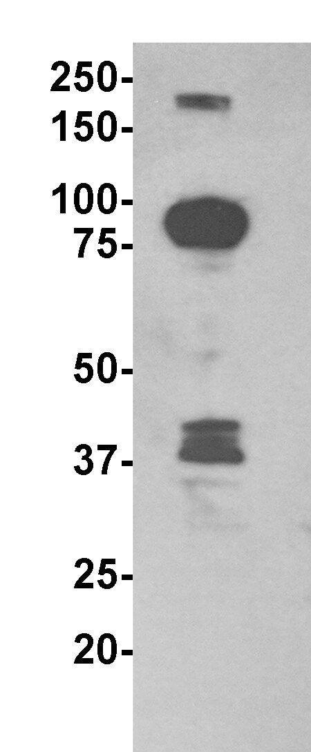
Western Blot: DNMT3A Antibody (64B1446) [NB120-13888] - NTERA-2 cells were treated with Zebularine as indicated for 24 hours. Cell lysates were prepared and separated on a 7.5% gel by SDS-PAGE. Protein was transferred to PVDF membrane and blocked in 5% non-fat milk. The membrane was probed with 2 ug/mL anti-Dnmt3a in 1% milk, and detected with an anti-mouse HRP secondary antibody using chemiluminescence. Note the decrease in Dnmt3a expression upon treatment with 500 uM Zebularine (arrowhead). Additional bands at 90 and 75 kDa can also be detected with this antibody and may represent alternative splice variants. Tubulin is shown as a loading control.![Flow Cytometry: DNMT3A Antibody (64B1446) - BSA Free [NB120-13888]](https://resources.rndsystems.com/images/products/DNMT3A-Antibody-64B1446-Flow-Cytometry-NB120-13888-img0023.jpg "Flow Cytometry: DNMT3A Antibody (64B1446) - BSA Free [NB120-13888]")
Flow Cytometry: DNMT3A Antibody (64B1446) - BSA Free [NB120-13888]
Flow Cytometry: DNMT3A Antibody (64B1446) [NB120-13888] - An intracellular stain was performed on HepG2 cells with DNMT3A (64B1446) NB120-13888APC (blue) and a matched isotype control (orange). Cells were fixed with 4% PFA and then permeablized with 0.1% saponin. Cells were incubated in an antibody dilution of 1 ug/mL for 30 minutes at room temperature. Both antibodies were conjugated to allophycocyanin (APC).![Flow Cytometry: DNMT3A Antibody (64B1446) - BSA Free [NB120-13888]](https://resources.rndsystems.com/images/products/DNMT3A-Antibody-64B1446-Flow-Cytometry-NB120-13888-img0024.jpg "Flow Cytometry: DNMT3A Antibody (64B1446) - BSA Free [NB120-13888]")
Flow Cytometry: DNMT3A Antibody (64B1446) - BSA Free [NB120-13888]
Flow Cytometry: DNMT3A Antibody (64B1446) [NB120-13888] - An intracellular stain was performed on HepG2 cells with DNMT3A (64B1446) NB120-13888AF647 (blue) and a matched isotype control (orange). Cells were fixed with 4% PFA and then permeablized with 0.1% saponin. Cells were incubated in an antibody dilution of 2.5 ug/mL for 30 minutes at room temperature. Both antibodies were conjugated to Alexa Fluor 647.![Flow Cytometry: DNMT3A Antibody (64B1446) - BSA Free [NB120-13888]](https://resources.rndsystems.com/images/products/DNMT3A-Antibody-64B1446-Flow-Cytometry-NB120-13888-img0025.jpg "Flow Cytometry: DNMT3A Antibody (64B1446) - BSA Free [NB120-13888]")
Flow Cytometry: DNMT3A Antibody (64B1446) - BSA Free [NB120-13888]
Flow Cytometry: DNMT3A Antibody (64B1446) [NB120-13888] - An intracellular stain was performed on HeLa cells with DNMT3A (64B1446) NB120-13888APC (blue) and a matched isotype control (orange). Cells were fixed with 4% PFA and then permeabilized with 0.1% saponin. Cells were incubated in an antibody dilution of 1 ug/mL for 30 minutes at room temperature. Both antibodies were conjugated to allophycocyanin (APC). - BSA Free [NB120-13888] -")
 - BSA Free [NB120-13888] -")
Flow Cytometry: DNMT3A Antibody (64B1446) - BSA Free [NB120-13888] -
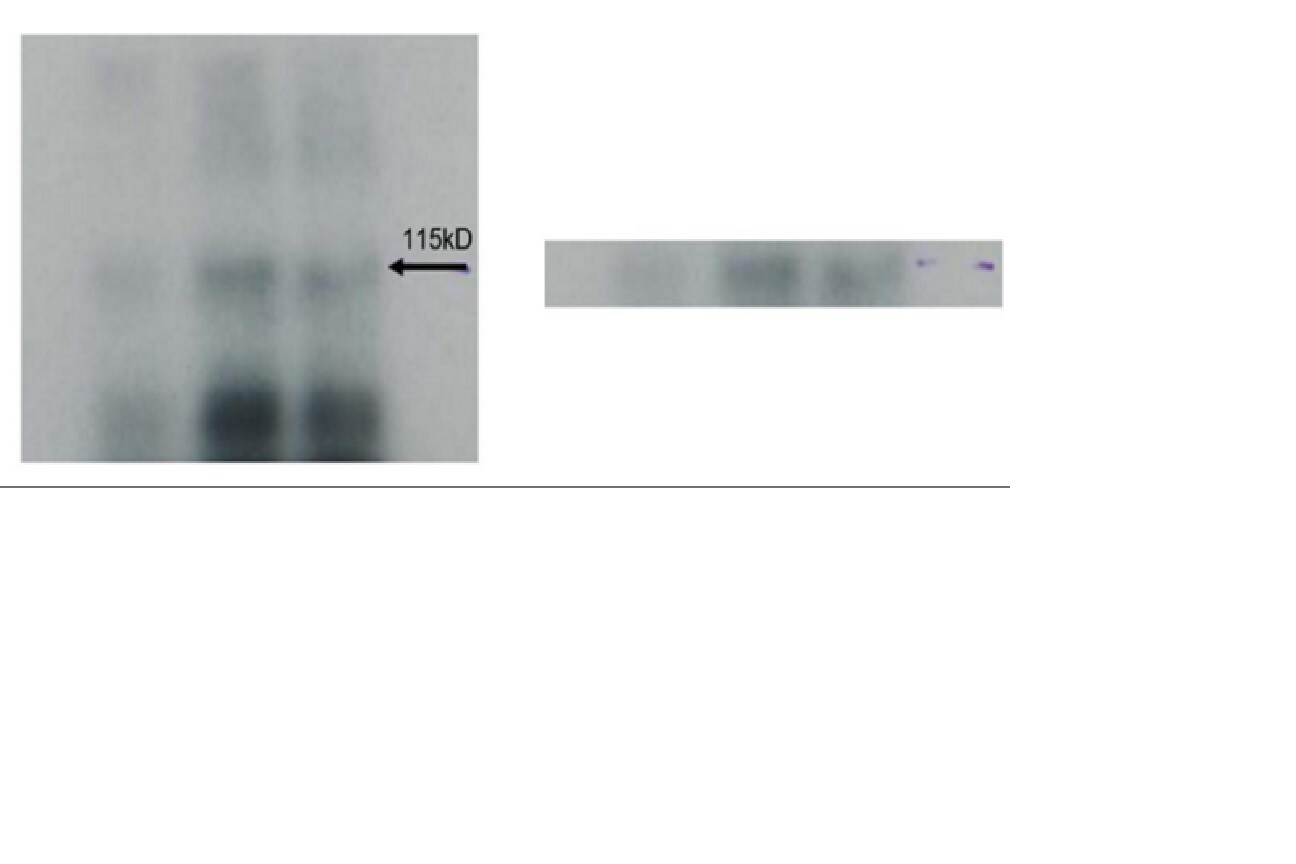
Flow Cytometry: DNMT3A Antibody (64B1446) - BSA Free [NB120-13888] - Protein levels of Dnmt3a & MeCP2 are reduced in inhibitory neurons of conditional knockout mice.(A) Western blot of half brain hemisphere from Dnmt3a or Mecp2 cKO mice demonstrating loss of each protein. (B) Immunofluorescence (IF) images of WT, Dnmt3a cKO or Mecp2 cKO mice probing for Dnmt3a or MeCP2 (red), the Sun1-sfGFP-myc-PA fusion protein that marks the nuclear envelope & is dependent on Cre expression (green), & DAPI to mark genomic DNA (blue). n = 3 mice per genotype (western), n = 3 mice (IF) representative images for two brain regions shown. Image collected & cropped by CiteAb from the following publication (https://pubmed.ncbi.nlm.nih.gov/32159514), licensed under a CC-BY license. Not internally tested by Novus Biologicals. - BSA Free [NB120-13888] -")
Immunocytochemistry/ Immunofluorescence: DNMT3A Antibody (64B1446) - BSA Free [NB120-13888] -
DNMT1 reveals a SCMC-like cortical localization with aberrant localization in Nlrp2tm1a/tm1a derived oocytes.(A) Whole mount immunofluorescence co-staining with anti-DNMT1 and DAPI on the same Nlrp2M+/Z+ embryos used to stain NLRP2 reveals a SCMC-like cortical localization of DNMT1 with a diffuse nuclear stain in later stage embryos. Scale bar represented is 200 μm. (B) DNMT1 staining in paraffin embedded oocyte sections reveals a less cortical, more cytoplasmic intense stain with nucleolar focus in oocytes from Nlrp2tm1a/tm1a dams compared to oocytes from Nlrp2+/+ dams. (C) Whole mount immunofluorescence for DNMT3A in unfertilized control oocytes reveals a characteristic metaphase associated localization and no difference is noted in oocytes derived from Nlrp2tm1a/tm1adams. (D) Whole mount immunofluorescence in unfertilized oocytes reveals that as expected, DNMT3B is not yet expressed in unfertilized control oocytes and no expression is noted in oocytes derived from Nlrp2tm1a/tm1adams. Image collected and cropped by CiteAb from the following open publication (https://pubmed.ncbi.nlm.nih.gov/28317850), licensed under a CC-BY license. Not internally tested by Novus Biologicals.Applications for DNMT3A Antibody (64B1446) - BSA Free
Application
Recommended Usage
Chromatin Immunoprecipitation
reported in scientific literature (PMID 24623306)
Flow Cytometry
1 ug per million cells
Immunocytochemistry/ Immunofluorescence
1:10-1:500
Immunohistochemistry
reported in multiple pieces of scientific literature
Immunohistochemistry Whole-Mount
reported in scientific literature (PMID 26507142)
Immunohistochemistry-Frozen
reported in scientific literature (PMID 22134929)
Immunohistochemistry-Paraffin
5 ug/ml. Use reported in scientific literature (PMID 22134929)
Knockdown Validated
Validated for Knockdown from CiteAb
Application Notes
Western Blot: Detects a band of approximately 120 kDa (predicted molecular weight: 102 kDa).
Staining of formalin-fixed tissues is enhanced by boiling tissue sections in 10 mM sodium citrate buffer, pH 6.0 for 10-20 min followed by cooling at RT for 20 min.
Staining of formalin-fixed tissues is enhanced by boiling tissue sections in 10 mM sodium citrate buffer, pH 6.0 for 10-20 min followed by cooling at RT for 20 min.
Reviewed Applications
Read 3 reviews rated 4.3 using NB120-13888 in the following applications:
Flow Cytometry Panel Builder
Bio-Techne Knows Flow Cytometry
Save time and reduce costly mistakes by quickly finding compatible reagents using the Panel Builder Tool.
Advanced Features
- Spectra Viewer - Custom analysis of spectra from multiple fluorochromes
- Spillover Popups - Visualize the spectra of individual fluorochromes
- Antigen Density Selector - Match fluorochrome brightness with antigen density

Formulation, Preparation, and Storage
Purification
Protein G purified
Formulation
PBS
Format
BSA Free
Preservative
0.05% Sodium Azide
Concentration
1.0 mg/ml
Shipping
The product is shipped with polar packs. Upon receipt, store it immediately at the temperature recommended below.
Stability & Storage
Store at 4C short term. Aliquot and store at -20C long term. Avoid freeze-thaw cycles.
Background: DNMT3A
References
1. Thakur, A., Mackin, S. J., Irwin, R. E., O'Neill, K. M., Pollin, G., & Walsh, C. (2016). Widespread recovery of methylation at gametic imprints in hypomethylated mouse stem cells following rescue with DNMT3A2. Epigenetics Chromatin, 9, 53. doi:10.1186/s13072-016-0104-2
2. Ravichandran, M., Lei, R., Tang, Q., Zhao, Y., Lee, J., Ma, L.,... Dawlaty, M. M. (2019). Rinf Regulates Pluripotency Network Genes and Tet Enzymes in Embryonic Stem Cells. Cell Rep, 28(8), 1993-2003.e1995. doi:10.1016/j.celrep.2019.07.080
3. Pang, Y., Liu, J., Li, X., Xiao, G., Wang, H., Yang, G.,... Ren, H. (2018). MYC and DNMT3A-mediated DNA methylation represses microRNA-200b in triple negative breast cancer. J Cell Mol Med, 22(12), 6262-6274. doi:10.1111/jcmm.13916
4. Xia, L., Huang, W., Bellani, M., Seidman, M. M., Wu, K., Fan, D.,... Baylin, S. B. (2017). CHD4 Has Oncogenic Functions in Initiating and Maintaining Epigenetic Suppression of Multiple Tumor Suppressor Genes. Cancer Cell, 31(5), 653-668.e657. doi:10.1016/j.ccell.2017.04.005
Long Name
DNA [Cytosine-5-]-Methyltransferase 3A
Alternate Names
DNA (cytosine-5-)-methyltransferase 3 alpha, DNA (cytosine-5)-methyltransferase 3A, DNA cytosine methyltransferase 3A2, DNA Methyltransferase 3 Alpha, DNA methyltransferase HsaIIIA, DNA MTase HsaIIIA, Dnmt3a, DNMT3A2, EC 2.1.1.37, M.HsaIIIA, TBRS, 64B1446 clone
Gene Symbol
DNMT3A
Additional DNMT3A Products
Product Documents for DNMT3A Antibody (64B1446) - BSA Free
Certificate of Analysis
To download a Certificate of Analysis, please enter a lot or batch number in the search box below.
Product Specific Notices for DNMT3A Antibody (64B1446) - BSA Free
This product is for research use only and is not approved for use in humans or in clinical diagnosis. Primary Antibodies are guaranteed for 1 year from date of receipt.
Related Research Areas
Citations for DNMT3A Antibody (64B1446) - BSA Free
 Powered by Bioz
Customer Reviews for DNMT3A Antibody (64B1446) - BSA Free (3)
4.3 out of 5
3 Customer Ratings
Have you used DNMT3A Antibody (64B1446) - BSA Free?
Submit a review and receive an Amazon gift card!
$25/€18/£15/$25CAN/¥2500 Yen for a review with an image
$10/€7/£6/$10CAN/¥1110 Yen for a review without an image
Submit a review
Customer Images
Showing
1
-
3 of
3 reviews
Showing All
Filter By:
-
Application: Western BlotSample Tested: cellSpecies: HumanVerified Customer | Posted 01/30/2019

-
Application: Western BlotSample Tested: recombinant purified proteinSpecies: HumanVerified Customer | Posted 01/23/2017DNMT3A protein1ug of recombinant DNMT3A protein. 4-12% Bis tris gel
-
Application: Western BlotSample Tested:Species: OtherVerified Customer | Posted 08/02/2016
There are no reviews that match your criteria.
Protocols
Find general support by application which include: protocols, troubleshooting, illustrated assays, videos and webinars.
- 7-Amino Actinomycin D (7-AAD) Cell Viability Flow Cytometry Protocol
- Antigen Retrieval Protocol (PIER)
- Antigen Retrieval for Frozen Sections Protocol
- Appropriate Fixation of IHC/ICC Samples
- Cellular Response to Hypoxia Protocols
- ChIP Protocol Video
- Chromatin Immunoprecipitation (ChIP) Protocol
- Chromatin Immunoprecipitation Protocol
- Chromogenic IHC Staining of Formalin-Fixed Paraffin-Embedded (FFPE) Tissue Protocol
- Chromogenic Immunohistochemistry Staining of Frozen Tissue
- ClariTSA™ Fluorophore Kits
- Detection & Visualization of Antibody Binding
- Extracellular Membrane Flow Cytometry Protocol
- Flow Cytometry Protocol for Cell Surface Markers
- Flow Cytometry Protocol for Staining Membrane Associated Proteins
- Flow Cytometry Staining Protocols
- Flow Cytometry Troubleshooting Guide
- Fluorescent IHC Staining of Frozen Tissue Protocol
- Graphic Protocol for Heat-induced Epitope Retrieval
- Graphic Protocol for the Preparation and Fluorescent IHC Staining of Frozen Tissue Sections
- Graphic Protocol for the Preparation and Fluorescent IHC Staining of Paraffin-embedded Tissue Sections
- Graphic Protocol for the Preparation of Gelatin-coated Slides for Histological Tissue Sections
- ICC Cell Smear Protocol for Suspension Cells
- ICC Immunocytochemistry Protocol Videos
- ICC for Adherent Cells
- IHC Sample Preparation (Frozen sections vs Paraffin)
- Immunocytochemistry (ICC) Protocol
- Immunocytochemistry Troubleshooting
- Immunofluorescence of Organoids Embedded in Cultrex Basement Membrane Extract
- Immunofluorescent IHC Staining of Formalin-Fixed Paraffin-Embedded (FFPE) Tissue Protocol
- Immunohistochemistry (IHC) and Immunocytochemistry (ICC) Protocols
- Immunohistochemistry Frozen Troubleshooting
- Immunohistochemistry Paraffin Troubleshooting
- Intracellular Flow Cytometry Protocol Using Alcohol (Methanol)
- Intracellular Flow Cytometry Protocol Using Detergents
- Intracellular Nuclear Staining Flow Cytometry Protocol Using Detergents
- Intracellular Staining Flow Cytometry Protocol Using Alcohol Permeabilization
- Intracellular Staining Flow Cytometry Protocol Using Detergents to Permeabilize Cells
- Preparing Samples for IHC/ICC Experiments
- Preventing Non-Specific Staining (Non-Specific Binding)
- Primary Antibody Selection & Optimization
- Propidium Iodide Cell Viability Flow Cytometry Protocol
- Protocol for Heat-Induced Epitope Retrieval (HIER)
- Protocol for Liperfluo
- Protocol for Making a 4% Formaldehyde Solution in PBS
- Protocol for VisUCyte™ HRP Polymer Detection Reagent
- Protocol for the Characterization of Human Th22 Cells
- Protocol for the Characterization of Human Th9 Cells
- Protocol for the Fluorescent ICC Staining of Cell Smears - Graphic
- Protocol for the Fluorescent ICC Staining of Cultured Cells on Coverslips - Graphic
- Protocol for the Preparation & Fixation of Cells on Coverslips
- Protocol for the Preparation and Chromogenic IHC Staining of Frozen Tissue Sections
- Protocol for the Preparation and Chromogenic IHC Staining of Frozen Tissue Sections - Graphic
- Protocol for the Preparation and Chromogenic IHC Staining of Paraffin-embedded Tissue Sections
- Protocol for the Preparation and Chromogenic IHC Staining of Paraffin-embedded Tissue Sections - Graphic
- Protocol for the Preparation and Fluorescent ICC Staining of Cells on Coverslips
- Protocol for the Preparation and Fluorescent ICC Staining of Non-adherent Cells
- Protocol for the Preparation and Fluorescent ICC Staining of Stem Cells on Coverslips
- Protocol for the Preparation and Fluorescent IHC Staining of Frozen Tissue Sections
- Protocol for the Preparation and Fluorescent IHC Staining of Paraffin-embedded Tissue Sections
- Protocol for the Preparation of Gelatin-coated Slides for Histological Tissue Sections
- Protocol for the Preparation of a Cell Smear for Non-adherent Cell ICC - Graphic
- Protocol: Annexin V and PI Staining by Flow Cytometry
- Protocol: Annexin V and PI Staining for Apoptosis by Flow Cytometry
- R&D Systems Quality Control Western Blot Protocol
- TUNEL and Active Caspase-3 Detection by IHC/ICC Protocol
- The Importance of IHC/ICC Controls
- Troubleshooting Guide: Fluorokine Flow Cytometry Kits
- Troubleshooting Guide: Immunohistochemistry
- Troubleshooting Guide: Western Blot Figures
- Western Blot Conditions
- Western Blot Protocol
- Western Blot Protocol for Cell Lysates
- Western Blot Troubleshooting
- Western Blot Troubleshooting Guide
- View all Protocols, Troubleshooting, Illustrated assays and Webinars
FAQs for DNMT3A Antibody (64B1446) - BSA Free
Showing
1
-
1 of
1 FAQ
Showing All
-
Q: Do you have any information about the epitope of your antibody, so I could check the chances for the antibody working in the honey bee?
A:
This antibody was raised against bacteria expressed recombinant mouse Dnmt3a. The epitope was found to lie near the C-terminus (a.a. 705-908), see Chen et (2002) for details. We haven't mapped the binding binding epitope although Chen et (2002) mapped it to a range between 705-908 amino acids: I am not sure what technique you are planning with the antibody, but if you are doing a western blot, we do have a western blot postive control ready to use lysate NBP2-25063 and I would l highly suggest using that.
Loading...