![Western Blot: DNMT3B Antibody [NB300-516]](https://resources.rndsystems.com/images/products/DNMT3B-Antibody-Western-Blot-NB300-516-img0012.jpg "Western Blot: DNMT3B Antibody [NB300-516]")
![Western Blot: DNMT3B Antibody [NB300-516]](https://resources.rndsystems.com/images/products/DNMT3B-Antibody-Western-Blot-NB300-516-img0014.jpg "Western Blot: DNMT3B Antibody [NB300-516]")
Loading...
Key Product Details
Validated by
Knockout/Knockdown, Biological Validation
Species Reactivity
Validated:
Human, Mouse, Rat
Cited:
Human, Mouse, Rat
Applications
Validated:
Immunohistochemistry, Immunohistochemistry-Paraffin, Western Blot, Simple Western, Chromatin Immunoprecipitation (ChIP), Knockdown Validated
Cited:
Western Blot, Chemotaxis
Label
Unconjugated
Antibody Source
Polyclonal Rabbit IgG
Loading...
Product Specifications
Immunogen
Synthetic peptide corresponding to the residues M(1) K G D S R H L N E E E G A(14) C of mouse DNMT3b
Reactivity Notes
Use in Rat reported in scientific literature (PMID:34874218) Human reactivity reported in scientific literature (PMID: 20847044). Mouse reactivity reported in scientific literature (PMID: 21149390)..
Localization
Nuclear
Specificity
This detects DNA methyltransferase 3b (Dnmt3b) from human and mouse tissues and cells as well as recombinant human Dnmt3b. This detects, to a lesser extent, full-length human recombinant Dnmt3a.
Clonality
Polyclonal
Host
Rabbit
Isotype
IgG
Scientific Data Images for DNMT3B Antibody
Western Blot: DNMT3B Antibody [NB300-516]
DNMT3B-Antibody-Western-Blot-NB300-516-img0014.jpg![Immunohistochemistry-Paraffin: DNMT3B Antibody [NB300-516]](https://resources.rndsystems.com/images/products/DNMT3B-Antibody-Immunohistochemistry-Paraffin-NB300-516-img0010.jpg "Immunohistochemistry-Paraffin: DNMT3B Antibody [NB300-516]")
Immunohistochemistry-Paraffin: DNMT3B Antibody [NB300-516]
Immunohistochemistry-Paraffin: DNMT3B Antibody [NB300-516] - Analysis showing positive staining in the nucleus and cytoplasm of Human breast carcinoma (right) compared with a negative control in the absence of primary antibody (left).![Western Blot: DNMT3B Antibody [NB300-516]](https://resources.rndsystems.com/images/products/DNMT3B-Antibody-Western-Blot-NB300-516-img0006.jpg "Western Blot: DNMT3B Antibody [NB300-516]")
Western Blot: DNMT3B Antibody [NB300-516]
Western Blot: DNMT3B Antibody [NB300-516] - Homogenized mouse brain cells. WB image submitted by a verified customer review.![Immunohistochemistry-Paraffin: DNMT3B Antibody [NB300-516]](https://resources.rndsystems.com/images/products/DNMT3B-Antibody-Immunohistochemistry-Paraffin-NB300-516-img0008.jpg "Immunohistochemistry-Paraffin: DNMT3B Antibody [NB300-516]")
Immunohistochemistry-Paraffin: DNMT3B Antibody [NB300-516]
Immunohistochemistry-Paraffin: DNMT3B Antibody [NB300-516] - Analysis showing positive staining in the nucleus of Human testis tissue (right) compared with a negative control in the absence of primary antibody (left).![Immunohistochemistry-Paraffin: DNMT3B Antibody [NB300-516]](https://resources.rndsystems.com/images/products/DNMT3B-Antibody-Immunohistochemistry-Paraffin-NB300-516-img0009.jpg "Immunohistochemistry-Paraffin: DNMT3B Antibody [NB300-516]")
Immunohistochemistry-Paraffin: DNMT3B Antibody [NB300-516]
Immunohistochemistry-Paraffin: DNMT3B Antibody [NB300-516] - Analysis showing positive staining in the nucleus and cytoplasm of Mouse brain tissue (right) compared with a negative control in the absence of primary antibody (left).![Simple Western: DNMT3B Antibody [NB300-516]](https://resources.rndsystems.com/images/products/DNMT3B-Antibody-Simple-Western-NB300-516-img0007.jpg "Simple Western: DNMT3B Antibody [NB300-516]")
Simple Western: DNMT3B Antibody [NB300-516]
Simple Western: DNMT3B Antibody [NB300-516] - Simple Western lane view shows a specific band for DNMT3B in 0.5 mg/mL of BG01V lysate. This experiment was performed under reducing conditions using the 12-230 kDa separation system. * Non-specific interaction with the 230 kDa Simple Western standard may be seen with this antibody![Simple Western: DNMT3B Antibody [NB300-516]](https://resources.rndsystems.com/images/products/DNMT3B-Antibody-Simple-Western-NB300-516-img0011.jpg "Simple Western: DNMT3B Antibody [NB300-516]")
Simple Western: DNMT3B Antibody [NB300-516]
Simple Western: DNMT3B Antibody [NB300-516] - Analysis of 25 ug of HeLa cell lysates and a molecular weight protein ladder.![Knockdown Validated: DNMT3B Antibody [NB300-516]](https://resources.rndsystems.com/images/products/DNMT3B-Antibody-Knockdown-Validated-NB300-516-img0013.jpg "Western Blot: DNMT3B Antibody [NB300-516]")

Chromatin Immunoprecipitation: DNMT3B Antibody [NB300-516] -
Epigenetic changes & protein binding at Ankrd26 promoter in eAT from mice upon 22 weeks of HFD.ChIP of DNMT1, DNMT3a, DNMT3b (a) & MBD2 (b) binding at Ankrd26 promoter region (−553 bp/−348 bp). (c) MNase for Nuc-2 (−257 bp/−198 bp) & Nuc-1 (−84 bp/−25 bp) occupancy at Ankrd26 promoter. (d) ChIP for acetyl-H4 enrichment at Nuc-2 & Nuc-1. (e) ChIP of RNA Pol II binding at Ankrd26 TSS (+16 bp/+159 bp). (a,b) & (d,e), ChIP enrichment is relative to Input chromatin. (a–e), results are mean ± SD from three independent experiments. **p < 0.01 & ***p < 0.001 vs STD. Image collected & cropped by CiteAb from the following publication (https://www.nature.com/articles/srep43526), licensed under a CC-BY license. Not internally tested by Novus Biologicals.
Western Blot: DNMT3B Antibody [NB300-516] -
Western Blot: DNMT3B Antibody [NB300-516] - DNMT3B is a direct target of miR-30a & miR-379. a The effect of miRNA mimics (PM-30a or PM-379, 20 nM) on the luciferase activities of the constructs containing the wild-type (wt-3′-UTR) or mutant-type 3′-UTR (mt-30a-3′-UTR or mt-379-3′-UTR) in OEC-M1 cells. The relative luciferase activity of each sample is measured at 48 h after transfection & normalized to Renilla luciferase activity. The data are represented as mean ± SD; ***p < 0.001 versus control mimics (PM-NC). b RT-PCR & Western blot analysis of DNMT3B level in SCC-15 & OEC-M1 cells after treatment with control mimics (NC), or miRNA mimics (PM-30a or PM-379, 20 nM) or miRNA inhibitor (AM-30a or AM-379, 20 nM) for 48 h. GAPDH & alpha -tubulin were used as internal control, respectively. c RT-PCR analysis of ADHFE1 & ALDH1A2 expression level after treatment with control mimics (NC, 20 nM), or miRNA mimics (PM-30a or PM-379, 20 nM) for 48 h in SCC-15 & OEC-M1 cells. GAPDH was used as internal control. d Relative miR-30a & miR-379 expression levels in 33 of adjacent normal tissues (N) compared with their own tumors (T). e Correlation analysis of DNMT3B & miR-30a or miR-379 in OSCC patients (n = 33) by qRT-PCR analysis. Pearson correlation coefficients & p-values were calculated as indicated. Red, tumor part; green, normal part Image collected & cropped by CiteAb from the following publication (https://pubmed.ncbi.nlm.nih.gov/32238162), licensed under a CC-BY license. Not internally tested by Novus Biologicals.
Western Blot: DNMT3B Antibody [NB300-516] -
Western Blot: DNMT3B Antibody [NB300-516] - Arecoline & NNK induced DNMT3B activity & repressed ADHFE1, ALDH1A2 & miRNAs expression. a qRT–PCR analysis of miR-30a & miR-379 expression level after treatment with arecoline (50 μM) or NNK (10 μM) for indicated days. b RT-PCR analysis of ADHFE1, ALDH1A2 level & western blot analysis of DNMT3B level in DOK cells after treatment with arecoline (50 μM) or NNK (10 μM) for indicated times. GAPDH & alpha -tubulin were used as internal control. c ChIP assay of ADHFE1 & ALDH1A2 promoter region was performed with DOK cells using anti-DNMT3B antibody after treatment with vehicle control (DMSO, 10 nM), arecoline (50 μM) or NNK (10 μM) for 5 days. Mouse IgG (mIgG) antibody was used as negative control. d RT-PCR analysis of ADHFE1 & ALDH1A2 level in DOK cells after treatment with arecoline (50 μM) or NNK (10 μM) alone or combined with 5-aza-dC (5 μM) for 5 days. GAPDH was used as internal control. e qRT-PCR analysis of ADHFE1 & ALDH1A2 level in DOK cells after treatment with vehicle control (C), or arecoline (50 μM) plus control mimics (NC), miR-30a (20 nM), miR-379 (20 nM), or NNK (10 μM) plus control mimics (NC), miR-30a (20 nM), miR-379 (20 nM) for 5 days. GAPDH was used as an internal control. All data are presented as mean ± SD; **p < 0.01; ***p < 0.001 Image collected & cropped by CiteAb from the following publication (https://pubmed.ncbi.nlm.nih.gov/32238162), licensed under a CC-BY license. Not internally tested by Novus Biologicals.
Western Blot: DNMT3B Antibody [NB300-516] -
Western Blot: DNMT3B Antibody [NB300-516] - Arecoline & NNK induced DNMT3B activity & repressed ADHFE1, ALDH1A2 & miRNAs expression. a qRT–PCR analysis of miR-30a & miR-379 expression level after treatment with arecoline (50 μM) or NNK (10 μM) for indicated days. b RT-PCR analysis of ADHFE1, ALDH1A2 level & western blot analysis of DNMT3B level in DOK cells after treatment with arecoline (50 μM) or NNK (10 μM) for indicated times. GAPDH & alpha -tubulin were used as internal control. c ChIP assay of ADHFE1 & ALDH1A2 promoter region was performed with DOK cells using anti-DNMT3B antibody after treatment with vehicle control (DMSO, 10 nM), arecoline (50 μM) or NNK (10 μM) for 5 days. Mouse IgG (mIgG) antibody was used as negative control. d RT-PCR analysis of ADHFE1 & ALDH1A2 level in DOK cells after treatment with arecoline (50 μM) or NNK (10 μM) alone or combined with 5-aza-dC (5 μM) for 5 days. GAPDH was used as internal control. e qRT-PCR analysis of ADHFE1 & ALDH1A2 level in DOK cells after treatment with vehicle control (C), or arecoline (50 μM) plus control mimics (NC), miR-30a (20 nM), miR-379 (20 nM), or NNK (10 μM) plus control mimics (NC), miR-30a (20 nM), miR-379 (20 nM) for 5 days. GAPDH was used as an internal control. All data are presented as mean ± SD; **p < 0.01; ***p < 0.001 Image collected & cropped by CiteAb from the following publication (https://pubmed.ncbi.nlm.nih.gov/32238162), licensed under a CC-BY license. Not internally tested by Novus Biologicals.Applications for DNMT3B Antibody
Application
Recommended Usage
Chromatin Immunoprecipitation (ChIP)
1:10-1:500
Immunohistochemistry
1:10 - 1:500
Immunohistochemistry-Paraffin
1:100 - 1:1000
Simple Western
20 ug/mL
Western Blot
2.0 ug/mL
Application Notes
May be Useful in WB. ChIP usage was reported in scientific literature. WB: Detects a 130 kDa band is seen in P19 nuclear extracts.
See Simple Western Antibody Database for Simple Western validation: Tested in BG01V lysate 0.5 mg/mL, separated by Size, antibody dilution of 20 ug/mL
See Simple Western Antibody Database for Simple Western validation: Tested in BG01V lysate 0.5 mg/mL, separated by Size, antibody dilution of 20 ug/mL
Reviewed Applications
Read 3 reviews rated 3.7 using NB300-516 in the following applications:
Formulation, Preparation, and Storage
Purification
Immunogen affinity purified
Formulation
PBS with 1 mg/ml BSA
Preservative
0.05% Sodium Azide
Concentration
1 mg/ml
Shipping
The product is shipped with polar packs. Upon receipt, store it immediately at the temperature recommended below.
Stability & Storage
Store at -20C. Avoid freeze-thaw cycles.
Background: DNMT3B
Long Name
DNA [Cytosine-5-]-Methyltransferase 3B
Alternate Names
ICF1, M.HsaIIIB
Gene Symbol
DNMT3B
UniProt
Additional DNMT3B Products
Product Documents for DNMT3B Antibody
Certificate of Analysis
To download a Certificate of Analysis, please enter a lot or batch number in the search box below.
Product Specific Notices for DNMT3B Antibody
This product is for research use only and is not approved for use in humans or in clinical diagnosis. Primary Antibodies are guaranteed for 1 year from date of receipt.
Related Research Areas
Citations for DNMT3B Antibody
 Powered by Bioz
Customer Reviews for DNMT3B Antibody (3)
3.7 out of 5
3 Customer Ratings
Have you used DNMT3B Antibody?
Submit a review and receive an Amazon gift card!
$25/€18/£15/$25CAN/¥2500 Yen for a review with an image
$10/€7/£6/$10CAN/¥1110 Yen for a review without an image
Submit a review
Customer Images
Showing
1
-
3 of
3 reviews
Showing All
Filter By:
-
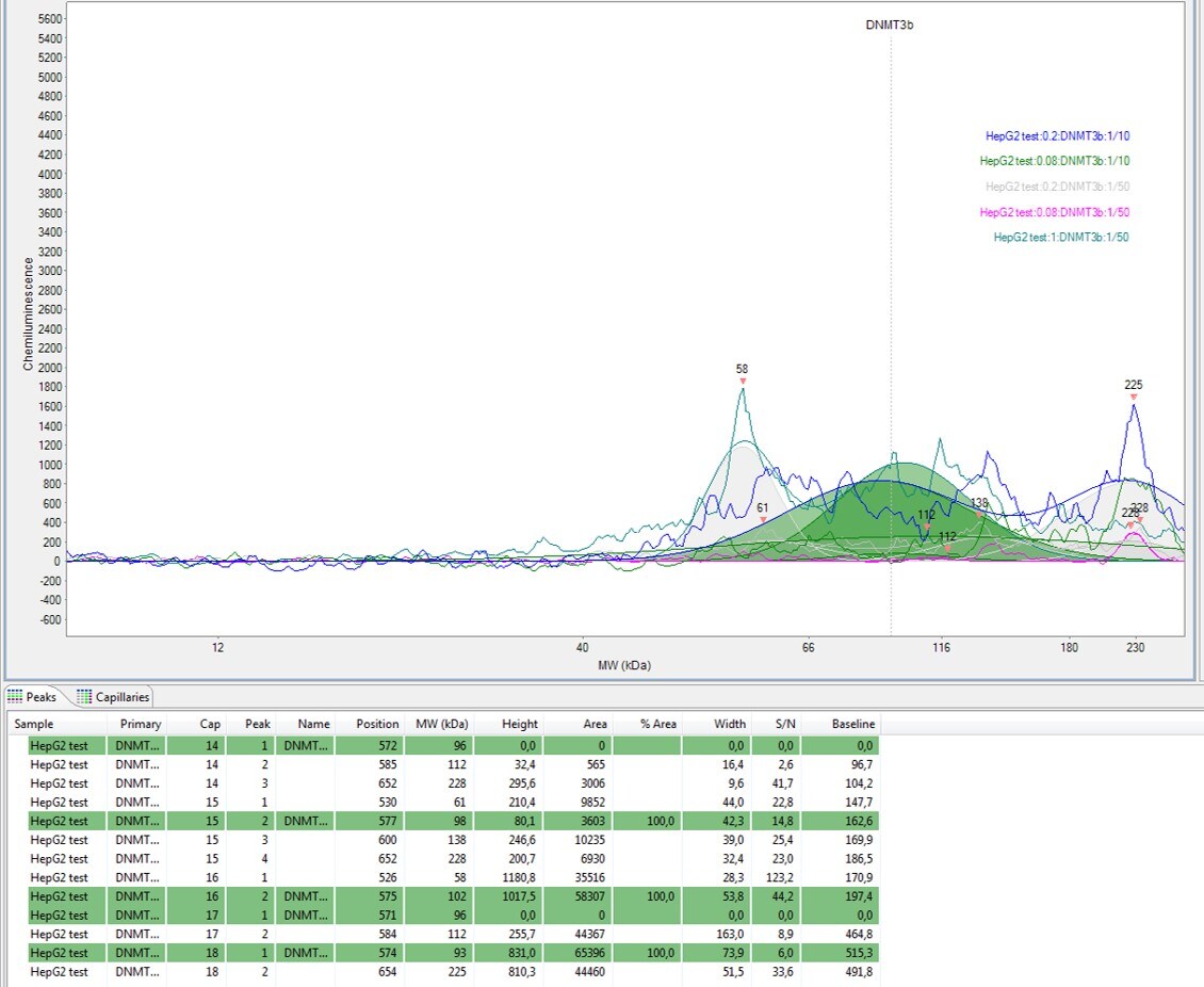
Application: Simple WesternSample Tested: HepG2 CELLSSpecies: HumanVerified Customer | Posted 02/03/2020materiel: 1x25 caps 12-230kDa échantillon : 0.08 à 1µg/µL anticorps : 1/250 à 1/10

-
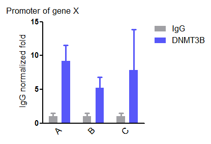
Application: Chromatin ImmunoprecipitationSample Tested: cellSpecies: HumanVerified Customer | Posted 01/30/2019
-
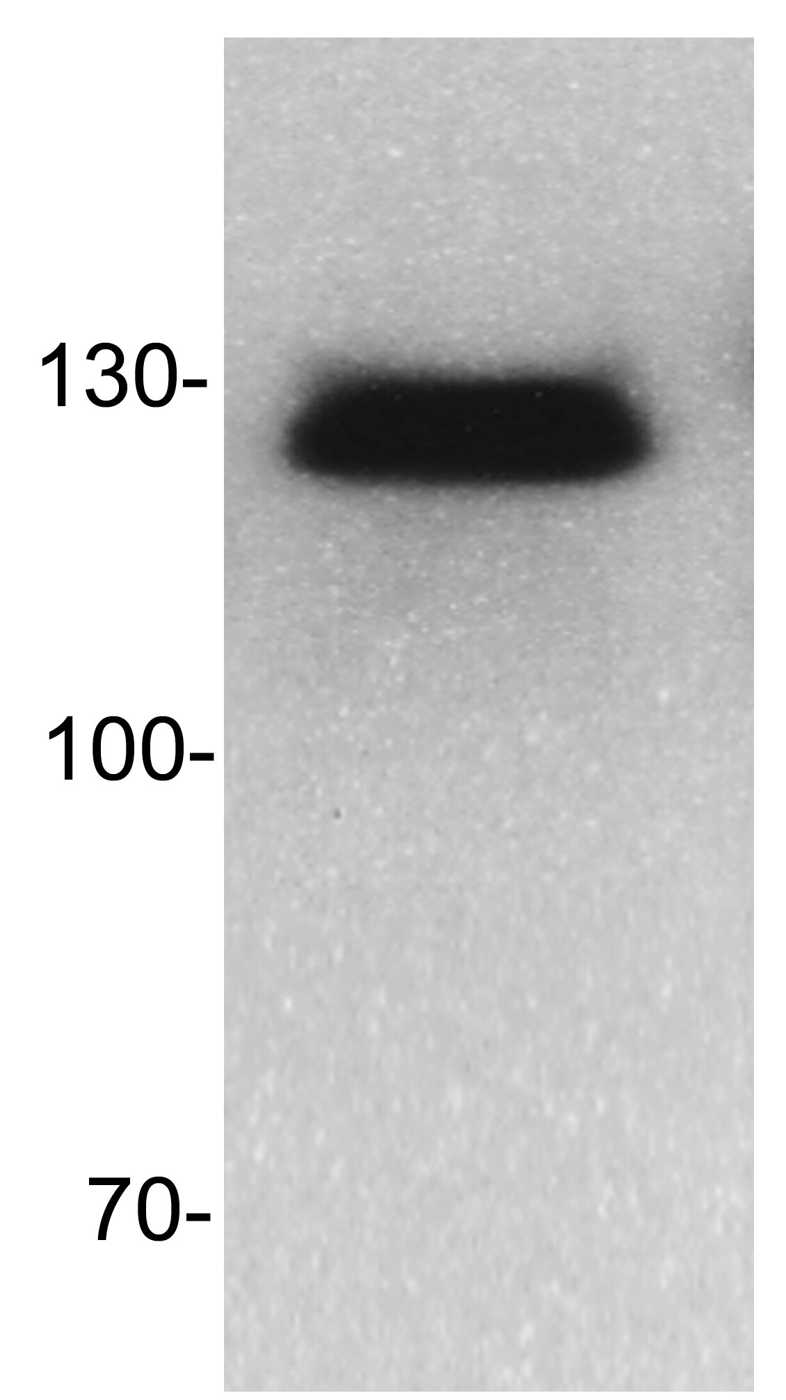
Application: Western BlotSample Tested: Lysate from homogenized mouse brain cellsSpecies: MouseVerified Customer | Posted 07/17/2014WB for Dnmt3b in mouse cells
There are no reviews that match your criteria.
Protocols
Find general support by application which include: protocols, troubleshooting, illustrated assays, videos and webinars.
- Antigen Retrieval Protocol (PIER)
- Antigen Retrieval for Frozen Sections Protocol
- Appropriate Fixation of IHC/ICC Samples
- Cellular Response to Hypoxia Protocols
- ChIP Protocol Video
- Chromatin Immunoprecipitation (ChIP) Protocol
- Chromatin Immunoprecipitation Protocol
- Chromogenic IHC Staining of Formalin-Fixed Paraffin-Embedded (FFPE) Tissue Protocol
- Chromogenic Immunohistochemistry Staining of Frozen Tissue
- ClariTSA™ Fluorophore Kits
- Detection & Visualization of Antibody Binding
- Fluorescent IHC Staining of Frozen Tissue Protocol
- Graphic Protocol for Heat-induced Epitope Retrieval
- Graphic Protocol for the Preparation and Fluorescent IHC Staining of Frozen Tissue Sections
- Graphic Protocol for the Preparation and Fluorescent IHC Staining of Paraffin-embedded Tissue Sections
- Graphic Protocol for the Preparation of Gelatin-coated Slides for Histological Tissue Sections
- IHC Sample Preparation (Frozen sections vs Paraffin)
- Immunofluorescent IHC Staining of Formalin-Fixed Paraffin-Embedded (FFPE) Tissue Protocol
- Immunohistochemistry (IHC) and Immunocytochemistry (ICC) Protocols
- Immunohistochemistry Frozen Troubleshooting
- Immunohistochemistry Paraffin Troubleshooting
- Preparing Samples for IHC/ICC Experiments
- Preventing Non-Specific Staining (Non-Specific Binding)
- Primary Antibody Selection & Optimization
- Protocol for Heat-Induced Epitope Retrieval (HIER)
- Protocol for Making a 4% Formaldehyde Solution in PBS
- Protocol for VisUCyte™ HRP Polymer Detection Reagent
- Protocol for the Preparation & Fixation of Cells on Coverslips
- Protocol for the Preparation and Chromogenic IHC Staining of Frozen Tissue Sections
- Protocol for the Preparation and Chromogenic IHC Staining of Frozen Tissue Sections - Graphic
- Protocol for the Preparation and Chromogenic IHC Staining of Paraffin-embedded Tissue Sections
- Protocol for the Preparation and Chromogenic IHC Staining of Paraffin-embedded Tissue Sections - Graphic
- Protocol for the Preparation and Fluorescent IHC Staining of Frozen Tissue Sections
- Protocol for the Preparation and Fluorescent IHC Staining of Paraffin-embedded Tissue Sections
- Protocol for the Preparation of Gelatin-coated Slides for Histological Tissue Sections
- R&D Systems Quality Control Western Blot Protocol
- TUNEL and Active Caspase-3 Detection by IHC/ICC Protocol
- The Importance of IHC/ICC Controls
- Troubleshooting Guide: Immunohistochemistry
- Troubleshooting Guide: Western Blot Figures
- Western Blot Conditions
- Western Blot Protocol
- Western Blot Protocol for Cell Lysates
- Western Blot Troubleshooting
- Western Blot Troubleshooting Guide
- View all Protocols, Troubleshooting, Illustrated assays and Webinars
Loading...