Key Product Details
Species Reactivity
Human
Applications
Immunohistochemistry, Immunohistochemistry-Paraffin, Western Blot, Immunocytochemistry/ Immunofluorescence
Label
Unconjugated
Antibody Source
Monoclonal Mouse IgG1 kappa Clone # SPM580
Loading...
Product Specifications
Immunogen
Recombinant human canine1/TMEM16A protein (Uniprot: Q5XX6)
Localization
Cell Surface and Cytoplasm
Marker
Marker for Gastrointestinal Stromal Tumors
Clonality
Monoclonal
Host
Mouse
Isotype
IgG1 kappa
Theoretical MW
114 kDa.
Disclaimer note: The observed molecular weight of the protein may vary from the listed predicted molecular weight due to post translational modifications, post translation cleavages, relative charges, and other experimental factors.
Disclaimer note: The observed molecular weight of the protein may vary from the listed predicted molecular weight due to post translational modifications, post translation cleavages, relative charges, and other experimental factors.
Description
200ug/ml of antibody purified from Bioreactor Concentrate by Protein A or G. Prepared in 10 mM PBS with 0.05% BSA & 0.05% azide. Also available WITHOUT BSA & azide at 1.0 mg/ml. (NBP2-34812)
Antibody with azide - store at 2 to 8C. Antibody without azide - store at -20 to -80C.
Antibody with azide - store at 2 to 8C. Antibody without azide - store at -20 to -80C.
Scientific Data Images for DOG1/TMEM16A Antibody (SPM580)
![Immunocytochemistry/ Immunofluorescence: DOG1/TMEM16A Antibody (SPM580) [NBP2-32841]](https://resources.rndsystems.com/images/products/DOG1-TMEM16A-Antibody-SPM580-Immunocytochemistry-Immunofluorescence-NBP2-32841-img0002.jpg "Immunocytochemistry/ Immunofluorescence: DOG1/TMEM16A Antibody (SPM580) [NBP2-32841]")
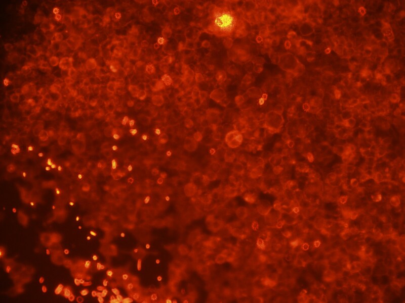
Immunocytochemistry/ Immunofluorescence: DOG1/TMEM16A Antibody (SPM580) [NBP2-32841]
Immunocytochemistry/Immunofluorescence: DOG1/TMEM16A Antibody (SPM580) [NBP2-32841] - HEK cells expressing Ano1-Myc. SPM580 gave more signal than the Myc Ab control and much more than the SP31 mAb. Negative control cells were negative. ICC image submitted by a verified customer review.![Immunohistochemistry-Paraffin: DOG1/TMEM16A Antibody (SPM580) [NBP2-32841]](https://resources.rndsystems.com/images/products/DOG1-TMEM16A-Antibody-SPM580-Immunohistochemistry-Paraffin-NBP2-32841-img0001.jpg "Immunohistochemistry-Paraffin: DOG1/TMEM16A Antibody (SPM580) [NBP2-32841]")
Immunohistochemistry-Paraffin: DOG1/TMEM16A Antibody (SPM580) [NBP2-32841]
Immunohistochemistry-Paraffin: DOG1/TMEM16A Antibody (SPM580) [NBP2-32841] - Formalin-fixed paraffin-embedded human GIST stained with DOG1 Monoclonal Antibody (SPM580).Applications for DOG1/TMEM16A Antibody (SPM580)
Application
Recommended Usage
Immunocytochemistry/ Immunofluorescence
0.5-1ug/ml
Immunohistochemistry-Paraffin
1-2 ug/ml
Western Blot
0.5-1.0ug/ml
Application Notes
Immunohistochemistry (Formalin-fixed): 1-2ug/ml for 30 minutes at RT. Staining of formalin-fixed tissues requires heating tissue sections in 10mM Tris with 1mM EDTA, pH 9.0, for 45 min at 95C followed by cooling at RT for 20 minutes.
Optimal dilution for a specific application should be determined.
Validated for ICC/IF & WB from a verified customer review.
Optimal dilution for a specific application should be determined.
Validated for ICC/IF & WB from a verified customer review.
Reviewed Applications
Read 2 reviews rated 3 using NBP2-32841 in the following applications:
Formulation, Preparation, and Storage
Purification
Protein A or G purified
Formulation
10 mM PBS with 0.05% BSA
Preservative
0.05% Sodium Azide
Concentration
0.2 mg/ml
Shipping
The product is shipped with polar packs. Upon receipt, store it immediately at the temperature recommended below.
Stability & Storage
Store at 4C.
Background: ANO1/TMEM16A
Long Name
Anoctamin 1
Alternate Names
DOG1, ORAOV2, TAOS2, TMEM16A
Gene Symbol
ANO1
UniProt
Additional ANO1/TMEM16A Products
Product Documents for DOG1/TMEM16A Antibody (SPM580)
Certificate of Analysis
To download a Certificate of Analysis, please enter a lot or batch number in the search box below.
Product Specific Notices for DOG1/TMEM16A Antibody (SPM580)
This product is for research use only and is not approved for use in humans or in clinical diagnosis. Primary Antibodies are guaranteed for 1 year from date of receipt.
Related Research Areas
Customer Reviews for DOG1/TMEM16A Antibody (SPM580) (2)
3 out of 5
2 Customer Ratings
Have you used DOG1/TMEM16A Antibody (SPM580)?
Submit a review and receive an Amazon gift card!
$25/€18/£15/$25CAN/¥2500 Yen for a review with an image
$10/€7/£6/$10CAN/¥1110 Yen for a review without an image
Submit a review
Customer Images
Showing
1
-
2 of
2 reviews
Showing All
Filter By:
-
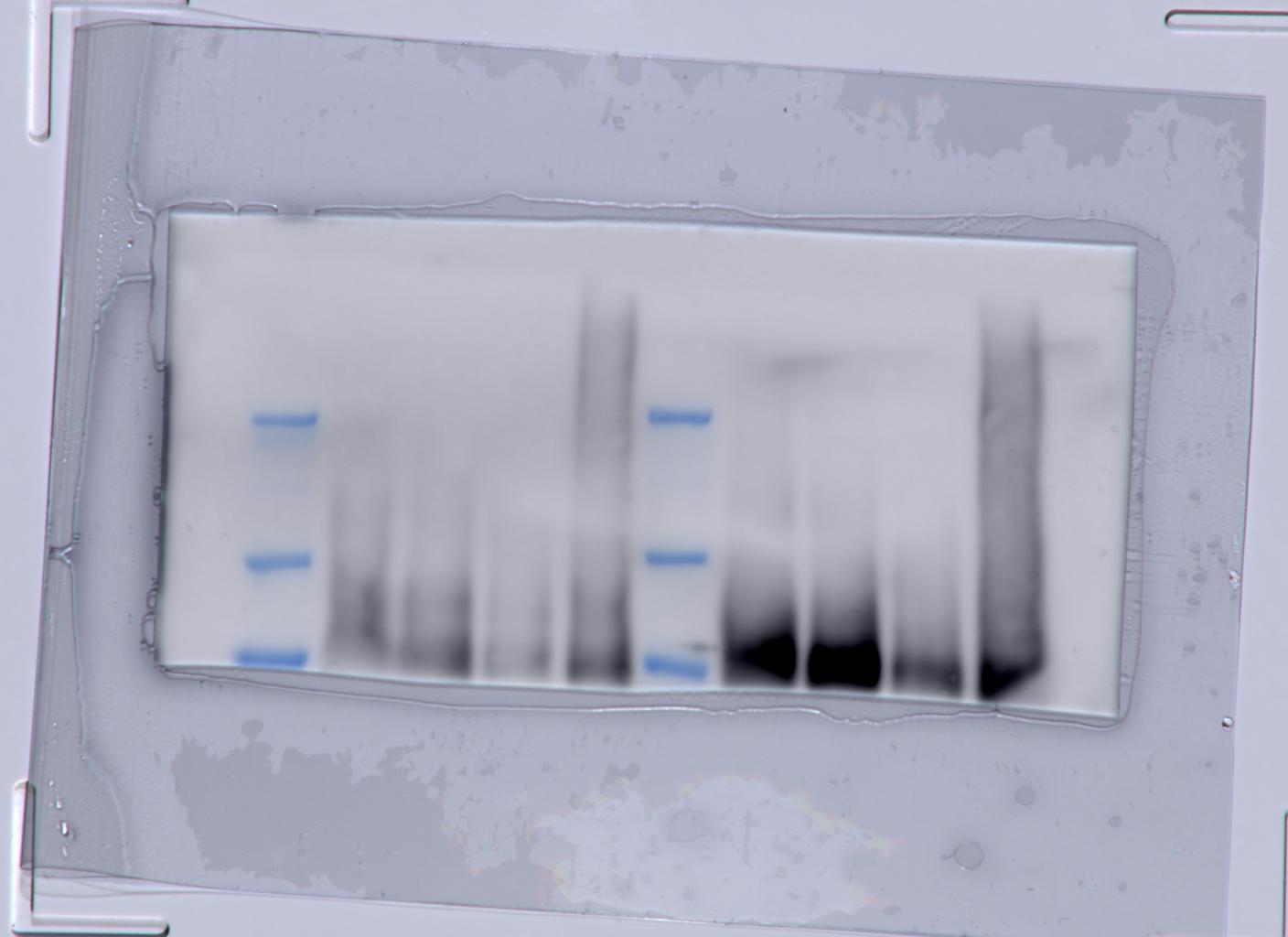
Application: Western BlotSample Tested: protein lysates and MDCK kidney protein lysateSpecies: MDCKVerified Customer | Posted 11/27/201950µg Protein load on normal SDS gel 20 min at 70V and then 1h at 120V Tank blotting for 3h at 120V in PVDF membrane Blocking for 1h with 5%MM/TBST Prim. AB 1:500 in 1%MM/TBST over night

-
Application: ImmunocytochemistrySample Tested: Hek 293TSpecies: HumanVerified Customer | Posted 02/01/2017HEK cells expressing Ano1-Myc. SPM580 gave more signal than the Myc Ab control and Much more than the SP31 mAb. Negative control cells were negative. The Santa Cruz mAb and pAb for Ano1 were negative on these cells.
There are no reviews that match your criteria.
Protocols
Find general support by application which include: protocols, troubleshooting, illustrated assays, videos and webinars.
- Antigen Retrieval Protocol (PIER)
- Antigen Retrieval for Frozen Sections Protocol
- Appropriate Fixation of IHC/ICC Samples
- Cellular Response to Hypoxia Protocols
- Chromogenic IHC Staining of Formalin-Fixed Paraffin-Embedded (FFPE) Tissue Protocol
- Chromogenic Immunohistochemistry Staining of Frozen Tissue
- ClariTSA™ Fluorophore Kits
- Detection & Visualization of Antibody Binding
- Fluorescent IHC Staining of Frozen Tissue Protocol
- Graphic Protocol for Heat-induced Epitope Retrieval
- Graphic Protocol for the Preparation and Fluorescent IHC Staining of Frozen Tissue Sections
- Graphic Protocol for the Preparation and Fluorescent IHC Staining of Paraffin-embedded Tissue Sections
- Graphic Protocol for the Preparation of Gelatin-coated Slides for Histological Tissue Sections
- ICC Cell Smear Protocol for Suspension Cells
- ICC Immunocytochemistry Protocol Videos
- ICC for Adherent Cells
- IHC Sample Preparation (Frozen sections vs Paraffin)
- Immunocytochemistry (ICC) Protocol
- Immunocytochemistry Troubleshooting
- Immunofluorescence of Organoids Embedded in Cultrex Basement Membrane Extract
- Immunofluorescent IHC Staining of Formalin-Fixed Paraffin-Embedded (FFPE) Tissue Protocol
- Immunohistochemistry (IHC) and Immunocytochemistry (ICC) Protocols
- Immunohistochemistry Frozen Troubleshooting
- Immunohistochemistry Paraffin Troubleshooting
- Preparing Samples for IHC/ICC Experiments
- Preventing Non-Specific Staining (Non-Specific Binding)
- Primary Antibody Selection & Optimization
- Protocol for Heat-Induced Epitope Retrieval (HIER)
- Protocol for Making a 4% Formaldehyde Solution in PBS
- Protocol for VisUCyte™ HRP Polymer Detection Reagent
- Protocol for the Fluorescent ICC Staining of Cell Smears - Graphic
- Protocol for the Fluorescent ICC Staining of Cultured Cells on Coverslips - Graphic
- Protocol for the Preparation & Fixation of Cells on Coverslips
- Protocol for the Preparation and Chromogenic IHC Staining of Frozen Tissue Sections
- Protocol for the Preparation and Chromogenic IHC Staining of Frozen Tissue Sections - Graphic
- Protocol for the Preparation and Chromogenic IHC Staining of Paraffin-embedded Tissue Sections
- Protocol for the Preparation and Chromogenic IHC Staining of Paraffin-embedded Tissue Sections - Graphic
- Protocol for the Preparation and Fluorescent ICC Staining of Cells on Coverslips
- Protocol for the Preparation and Fluorescent ICC Staining of Non-adherent Cells
- Protocol for the Preparation and Fluorescent ICC Staining of Stem Cells on Coverslips
- Protocol for the Preparation and Fluorescent IHC Staining of Frozen Tissue Sections
- Protocol for the Preparation and Fluorescent IHC Staining of Paraffin-embedded Tissue Sections
- Protocol for the Preparation of Gelatin-coated Slides for Histological Tissue Sections
- Protocol for the Preparation of a Cell Smear for Non-adherent Cell ICC - Graphic
- R&D Systems Quality Control Western Blot Protocol
- TUNEL and Active Caspase-3 Detection by IHC/ICC Protocol
- The Importance of IHC/ICC Controls
- Troubleshooting Guide: Immunohistochemistry
- Troubleshooting Guide: Western Blot Figures
- Western Blot Conditions
- Western Blot Protocol
- Western Blot Protocol for Cell Lysates
- Western Blot Troubleshooting
- Western Blot Troubleshooting Guide
- View all Protocols, Troubleshooting, Illustrated assays and Webinars
Loading...