DOT1L Antibody
Novus Biologicals | Catalog # NB100-40845
![Western Blot: DOT1L Antibody [NB100-40845]](https://resources.rndsystems.com/images/products/DOT1L-Antibody-Western-Blot-NB100-40845-img0014.jpg "Western Blot: DOT1L Antibody [NB100-40845]")
Loading...
Key Product Details
Validated by
Independent Antibodies, Biological Validation
Species Reactivity
Validated:
Human, Mouse
Cited:
Human, Mouse
Applications
Validated:
Immunohistochemistry, Immunohistochemistry-Paraffin, Western Blot, Immunoprecipitation, Chromatin Immunoprecipitation (ChIP), Chromatin Immunoprecipitation Sequencing, Knockdown Validated
Cited:
Western Blot, Immunocytochemistry/ Immunofluorescence, Chemotaxis, Chip Cytometry, IF/IHC, Knockdown Validated
Label
Unconjugated
Antibody Source
Polyclonal Rabbit IgG
Loading...
Product Specifications
Immunogen
The immunogen recognized by this antibody maps to a region between residue1000 and 1050 of human DOT1-Like using the numbering given in entry NP_115871.1 (GeneID 84444).
Reactivity Notes
Mouse reactivity reported in scientific literature (PMID: 23861670).
Clonality
Polyclonal
Host
Rabbit
Isotype
IgG
Theoretical MW
185 kDa.
Disclaimer note: The observed molecular weight of the protein may vary from the listed predicted molecular weight due to post translational modifications, post translation cleavages, relative charges, and other experimental factors.
Disclaimer note: The observed molecular weight of the protein may vary from the listed predicted molecular weight due to post translational modifications, post translation cleavages, relative charges, and other experimental factors.
Scientific Data Images for DOT1L Antibody
Western Blot: DOT1L Antibody [NB100-40845]
DOT1L-Antibody-Western-Blot-NB100-40845-img0014.jpg![Immunohistochemistry-Paraffin: DOT1L Antibody [NB100-40845]](https://resources.rndsystems.com/images/products/DOT1L-Antibody-Immunohistochemistry-Paraffin-NB100-40845-img0009.jpg "Immunohistochemistry-Paraffin: DOT1L Antibody [NB100-40845]")
Immunohistochemistry-Paraffin: DOT1L Antibody [NB100-40845]
Immunohistochemistry-Paraffin: DOT1L Antibody [NB100-40845] - Human lung carcinoma. Antibody: Affinity purified rabbit anti-DOT1L used at a dilution of 1:200 (1ug/ml). Detection: DAB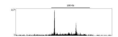
Chromatin Immunoprecipitation: DOT1L Antibody [NB100-40845] - Chromatin from KOPN-8 cell line was immunoprecipitated with anti-DOT1L antibody and analyzed by DNA sequencing. The figure illustrates the peak distribution of DOT1L binding within a 250 Kb region of chromosome 10 as detected using anti-DOT1L antibody.
![Western Blot: DOT1L Antibody [NB100-40845]](https://resources.rndsystems.com/images/products/DOT1L-Antibody-Western-Blot-NB100-40845-img0007.jpg "Western Blot: DOT1L Antibody [NB100-40845]")
Western Blot: DOT1L Antibody [NB100-40845]
Western Blot: DOT1L Antibody [NB100-40845] - Whole cell lysate (5, 15 and 50 ug for WB; 1 mg for IP, 20% of IP loaded) from HeLa cells. Affinity purified rabbit anti-DOT1L antibody NB100-40845 used for WB at 0.04 ug/ml (A) and 1 ug/ml (B) and used for IP at 3 ug/mg lysate (B).![Western Blot: DOT1L Antibody [NB100-40845]](https://resources.rndsystems.com/images/products/DOT1L-Antibody-Western-Blot-NB100-40845-img0015.jpg "Western Blot: DOT1L Antibody [NB100-40845]")

Western Blot: DOT1L Antibody [NB100-40845] -
siRNA mediated gene silencing of DOT1L validated by Q-PCR and western blot. The results show a down regulation of DOT1L mRNA expression (a) and a decrease of DOT1L protein level (b). Empty bars are untreated cells and solid bars are transfected cells. Results from HSkMSC cultures derived from three different experiments on cells at passage three, Graph is presenting means ± SEM. *, p-value < 0.05 siRNA treated cells compared to controlsApplications for DOT1L Antibody
Application
Recommended Usage
Immunohistochemistry
1:200-1:1000
Immunohistochemistry-Paraffin
1:200-1:1000
Immunoprecipitation
2-5 ug/mg lysate
Western Blot
1:2000-1:10000
Application Notes
Chromatin Immunoprecipitation was reported in scientific literature (PMID:19734945). For ChIP-Seq: 4 ug/30 ug chromatin. Epitope retrieval with Tris-EDTA pH9.0 is recommended for FFPE tissue sections.
Formulation, Preparation, and Storage
Purification
Immunogen affinity purified
Formulation
TBS and 0.1% BSA
Preservative
0.09% Sodium Azide
Concentration
0.2 mg/ml
Shipping
The product is shipped with polar packs. Upon receipt, store it immediately at the temperature recommended below.
Stability & Storage
Store at 4C. Do not freeze.
Background: DOT1L
Long Name
Disruptor of Telomeric Silencing 1-like
Alternate Names
H3-K79-HMTase, KMT4
Entrez Gene IDs
84444 (Human)
Gene Symbol
DOT1L
UniProt
Additional DOT1L Products
Product Documents for DOT1L Antibody
Certificate of Analysis
To download a Certificate of Analysis, please enter a lot or batch number in the search box below.
Product Specific Notices for DOT1L Antibody
This product is for research use only and is not approved for use in humans or in clinical diagnosis. Primary Antibodies are guaranteed for 1 year from date of receipt.
Related Research Areas
Citations for DOT1L Antibody
 Powered by Bioz
Customer Reviews for DOT1L Antibody
There are currently no reviews for this product. Be the first to review DOT1L Antibody and earn rewards!
Have you used DOT1L Antibody?
Submit a review and receive an Amazon gift card!
$25/€18/£15/$25CAN/¥2500 Yen for a review with an image
$10/€7/£6/$10CAN/¥1110 Yen for a review without an image
Submit a review
Protocols
Find general support by application which include: protocols, troubleshooting, illustrated assays, videos and webinars.
- Antigen Retrieval Protocol (PIER)
- Antigen Retrieval for Frozen Sections Protocol
- Appropriate Fixation of IHC/ICC Samples
- Cellular Response to Hypoxia Protocols
- ChIP Protocol Video
- Chromatin Immunoprecipitation (ChIP) Protocol
- Chromatin Immunoprecipitation Protocol
- Chromogenic IHC Staining of Formalin-Fixed Paraffin-Embedded (FFPE) Tissue Protocol
- Chromogenic Immunohistochemistry Staining of Frozen Tissue
- ClariTSA™ Fluorophore Kits
- Detection & Visualization of Antibody Binding
- Fluorescent IHC Staining of Frozen Tissue Protocol
- Graphic Protocol for Heat-induced Epitope Retrieval
- Graphic Protocol for the Preparation and Fluorescent IHC Staining of Frozen Tissue Sections
- Graphic Protocol for the Preparation and Fluorescent IHC Staining of Paraffin-embedded Tissue Sections
- Graphic Protocol for the Preparation of Gelatin-coated Slides for Histological Tissue Sections
- IHC Sample Preparation (Frozen sections vs Paraffin)
- Immunofluorescent IHC Staining of Formalin-Fixed Paraffin-Embedded (FFPE) Tissue Protocol
- Immunohistochemistry (IHC) and Immunocytochemistry (ICC) Protocols
- Immunohistochemistry Frozen Troubleshooting
- Immunohistochemistry Paraffin Troubleshooting
- Immunoprecipitation Protocol
- Preparing Samples for IHC/ICC Experiments
- Preventing Non-Specific Staining (Non-Specific Binding)
- Primary Antibody Selection & Optimization
- Protocol for Heat-Induced Epitope Retrieval (HIER)
- Protocol for Making a 4% Formaldehyde Solution in PBS
- Protocol for VisUCyte™ HRP Polymer Detection Reagent
- Protocol for the Preparation & Fixation of Cells on Coverslips
- Protocol for the Preparation and Chromogenic IHC Staining of Frozen Tissue Sections
- Protocol for the Preparation and Chromogenic IHC Staining of Frozen Tissue Sections - Graphic
- Protocol for the Preparation and Chromogenic IHC Staining of Paraffin-embedded Tissue Sections
- Protocol for the Preparation and Chromogenic IHC Staining of Paraffin-embedded Tissue Sections - Graphic
- Protocol for the Preparation and Fluorescent IHC Staining of Frozen Tissue Sections
- Protocol for the Preparation and Fluorescent IHC Staining of Paraffin-embedded Tissue Sections
- Protocol for the Preparation of Gelatin-coated Slides for Histological Tissue Sections
- R&D Systems Quality Control Western Blot Protocol
- TUNEL and Active Caspase-3 Detection by IHC/ICC Protocol
- The Importance of IHC/ICC Controls
- Troubleshooting Guide: Immunohistochemistry
- Troubleshooting Guide: Western Blot Figures
- Western Blot Conditions
- Western Blot Protocol
- Western Blot Protocol for Cell Lysates
- Western Blot Troubleshooting
- Western Blot Troubleshooting Guide
- View all Protocols, Troubleshooting, Illustrated assays and Webinars
Loading...