DRP1 Antibody - BSA Free
Novus Biologicals | Catalog # NB110-55288
![Western Blot: DRP1 AntibodyBSA Free [NB110-55288]](https://resources.rndsystems.com/images/products/DRP1-Antibody-Western-Blot-NB110-55288-img0012.jpg "Western Blot: DRP1 AntibodyBSA Free [NB110-55288]")
Key Product Details
Validated by
Knockout/Knockdown
Species Reactivity
Validated:
Human, Mouse, Rat, Fish, Primate
Cited:
Human, Mouse, Rat, Fish
Predicted:
Bovine (100%). Backed by our 100% Guarantee.
Applications
Validated:
Immunohistochemistry, Immunohistochemistry-Paraffin, Western Blot, Flow Cytometry, Flow (Intracellular), Immunocytochemistry/ Immunofluorescence, Simple Western, Immunoprecipitation
Cited:
Immunohistochemistry-Paraffin, Western Blot, Immunocytochemistry/ Immunofluorescence, Simple Western, Immunoprecipitation, IF/IHC
Label
Unconjugated
Antibody Source
Polyclonal Rabbit IgG
Format
BSA Free
Loading...
Product Specifications
Immunogen
A synthetic peptide made to an internal region within residues 500-600 of the human DRP1 protein. [Swiss-Prot# O00429]
Reactivity Notes
Use in Rat reported in scientific literature (PMID:34622072). Fish reactivity reported in scientific literature (PMID: 25008790).
Localization
Cytoplasm; cytosol. Intracytoplasmic membrane; peripheral mambrane protein. Mainly cytosolic. Also membrane-associated. Localizes to mitochondia at spots of division events. Associated with peroxisomal membranes, it is recruited in part by PEX11B.
Clonality
Polyclonal
Host
Rabbit
Isotype
IgG
Theoretical MW
81 kDa.
Disclaimer note: The observed molecular weight of the protein may vary from the listed predicted molecular weight due to post translational modifications, post translation cleavages, relative charges, and other experimental factors.
Disclaimer note: The observed molecular weight of the protein may vary from the listed predicted molecular weight due to post translational modifications, post translation cleavages, relative charges, and other experimental factors.
Scientific Data Images for DRP1 Antibody - BSA Free
Western Blot: DRP1 AntibodyBSA Free [NB110-55288]
Western Blot: DRP1 Antibody [NB110-55288] - Total protein from human HeLa and MCF7 cells, mouse 3T3 cells and rat PC12 cells was separated on a 7.5 % gel by SDS-PAGE, transferred to PVDF membrane and blocked in 5% non-fat milk in TBST. The membrane was probed with 2.0 ug/mL anti-DRP1 in blocking buffer and detected with an anti-rabbit HRP secondary antibody using chemiluminescence.![Immunohistochemistry-Paraffin: DRP1 Antibody - BSA Free [NB110-55288]](https://resources.rndsystems.com/images/products/DRP1-Antibody-Immunohistochemistry-Paraffin-NB110-55288-img0013.jpg "Immunohistochemistry-Paraffin: DRP1 Antibody - BSA Free [NB110-55288]")
Immunohistochemistry-Paraffin: DRP1 Antibody - BSA Free [NB110-55288]
Immunohistochemistry-Paraffin: DRP1 Antibody [NB110-55288] - Analysis of FFPE tissue section of mouse kidney using DRP1 antibody #NB110-55288 at 1:300. The primary antibody bound to DRP1 protein in the tissue section was detected using a HRP labeled secondary antibody and DAB reagent. Nuclei of the cells were counterstained with hematoxylin. This antibody generated a diffused cytoplasmic staining of DRP1 in the epithelial cells of various tubules and in the cells of glomeruli.![Western Blot: DRP1 AntibodyBSA Free [NB110-55288]](https://resources.rndsystems.com/images/products/DRP1-Antibody-Western-Blot-NB110-55288-img0004.jpg "Western Blot: DRP1 AntibodyBSA Free [NB110-55288]")
![Immunocytochemistry/ Immunofluorescence: DRP1 Antibody - BSA Free [NB110-55288]](https://resources.rndsystems.com/images/products/DRP1-Antibody-Immunocytochemistry-Immunofluorescence-NB110-55288-img0015.jpg "Immunocytochemistry/ Immunofluorescence: DRP1 Antibody - BSA Free [NB110-55288]")
Immunocytochemistry/ Immunofluorescence: DRP1 Antibody - BSA Free [NB110-55288]
Immunocytochemistry/Immunofluorescence: DRP1 Antibody [NB110-55288] - HeLa cells were fixed for 10 minutes using 10% formalin and then permeabilized for 5 minutes using 1X PBS + 0.5% Triton X-100. The cells were incubated with anti-DRP1 at 5 ug/mL overnight at 4C and detected with an anti-rabbit Dylight 488 (Green) at a 1:500 dilution. Nuclei were counterstained with DAPI (Blue). Cells were imaged using a 40X objective.![Immunohistochemistry: DRP1 Antibody - BSA Free [NB110-55288]](https://resources.rndsystems.com/images/products/DRP1-Antibody-Immunohistochemistry-NB110-55288-img0005.jpg "Immunohistochemistry: DRP1 Antibody - BSA Free [NB110-55288]")
Immunohistochemistry: DRP1 Antibody - BSA Free [NB110-55288]
Immunohistochemistry: DRP1 Antibody [NB110-55288] - Staining of renal tubular epithelium and visceral epithelial cells of the glomerulus. Human kidney cortex, 40X magnification.![Western Blot: DRP1 AntibodyBSA Free [NB110-55288]](https://resources.rndsystems.com/images/products/DRP1-Antibody-Western-Blot-NB110-55288-img0019.jpg "Western Blot: DRP1 AntibodyBSA Free [NB110-55288]")
Western Blot: DRP1 AntibodyBSA Free [NB110-55288]
DRP1-Antibody-Western-Blot-NB110-55288-img0019.jpg![Immunocytochemistry/ Immunofluorescence: DRP1 Antibody - BSA Free [NB110-55288]](https://resources.rndsystems.com/images/products/DRP1-Antibody-Immunocytochemistry-Immunofluorescence-NB110-55288-img0017.jpg "Immunocytochemistry/ Immunofluorescence: DRP1 Antibody - BSA Free [NB110-55288]")
Immunocytochemistry/ Immunofluorescence: DRP1 Antibody - BSA Free [NB110-55288]
Immunocytochemistry/Immunofluorescence: DRP1 Antibody [NB110-55288] - PC12 cells were fixed for 10 minutes using 10% formalin and then permeabilized for 5 minutes using 1X PBS + 0.05% Triton-X100. The cells were incubated with anti-DRP1 Antibody at 2 ug/ml overnight at 4C and detected with an anti-rabbit Dylight 488 (Green) at a 1:500 dilution. Nuclei were counterstained with DAPI (Blue). Cells were imaged using a 40X objective.![Flow Cytometry: DRP1 Antibody - BSA Free [NB110-55288]](https://resources.rndsystems.com/images/products/DRP1-Antibody-Flow-Cytometry-NB110-55288-img0018.jpg "Flow Cytometry: DRP1 Antibody - BSA Free [NB110-55288]")
Flow Cytometry: DRP1 Antibody - BSA Free [NB110-55288]
Flow Cytometry: DRP1 Antibody [NB110-55288] - An intracellular stain was performed on HeLa cells with DRP1 Antibody NB110-55288AF488 (blue) and a matched isotype control (orange). Cells were fixed with 4% PFA and then permeabilized with 0.1% saponin. Cells were incubated in an antibody dilution of 5 ug/mL for 30 minutes at room temperature. Both antibodies were conjugated to Alexa Fluor 488.![Western Blot: DRP1 AntibodyBSA Free [NB110-55288]](https://resources.rndsystems.com/images/products/DRP1-Antibody-Western-Blot-NB110-55288-img0007.jpg "Western Blot: DRP1 AntibodyBSA Free [NB110-55288]")
Western Blot: DRP1 AntibodyBSA Free [NB110-55288]
Western Blot: DRP1 Antibody [NB110-55288] - Rat spinal cord-DRP1 (81kda). Image from verified customer review.![Immunocytochemistry/ Immunofluorescence: DRP1 Antibody - BSA Free [NB110-55288]](https://resources.rndsystems.com/images/products/DRP1-Antibody-Immunocytochemistry-Immunofluorescence-NB110-55288-img0009.jpg "Immunocytochemistry/ Immunofluorescence: DRP1 Antibody - BSA Free [NB110-55288]")
Immunocytochemistry/ Immunofluorescence: DRP1 Antibody - BSA Free [NB110-55288]
Immunocytochemistry/Immunofluorescence: DRP1 Antibody [NB110-55288] - HeLa cells were fixed for 10 minutes using 10% formalin and then permeabilized for 5 minutes using 1X TBS + 0.5% Triton X-100. The cells were incubated with anti-DRP1 at 5.0 ug/mL overnight at 4C and detected with an anti-rabbit Dylight 488 (Green) at 1:500. Alpha tubulin (DM1A) NB100-690 was used as a co-stain at 1:1000 and detected with an anti-mouse Dylight 550 (Red) at 1:500. Nuclei were counterstained with DAPI (Blue). Cells were imaged using a 40X objective.![Immunocytochemistry/ Immunofluorescence: DRP1 Antibody - BSA Free [NB110-55288]](https://resources.rndsystems.com/images/products/DRP1-Antibody-Immunocytochemistry-Immunofluorescence-NB110-55288-img0016.jpg "Immunocytochemistry/ Immunofluorescence: DRP1 Antibody - BSA Free [NB110-55288]")
Immunocytochemistry/ Immunofluorescence: DRP1 Antibody - BSA Free [NB110-55288]
Immunocytochemistry/Immunofluorescence: DRP1 Antibody [NB110-55288] - NIH3T3 cells were fixed for 10 minutes using 10% formalin and then permeabilized for 5 minutes using 1X PBS + 0.05% Triton-X100. The cells were incubated with anti-DRP1 Antibody at 2 ug/ml overnight at 4C and detected with an anti-rabbit Dylight 488 (Green) at a 1:500 dilution. Nuclei were counterstained with DAPI (Blue). Cells were imaged using a 40X objective.![Flow Cytometry: DRP1 Antibody - BSA Free [NB110-55288]](https://resources.rndsystems.com/images/products/DRP1-Antibody-Flow-Cytometry-NB110-55288-img0014.jpg "Flow Cytometry: DRP1 Antibody - BSA Free [NB110-55288]")
Flow Cytometry: DRP1 Antibody - BSA Free [NB110-55288]
Flow Cytometry: DRP1 Antibody [NB110-55288] - An intracellular stain was performed on HeLa cells with NB110-55288 and a matched isotype control. Cells were fixed with 4% PFA and then permeablized with 0.1% saponin. Cells were incubated in an antibody dilution of 2.5 ug/mL for 30 minutes at room temperature, followed by Rabbit IgG (H+L) Cross-Adsorbed Secondary Antibody, Dylight 550.![Simple Western: DRP1 AntibodyBSA Free [NB110-55288]](https://resources.rndsystems.com/images/products/DRP1-Antibody-Simple-Western-NB110-55288-img0008.jpg "Simple Western: DRP1 AntibodyBSA Free [NB110-55288]")
Simple Western: DRP1 AntibodyBSA Free [NB110-55288]
Simple Western: DRP1 Antibody [NB110-55288] - Image shows a specific band for DUX4 in 0.5 mg/mL of HeLa lysate. This experiment was performed under reducing conditions using the 12-230 kDa separation system.![Simple Western: DRP1 AntibodyBSA Free [NB110-55288]](https://resources.rndsystems.com/images/products/DRP1-Antibody---BSA-Free-Simple-Western-NB110-55288-img0020.jpg "Simple Western: DRP1 AntibodyBSA Free [NB110-55288]")
Simple Western: DRP1 AntibodyBSA Free [NB110-55288]
Simple Western: DRP1 Antibody - BSA Free [NB110-55288] - Image shows a specific band for DRP1 in 0.1 ug/uL mouse hippocampus tissue lysate. Primary antibody dilution: 1:200. Image from verified customer review.
Western Blot: DRP1 Antibody - BSA Free [NB110-55288] -
Assessment of skeletal muscle mitochondrial dynamic markers. (A) Total Drp1 protein content, (B) phospho-Drp1Ser616 protein content, (C) percent difference in total and phospho-Drp1Ser616 in ET participants relative to SED participants, (D) total Mfn1 protein content, (E) total Mfn2 protein content. (F) Representative western blots. Significant main effect of training: #, p < 0.05; ##, p < 0.01. Significant post-hoc training effect: *, p < 0.05; **, p < 0.01. Data are presented as mean ± SEM.
Detection of DRP1 in SJCRH30 Human Cell Line by Flow Cytometry.
An intracellular stain was performed on SJCRH30 human Rhabdomyosarcoma cell line with Rabbit anti-DRP1 Affinity-purified Polyclonal Antibody conjugated to FITC (Catalog # NB110-55288F, blue histogram) or matched control antibody (NBP2-24892, orange histogram) at 5 µg/mL for 30 minutes at RT.Applications for DRP1 Antibody - BSA Free
Application
Recommended Usage
Flow (Intracellular)
1.0 ug/ml
Flow Cytometry
1:1000
Immunocytochemistry/ Immunofluorescence
1:500
Immunohistochemistry
2.5 ug/ml
Immunohistochemistry-Paraffin
2.5 ug/ml
Immunoprecipitation
reported in scientific literature
Simple Western
1:50
Western Blot
1:500
Application Notes
In Western blot a band is seen at ~81 kDa.
In Simple Western only 10 - 15 uL of the recommended dilution is used per data point.
See Simple Western Antibody Database for Simple Western validation: Tested in Human Brain lysate 0.5 mg/mL, separated by Size, antibody dilution of 1:50, apparent MW was 89 kDa. Separated by Size-Wes, Sally Sue/Peggy Sue.
The observed molecular weight of the protein may vary from the listed predicted molecular weight due to post translational modifications, post translation cleavages, relative charges, and other experimental factors.
Reviewed Applications
Read 4 reviews rated 5 using NB110-55288 in the following applications:
Flow Cytometry Panel Builder
Bio-Techne Knows Flow Cytometry
Save time and reduce costly mistakes by quickly finding compatible reagents using the Panel Builder Tool.
Advanced Features
- Spectra Viewer - Custom analysis of spectra from multiple fluorochromes
- Spillover Popups - Visualize the spectra of individual fluorochromes
- Antigen Density Selector - Match fluorochrome brightness with antigen density

Formulation, Preparation, and Storage
Purification
Immunogen affinity purified
Formulation
PBS
Format
BSA Free
Preservative
0.02% Sodium Azide
Concentration
1 mg/ml
Shipping
The product is shipped with polar packs. Upon receipt, store it immediately at the temperature recommended below.
Stability & Storage
Aliquot and store at -20C or -80C. Avoid freeze-thaw cycles.
Background: DRP1
Long Name
Dynamin-1-like protein
Alternate Names
DLP1, DNM1L, DVLP, Dymple, HdynIV
Gene Symbol
DNM1L
UniProt
Additional DRP1 Products
Product Documents for DRP1 Antibody - BSA Free
Certificate of Analysis
To download a Certificate of Analysis, please enter a lot or batch number in the search box below.
Product Specific Notices for DRP1 Antibody - BSA Free
This product is for research use only and is not approved for use in humans or in clinical diagnosis. Primary Antibodies are guaranteed for 1 year from date of receipt.
Citations for DRP1 Antibody - BSA Free
 Powered by Bioz
Customer Reviews for DRP1 Antibody - BSA Free (4)
5 out of 5
4 Customer Ratings
Have you used DRP1 Antibody - BSA Free?
Submit a review and receive an Amazon gift card!
$25/€18/£15/$25CAN/¥2500 Yen for a review with an image
$10/€7/£6/$10CAN/¥1110 Yen for a review without an image
Submit a review
Customer Images

-(01-ml)_NB110-55288_6956.jpg)
Showing
1
-
4 of
4 reviews
Showing All
Filter By:
-
Application: Simple WesternSample Tested: Brain (hippocampus) tissueSpecies: MouseVerified Customer | Posted 02/06/20230.1ug/ul hippocampus tissue lysate with DRP1 antibody (1:200)

-
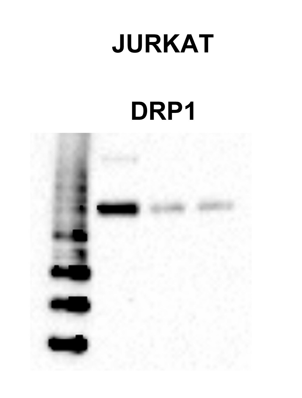
Application: Western BlotSample Tested: Jurkat cell lineSpecies: HumanVerified Customer | Posted 10/05/2018
-
Application: Western BlotSample Tested: PC12 whole cell lysateSpecies: OtherVerified Customer | Posted 06/09/2014
-
Application: Western BlotSample Tested: RatSpecies: RatVerified Customer | Posted 04/25/2014rat spinal cord-DRP1(81kda)
There are no reviews that match your criteria.
Protocols
View specific protocols for DRP1 Antibody - BSA Free (NB110-55288):
Protocol for Flow Cytometry Intracellular Staining
Sample Preparation.
1. Grow cells to 60-85% confluency. Flow cytometry requires between 2 x 10⁵ and 1 x 10⁶ cells for optimal performance.
2. If cells are adherent, harvest gently by washing once with staining buffer and then scraping. Avoid using trypsin as this can disrupt certain epitopes of interest. If enzymatic harvest is required, use Accutase, Collagenase, or TrypLE Express for a less damaging option.
3. Reserve 100 uL for counting, then transfer cell volume into a 50 mL conical tube and centrifuge for 8 minutes at 400 RCF.
a. Count cells using a hemocytometer and a 1:1 trypan blue exclusion stain to determine cell viability before starting the flow protocol. If cells appear blue, do not proceed.
4. Re-suspend cells to a concentration of 1 x 10⁶ cells/mL in staining buffer (NBP2-26247).
5. Aliquot out 100 uL samples in accordance with your experimental samples.
Tip: When cell surface and intracellular staining are required in the same sample, it is advisable that the cell surface staining be performed first since the fixation and permeabilization steps might reduce the availability of surface antigens.
Intracellular Staining.
Tip: When performing intracellular staining, it is important to use appropriate fixation and permeabilization reagents based upon the target and its subcellular location. Generally, our Intracellular Flow Assay Kit (NBP2-29450) is a good place to start as it contains an optimized combination of reagents for intracellular staining as well as an inhibitor of intracellular protein transport (necessary if staining secreted proteins). Certain targets may require more gentle or transient permeabilization protocols such as the commonly employed methanol or saponin-based methods.
Protocol for Cytoplasmic Targets:
1. Fix the cells by adding 100 uL fixation solution (such as 4% PFA) to each sample for 10-15 minutes.
2. Permeabilize cells by adding 100 uL of a permeabilization buffer to every 1 x 10⁶ cells present in the sample. Mix well and incubate at room temperature for 15 minutes.
a. For cytoplasmic targets, use a gentle permeabilization solution such as 1X PBS + 0.5% Saponin or 1X PBS + 0.5% Tween-20.
b. To maintain the permeabilized state throughout your experiment, use staining buffer + 0.1% of the permeabilization reagent (i.e. 0.1% Tween-20 or 0.1% Saponin).
3. Following the 15 minute incubation, add 2 mL of the staining buffer + 0.1% permeabilizer to each sample.
4. Centrifuge for 1 minute at 400 RCF.
5. Discard supernatant and re-suspend in 100 uL of staining buffer + 0.1% permeabilizer.
6. Add appropriate amount of each antibody (eg. 1 test or 1 ug per sample, as experimentally determined).
7. Mix well and incubate at room temperature for 30 minutes- 1 hour. Gently mix samples every 10-15 minutes.
8. Following the primary/conjugate incubation, add 1-2 mL/sample of staining buffer +0.1% permeabilizer and centrifuge for 1 minute at 400 RCF.
9. Wash twice by re-suspending cells in staining buffer (2 mL for tubes or 200 uL for wells) and centrifuging at 400 RCF for 5 minutes. Discard supernatant.
10. Add appropriate amount of secondary antibody (as experimentally determined) to each sample.
11. Incubate at room temperature in dark for 20 minutes.
12. Add 1-2 mL of staining buffer and centrifuge at 400 RCF for 1 minute and discard supernatant.
13. Wash twice by re-suspending cells in staining buffer (2 mL for tubes or 200 uL for wells) and centrifuging at 400 RCF for 5 minutes. Discard supernatant.
14. Resuspend in an appropriate volume of staining buffer (usually 500 uL per sample) and proceed with analysis on your flow cytometer.
Immunocytochemistry Protocol
Culture cells to appropriate density in 35 mm culture dishes or 6-well plates.
1. Remove culture medium and add 10% formalin to the dish. Fix at room temperature for 30 minutes.
2. Remove the formalin and add ice cold methanol. Incubate for 5-10 minutes.
3. Remove methanol and add washing solution (i.e. PBS). Be sure to not let the specimen dry out. Wash three times for 10 minutes.
4. To block nonspecific antibody binding incubate in 10% normal goat serum from 1 hour to overnight at room temperature.
5. Add primary antibody at appropriate dilution and incubate at room temperature from 2 hours to overnight at room temperature.
6. Remove primary antibody and replace with washing solution. Wash three times for 10 minutes.
7. Add secondary antibody at appropriate dilution. Incubate for 1 hour at room temperature.
8. Remove antibody and replace with wash solution, then wash for 10 minutes. Add Hoechst 33258 to wash solution at 1:25,0000 and incubate for 10 minutes. Wash a third time for 10 minutes.
9. Cells can be viewed directly after washing. The plates can also be stored in PBS containing Azide covered in Parafilm (TM). Cells can also be cover-slipped using Fluoromount, with appropriate sealing.
*The above information is only intended as a guide. The researcher should determine what protocol best meets their needs. Please follow safe laboratory procedures.
Immunohistochemistry-Paraffin Embedded Sections
Antigen Unmasking:
Bring slides to a boil in 10 mM sodium citrate buffer (pH 6.0) then maintain at a sub-boiling temperature for 10 minutes. Cool slides on bench-top for 30 minutes (keep slides in the sodium citrate buffer all the time).
Staining:
1. Wash sections in deionized water three times for 5 minutes each.
2. Wash sections in PBS for 5 minutes.
3. Block each section with 100-400 ul blocking solution (1% BSA in PBS) for 1 hour at room temperature.
4. Remove blocking solution and add 100-400 ul diluted primary antibody. Incubate overnight at 4 C.
5. Remove antibody solution and wash sections in wash buffer three times for 5 minutes each.
6. Add 100-400 ul HRP polymer conjugated secondary antibody. Incubate 30 minutes at room temperature.
7. Wash sections three times in wash buffer for 5 minutes each.
8. Add 100-400 ul DAB substrate to each section and monitor staining closely.
9. As soon as the sections develop, immerse slides in deionized water.
10. Counterstain sections in hematoxylin.
11. Wash sections in deionized water two times for 5 minutes each.
12. Dehydrate sections.
13. Mount coverslips.
Western Blot Protocol
1. Perform SDS-PAGE (4-12%) on samples to be analyzed, loading 30 ug of total protein per lane.
2. Transfer proteins to Nitrocellulose according to the instructions provided by the manufacturer of the transfer
apparatus.
3. Rinse membrane with dH2O and then stain the blot using Ponceau S for 1-2 minutes to access the transfer of proteins onto the nitrocellulose membrane. Rinse the blot in water to remove excess stain and mark the lane locations and locations of molecular weight markers using a pencil.
4. Rinse the blot in TBS for approximately 5 minutes.
5. Block the membrane using 5% non-fat dry milk + 1% BSA in TBS, 1 hour at room temperature.
6. Rinse the membrane in dH2O and then wash the membrane in wash buffer [TBS + 0.1% Tween] 3 times for 10 minutes each.
7. Dilute the rabbit anti-DRP1 primary antibody (NB 110-55288) in blocking buffer and incubate 2 hours at room temperature.
8. Rinse the membrane in dH2O and then wash the membrane in wash buffer [TBS + 0.1% Tween] 3 times for 10 minutes each.
9. Apply the diluted rabbit-IgG HRP-conjugated secondary antibody in blocking buffer (as per manufacturers
instructions) and incubate 1 hour at room temperature.
10. Wash the blot in wash buffer [TBS + 0.1% Tween] 3 times for 10 minutes each (this step can be repeated as required to reduce background).
11. Apply the detection reagent of choice in accordance with the manufacturers instructions (Pierce, ECL).
Note: Tween-20 can be added to the blocking or antibody diultion buffer at a final concentration of 0.05-0.2%, provided it does not interfere with antibody-antigen binding.
IHC-FFPE sectionsI. Deparaffinization:
A. Treat slides with Xylene: 3 changes for 5 minutes each. Drain slides for 10 seconds between changes.
B. Treat slides with 100% Reagent Alcohol: 3 changes for 5 minutes each. Drain slides for 10 seconds between changes.
II. Quench Endogenous Peroxidase:
A.Place slides in peroxidase quenching solution: 15-30 minutes.
To Prepare 200 ml of Quenching Solution:
-Add 3 ml of 30% Hydrogen Peroxide to 200 ml of Methanol.
-Use within 4 hours of preparation
B. Place slides in distilled water: 2 changes for 2 minutes each.
III. Retrieve Epitopes:
A. Preheat Citrate Buffer. Place 200 ml of Citrate Buffer Working Solution into container, cover and place into steamer. Heat to 90-96 degrees Celcius.
B. Place rack of slides into hot Citrate Buffer for 20 minutes. Cover.
C.Carefully remove container with slides from steamer and cool on bench, uncovered, for 20 minutes.
D. Slowly add distilled water to further cool for 5 minutes.
E. Rinse slides with distilled water. 2 changes for 2 minutes each.
IV. Immunostaining Procedure:
A. Remove each slide from rack and circle tissue section with a hydrophobic barrier pen (e.g. Liquid Blocker-Super Pap-Pen).
B. Flood slide with Wash Solution. Do not allow tissue sections to dry for the rest of the procedure.
C. Drain wash solution and apply 4 drops of Blocking Reagent to each slide and incubate for 15 minutes.
D. Drain Blocking Reagent (do not wash off the Blocking Reagent), apply 200 ul of Primary Antibody solution to each slide, and incubate for 1 hour.
E. Wash slides with Wash Solution: 3 changes for 5 minutes each.
F. Drain wash solution, apply 4 drops of Secondary antibody to each slide and incubate for 1 hour.
G. Wash slides with Wash Solution: 3 changes for 5 minutes each.
H. Drain wash solution, apply 4 drops of DAB Substrate to each slide and develop for 5-10 minutes. Check development with microscope.
I. Wash slides with Wash Solution: 3 changes for 5 minutes each.Wash slides with Wash Solution: 3 changes for 5 minutes each
J. Drain wash solution, apply 4 drops of Hematoxylin to each slide and stain for 1-3 minutes. Increase time if darker counterstaining is desired.
K. Wash slides with Wash Solution: 2-3 changes for 2 minutes each.
L. Drain wash solution and apply 4 drops of Bluing Solution to each slide for 1-2 minutes.
M. Rinse slides in distilled water.
N. Soak slides in 70% reagent alcohol: 3 minutes with intermittent agitation.
O. Soak slides in 95% reagent alcohol: 2 changes for 3 minutes each with intermittent agitation.
P. Soak slides in 100% reagent alcohol: 3 changes for 3 minutes each with intermittent agitation. Drain slides for 10 seconds between each change.
Q. Soak slides in Xylene: 3 changes for 3 minutes each with intermittent agitation. Drain slides for 10 seconds between each change.
R. Apply 2-3 drops of non-aqueous mounting media to each slide and mount coverslip.
S. Lay slides on a flat surface to dry prior to viewing under microscope.
NOTES:
-Use treated slides (e.g. HistoBond) to assure adherence of FFPE sections to slide.
-Prior to deparaffinization, heat slides overnight in a 60 degrees Celcius oven.
-All steps in which Xylene is used should be performed in a fume hood.
-For Epitope Retrieval, a microwave or pressure cooker may be substituted for the steamer method. Adjust times as necessary depending on conditions.
-For the initial IHC run with a new primary antibody, test tissues with and without Epitope Retrieval. In some instances, Epitope Retrieval may not be necessary.
-200 ul is the recommended maximum volume to apply to a slide for full coverage. Using more than 200 ul may allow solutions to wick off the slide and create drying artifacts. For small tissue sections less than 200 ul may be used.
-5 minutes of development with DAB Substrate should be sufficient. Do not develop for more than 10 minutes. If 5 minutes of development causes background staining, further dilution of the primary antibody may be necessary.
-Hematoxylin should produce a light nuclear counterstain so as not to obscure the DAB staining. Counterstain for 1-1.5 minutes for nuclear antigens. Counterstain for 2-3 minutes for cytoplasmic and membranous antigens. If darker counterstaining is desired increase time (up to 10 minutes).
Find general support by application which include: protocols, troubleshooting, illustrated assays, videos and webinars.
- 7-Amino Actinomycin D (7-AAD) Cell Viability Flow Cytometry Protocol
- Antigen Retrieval Protocol (PIER)
- Antigen Retrieval for Frozen Sections Protocol
- Appropriate Fixation of IHC/ICC Samples
- Cellular Response to Hypoxia Protocols
- Chromogenic IHC Staining of Formalin-Fixed Paraffin-Embedded (FFPE) Tissue Protocol
- Chromogenic Immunohistochemistry Staining of Frozen Tissue
- ClariTSA™ Fluorophore Kits
- Detection & Visualization of Antibody Binding
- Extracellular Membrane Flow Cytometry Protocol
- Flow Cytometry Protocol for Cell Surface Markers
- Flow Cytometry Protocol for Staining Membrane Associated Proteins
- Flow Cytometry Staining Protocols
- Flow Cytometry Troubleshooting Guide
- Fluorescent IHC Staining of Frozen Tissue Protocol
- Graphic Protocol for Heat-induced Epitope Retrieval
- Graphic Protocol for the Preparation and Fluorescent IHC Staining of Frozen Tissue Sections
- Graphic Protocol for the Preparation and Fluorescent IHC Staining of Paraffin-embedded Tissue Sections
- Graphic Protocol for the Preparation of Gelatin-coated Slides for Histological Tissue Sections
- ICC Cell Smear Protocol for Suspension Cells
- ICC Immunocytochemistry Protocol Videos
- ICC for Adherent Cells
- IHC Sample Preparation (Frozen sections vs Paraffin)
- Immunocytochemistry (ICC) Protocol
- Immunocytochemistry Troubleshooting
- Immunofluorescence of Organoids Embedded in Cultrex Basement Membrane Extract
- Immunofluorescent IHC Staining of Formalin-Fixed Paraffin-Embedded (FFPE) Tissue Protocol
- Immunohistochemistry (IHC) and Immunocytochemistry (ICC) Protocols
- Immunohistochemistry Frozen Troubleshooting
- Immunohistochemistry Paraffin Troubleshooting
- Immunoprecipitation Protocol
- Intracellular Flow Cytometry Protocol Using Alcohol (Methanol)
- Intracellular Flow Cytometry Protocol Using Detergents
- Intracellular Nuclear Staining Flow Cytometry Protocol Using Detergents
- Intracellular Staining Flow Cytometry Protocol Using Alcohol Permeabilization
- Intracellular Staining Flow Cytometry Protocol Using Detergents to Permeabilize Cells
- Preparing Samples for IHC/ICC Experiments
- Preventing Non-Specific Staining (Non-Specific Binding)
- Primary Antibody Selection & Optimization
- Propidium Iodide Cell Viability Flow Cytometry Protocol
- Protocol for Heat-Induced Epitope Retrieval (HIER)
- Protocol for Liperfluo
- Protocol for Making a 4% Formaldehyde Solution in PBS
- Protocol for VisUCyte™ HRP Polymer Detection Reagent
- Protocol for the Characterization of Human Th22 Cells
- Protocol for the Characterization of Human Th9 Cells
- Protocol for the Fluorescent ICC Staining of Cell Smears - Graphic
- Protocol for the Fluorescent ICC Staining of Cultured Cells on Coverslips - Graphic
- Protocol for the Preparation & Fixation of Cells on Coverslips
- Protocol for the Preparation and Chromogenic IHC Staining of Frozen Tissue Sections
- Protocol for the Preparation and Chromogenic IHC Staining of Frozen Tissue Sections - Graphic
- Protocol for the Preparation and Chromogenic IHC Staining of Paraffin-embedded Tissue Sections
- Protocol for the Preparation and Chromogenic IHC Staining of Paraffin-embedded Tissue Sections - Graphic
- Protocol for the Preparation and Fluorescent ICC Staining of Cells on Coverslips
- Protocol for the Preparation and Fluorescent ICC Staining of Non-adherent Cells
- Protocol for the Preparation and Fluorescent ICC Staining of Stem Cells on Coverslips
- Protocol for the Preparation and Fluorescent IHC Staining of Frozen Tissue Sections
- Protocol for the Preparation and Fluorescent IHC Staining of Paraffin-embedded Tissue Sections
- Protocol for the Preparation of Gelatin-coated Slides for Histological Tissue Sections
- Protocol for the Preparation of a Cell Smear for Non-adherent Cell ICC - Graphic
- Protocol: Annexin V and PI Staining by Flow Cytometry
- Protocol: Annexin V and PI Staining for Apoptosis by Flow Cytometry
- R&D Systems Quality Control Western Blot Protocol
- TUNEL and Active Caspase-3 Detection by IHC/ICC Protocol
- The Importance of IHC/ICC Controls
- Troubleshooting Guide: Fluorokine Flow Cytometry Kits
- Troubleshooting Guide: Immunohistochemistry
- Troubleshooting Guide: Western Blot Figures
- Western Blot Conditions
- Western Blot Protocol
- Western Blot Protocol for Cell Lysates
- Western Blot Troubleshooting
- Western Blot Troubleshooting Guide
- View all Protocols, Troubleshooting, Illustrated assays and Webinars
Loading...