Dynamin 2 Antibody
Novus Biologicals | Catalog # NB300-617
![Western Blot: Dynamin 2 Antibody [NB300-617]](https://resources.rndsystems.com/images/products/Dynamin-2-Antibody-Western-Blot-NB300-617-img0006.jpg "Western Blot: Dynamin 2 Antibody [NB300-617]")
Loading...
Key Product Details
Species Reactivity
Validated:
Human, Mouse, Rat, Primate
Cited:
Human
Applications
Validated:
Immunohistochemistry, Western Blot, Immunocytochemistry/ Immunofluorescence
Cited:
Western Blot
Label
Unconjugated
Antibody Source
Polyclonal Rabbit IgG
Loading...
Product Specifications
Immunogen
Synthetic Peptide: C S(760) P T P Q R R P V S S I H P P G R P P A(779)
Reactivity Notes
Primate reactivity reported in scientific literature (PMID: 15039342).
Specificity
Dynamin 2 - Synapse
Clonality
Polyclonal
Host
Rabbit
Isotype
IgG
Scientific Data Images for Dynamin 2 Antibody
Western Blot: Dynamin 2 Antibody [NB300-617]
Western Blot: Dynamin 2 Antibody [NB300-617] - Western blot analysis of Dynamin II was performed using whole cell lysates and tissue lysate of SH-SY5Y (Lane 1), PC-12 (Lane 2), Neuro-2a (Lane 3), SK-N-AS (Lane 4), U- 87 MG (Lane 5) and Rat Brain (Lane 6). The blot was probed with Anti-Dynamin II Rabbit polyclonal Antibody and detected by chemiluminescence using Goat anti-Rabbit IgG (H+L) )Secondary Antibody, HRP conjugate. A band ~98 kDa corresponding to Dynamin II was observed across cell lines and tissue lysate tested. A non specific band of ~48 KDa was observed across cell lines and tissue lysate tested except for Neuro-2a cell line.![Immunocytochemistry/ Immunofluorescence: Dynamin 2 Antibody [NB300-617]](https://resources.rndsystems.com/images/products/Dynamin-2-Antibody-Immunocytochemistry-Immunofluorescence-NB300-617-img0004.jpg "Immunocytochemistry/ Immunofluorescence: Dynamin 2 Antibody [NB300-617]")
Immunocytochemistry/ Immunofluorescence: Dynamin 2 Antibody [NB300-617]
Immunocytochemistry/Immunofluorescence: Dynamin 2 Antibody [NB300-617] - Immunofluorescence analysis of Dynamin 2 was performed using 70% confluent log phase SH-SY5Y cells. The cells were fixed with 4% paraformaldehyde for 10 minutes, permeabilized with 0.1% Trito(TM) X-100 for 15 minutes, and blocked with 2% BSA for 45 minutes at room temperature. The cells were labeled with Dynamin 2 Polyclonal Antibody in 0.1% BSA, incubated at 4 degree celsius overnight and then labeled with Goat anti-Rabbit IgG (H+L) Recombinant Secondary Antibody, Alexa Fluor(R) 488 conjugate, for 45 minutes at room temperature (Panel a: Green). Nuclei (Panel b:Blue) were stained with ProLong(TM) Diamond Antifade Mountant with DAPI F-actin (Panel c: Red) was stained with Rhodamine Phalloidin. Panel d represents the merged image showing Cytoskeletal and cytoplasmic localization. Panel e represents control cells with no primary antibody to assess background.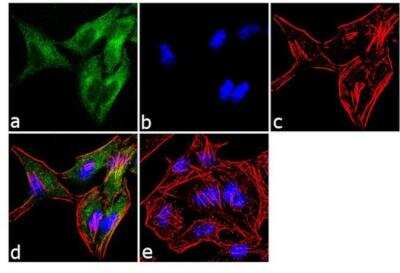
Dynamin 2 Antibody [NB300-617] - Immunofluorescence analysis of Dynamin II was performed using 70% confluent log phase HeLa cells. The cells were fixed with 4% paraformaldehyde for 10 minutes, permeabilized with 0.1% Triton(TM) X-100 for 10 minutes, and blocked with 1% BSA for 1 hour at room temperature. The cells were labeled with Dynamin II Rabbit Polyclonal Antibody at 2 ug/mL in 0.1% BSA and incubated for 3 hours at room temperature and then labeled with Goat anti-Rabbit IgG (H+L) Superclonal(TM) Secondary Antibody, Alexa Fluor(R) 488 conjugate for 45 minutes at room temperature (Panel a: green). Nuclei (Panel b: blue) were stained with SlowFade(R) Gold Antifade Mountant with DAPI. F-actin (Panel c: red) was stained with Alexa Fluor(R) 555 Rhodamine Phalloidin. Panel d represents the merged image showing cytoplasmic localization. Panel e shows the no primary antibody control.
Applications for Dynamin 2 Antibody
Application
Recommended Usage
Western Blot
1:1000
Application Notes
WB: Detects an approx. 100 kDa protein representing Dynamin 2 from HeLa cell lysate.
Formulation, Preparation, and Storage
Purification
Protein G purified
Formulation
PBS with 1 mg/ml BSA
Preservative
0.05% Sodium Azide
Concentration
1 mg/ml
Shipping
The product is shipped with polar packs. Upon receipt, store it immediately at the temperature recommended below.
Stability & Storage
Store at -20C. Avoid freeze-thaw cycles.
Background: Dynamin 2
Long Name
Dynamin-2
Alternate Names
DNM2, DYN2
Gene Symbol
DNM2
Additional Dynamin 2 Products
Product Documents for Dynamin 2 Antibody
Certificate of Analysis
To download a Certificate of Analysis, please enter a lot or batch number in the search box below.
Product Specific Notices for Dynamin 2 Antibody
This product is for research use only and is not approved for use in humans or in clinical diagnosis. Primary Antibodies are guaranteed for 1 year from date of receipt.
Citations for Dynamin 2 Antibody
 Powered by Bioz
Customer Reviews for Dynamin 2 Antibody
There are currently no reviews for this product. Be the first to review Dynamin 2 Antibody and earn rewards!
Have you used Dynamin 2 Antibody?
Submit a review and receive an Amazon gift card!
$25/€18/£15/$25CAN/¥2500 Yen for a review with an image
$10/€7/£6/$10CAN/¥1110 Yen for a review without an image
Submit a review
Protocols
Find general support by application which include: protocols, troubleshooting, illustrated assays, videos and webinars.
- Antigen Retrieval Protocol (PIER)
- Antigen Retrieval for Frozen Sections Protocol
- Appropriate Fixation of IHC/ICC Samples
- Cellular Response to Hypoxia Protocols
- Chromogenic IHC Staining of Formalin-Fixed Paraffin-Embedded (FFPE) Tissue Protocol
- Chromogenic Immunohistochemistry Staining of Frozen Tissue
- ClariTSA™ Fluorophore Kits
- Detection & Visualization of Antibody Binding
- Fluorescent IHC Staining of Frozen Tissue Protocol
- Graphic Protocol for Heat-induced Epitope Retrieval
- Graphic Protocol for the Preparation and Fluorescent IHC Staining of Frozen Tissue Sections
- Graphic Protocol for the Preparation and Fluorescent IHC Staining of Paraffin-embedded Tissue Sections
- Graphic Protocol for the Preparation of Gelatin-coated Slides for Histological Tissue Sections
- ICC Cell Smear Protocol for Suspension Cells
- ICC Immunocytochemistry Protocol Videos
- ICC for Adherent Cells
- IHC Sample Preparation (Frozen sections vs Paraffin)
- Immunocytochemistry (ICC) Protocol
- Immunocytochemistry Troubleshooting
- Immunofluorescence of Organoids Embedded in Cultrex Basement Membrane Extract
- Immunofluorescent IHC Staining of Formalin-Fixed Paraffin-Embedded (FFPE) Tissue Protocol
- Immunohistochemistry (IHC) and Immunocytochemistry (ICC) Protocols
- Immunohistochemistry Frozen Troubleshooting
- Immunohistochemistry Paraffin Troubleshooting
- Preparing Samples for IHC/ICC Experiments
- Preventing Non-Specific Staining (Non-Specific Binding)
- Primary Antibody Selection & Optimization
- Protocol for Heat-Induced Epitope Retrieval (HIER)
- Protocol for Making a 4% Formaldehyde Solution in PBS
- Protocol for VisUCyte™ HRP Polymer Detection Reagent
- Protocol for the Fluorescent ICC Staining of Cell Smears - Graphic
- Protocol for the Fluorescent ICC Staining of Cultured Cells on Coverslips - Graphic
- Protocol for the Preparation & Fixation of Cells on Coverslips
- Protocol for the Preparation and Chromogenic IHC Staining of Frozen Tissue Sections
- Protocol for the Preparation and Chromogenic IHC Staining of Frozen Tissue Sections - Graphic
- Protocol for the Preparation and Chromogenic IHC Staining of Paraffin-embedded Tissue Sections
- Protocol for the Preparation and Chromogenic IHC Staining of Paraffin-embedded Tissue Sections - Graphic
- Protocol for the Preparation and Fluorescent ICC Staining of Cells on Coverslips
- Protocol for the Preparation and Fluorescent ICC Staining of Non-adherent Cells
- Protocol for the Preparation and Fluorescent ICC Staining of Stem Cells on Coverslips
- Protocol for the Preparation and Fluorescent IHC Staining of Frozen Tissue Sections
- Protocol for the Preparation and Fluorescent IHC Staining of Paraffin-embedded Tissue Sections
- Protocol for the Preparation of Gelatin-coated Slides for Histological Tissue Sections
- Protocol for the Preparation of a Cell Smear for Non-adherent Cell ICC - Graphic
- R&D Systems Quality Control Western Blot Protocol
- TUNEL and Active Caspase-3 Detection by IHC/ICC Protocol
- The Importance of IHC/ICC Controls
- Troubleshooting Guide: Immunohistochemistry
- Troubleshooting Guide: Western Blot Figures
- Western Blot Conditions
- Western Blot Protocol
- Western Blot Protocol for Cell Lysates
- Western Blot Troubleshooting
- Western Blot Troubleshooting Guide
- View all Protocols, Troubleshooting, Illustrated assays and Webinars
Loading...