EDNRA/Endothelin R Type A Antibody - Azide and BSA Free
Novus Biologicals | Catalog # NB600-836
![Western Blot: EDNRA/Endothelin R Type A Antibody [NB600-836]](https://resources.rndsystems.com/images/products/EDNRA-Endothelin-R-Type-A-Antibody-Western-Blot-NB600-836-img0006.jpg "Western Blot: EDNRA/Endothelin R Type A Antibody [NB600-836]")
Loading...
Key Product Details
Species Reactivity
Validated:
Mouse
Cited:
Mouse
Applications
Validated:
Immunohistochemistry, Immunohistochemistry-Paraffin, Western Blot, Immunocytochemistry/ Immunofluorescence, Immunoprecipitation
Cited:
Immunocytochemistry/ Immunofluorescence, IF/IHC
Label
Unconjugated
Antibody Source
Polyclonal Sheep IgG
Format
Azide and BSA Free
Loading...
Product Specifications
Immunogen
Synthetic peptide corresponding to amino acids 410-422 (QEQNHNTERSSHK) of Rat Endothelin A Receptor, conjugated to KLH.
Localization
Plasma membrane
Specificity
This recognizes epitopes on the cytoplasmic side of the receptor and will not block the binding of endothelins. Therefore, the cell membrane must be permeable to the.
Clonality
Polyclonal
Host
Sheep
Isotype
IgG
Scientific Data Images for EDNRA/Endothelin R Type A Antibody - Azide and BSA Free
Western Blot: EDNRA/Endothelin R Type A Antibody [NB600-836]
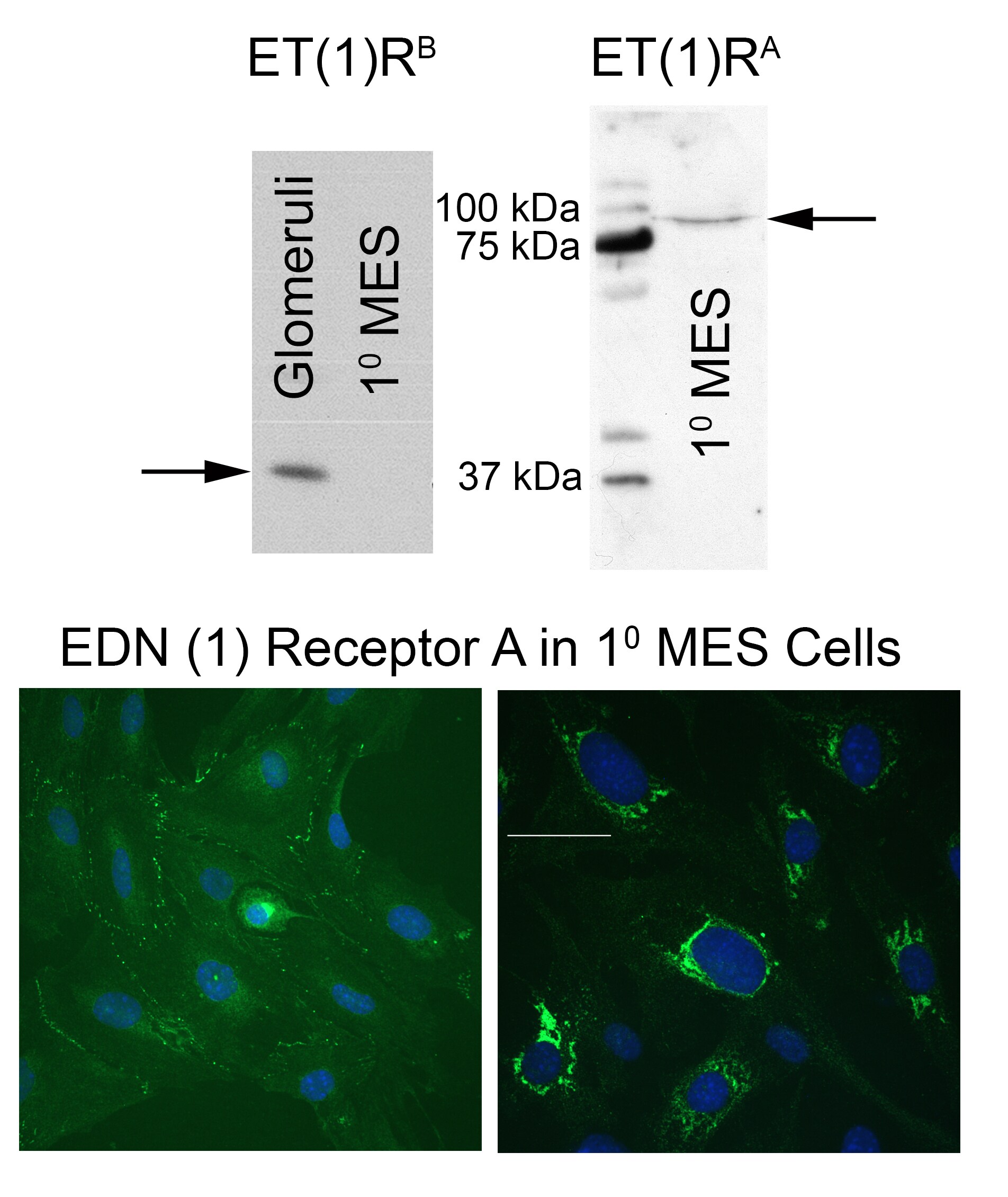
Western Blot: EDNRA/Endothelin R Type A Antibody [NB600-836] - EDN 1 Receptor A in 1 MES Cells. Western Provided by verified customer review.![Immunocytochemistry/ Immunofluorescence: EDNRA/Endothelin R Type A Antibody [NB600-836]](https://resources.rndsystems.com/images/products/EDNRA-Endothelin-R-Type-A-Antibody-Immunocytochemistry-Immunofluorescence-NB600-836-img0004.jpg "Immunocytochemistry/ Immunofluorescence: EDNRA/Endothelin R Type A Antibody [NB600-836]")
Immunocytochemistry/ Immunofluorescence: EDNRA/Endothelin R Type A Antibody [NB600-836]
Immunocytochemistry/Immunofluorescence: EDNRA/Endothelin R Type A Antibody [NB600-836] - EDN 1 Receptor A in 1 MES Cells. Immunofluorescent staining image provided by verified customer review.![Western Blot: EDNRA/Endothelin R Type A Antibody [NB600-836]](https://resources.rndsystems.com/images/products/EDNRA-Endothelin-R-Type-A-Antibody-Western-Blot-NB600-836-img0005.jpg "Western Blot: EDNRA/Endothelin R Type A Antibody [NB600-836]")
Western Blot: EDNRA/Endothelin R Type A Antibody [NB600-836]
Western Blot: EDNRA/Endothelin R Type A Antibody [NB600-836] - ET-1 A & B Receptor Western provided by verified customer review.Applications for EDNRA/Endothelin R Type A Antibody - Azide and BSA Free
Application
Recommended Usage
Immunocytochemistry/ Immunofluorescence
1:10-1:500
Immunohistochemistry
10-20 ug/ml
Immunohistochemistry-Paraffin
10-20 ug/ml
Immunoprecipitation
20-40 ug/ml
Western Blot
1:100-1:2000
Application Notes
EDNRA/Endothelin R Type A antibody validated for ICC/IF, WB from verified customer reviews.
Reviewed Applications
Read 1 review rated 4 using NB600-836 in the following applications:
Formulation, Preparation, and Storage
Purification
Protein G purified
Formulation
PBS
Format
Azide and BSA Free
Preservative
No Preservative
Concentration
Please see the vial label for concentration. If unlisted please contact technical services.
Shipping
The product is shipped with polar packs. Upon receipt, store it immediately at the temperature recommended below.
Stability & Storage
Aliquot and store at -20C or -80C. Avoid freeze-thaw cycles.
Background: EDNRA/Endothelin R Type A
Long Name
Endothelin Receptor Type A
Alternate Names
EDNRA, ETAR, ETRA
Gene Symbol
EDNRA
Additional EDNRA/Endothelin R Type A Products
Product Documents for EDNRA/Endothelin R Type A Antibody - Azide and BSA Free
Certificate of Analysis
To download a Certificate of Analysis, please enter a lot or batch number in the search box below.
Product Specific Notices for EDNRA/Endothelin R Type A Antibody - Azide and BSA Free
This product is for research use only and is not approved for use in humans or in clinical diagnosis. Primary Antibodies are guaranteed for 1 year from date of receipt.
Related Research Areas
Citations for EDNRA/Endothelin R Type A Antibody - Azide and BSA Free
 Powered by Bioz
Customer Reviews for EDNRA/Endothelin R Type A Antibody - Azide and BSA Free (1)
4 out of 5
1 Customer Rating
Have you used EDNRA/Endothelin R Type A Antibody - Azide and BSA Free?
Submit a review and receive an Amazon gift card!
$25/€18/£15/$25CAN/¥2500 Yen for a review with an image
$10/€7/£6/$10CAN/¥1110 Yen for a review without an image
Submit a review
Customer Images
Showing
1
-
1 of
1 review
Showing All
Filter By:
-
Application: Western BlotSample Tested: mesangial cell lysate glomerular lysateSpecies: MouseVerified Customer | Posted 02/11/2014Application: Western Blot, Sample Tested:mesangial cell lysate glomerular lysate, Catalog Num:NB600-836

There are no reviews that match your criteria.
Protocols
Find general support by application which include: protocols, troubleshooting, illustrated assays, videos and webinars.
- Antigen Retrieval Protocol (PIER)
- Antigen Retrieval for Frozen Sections Protocol
- Appropriate Fixation of IHC/ICC Samples
- Cellular Response to Hypoxia Protocols
- Chromogenic IHC Staining of Formalin-Fixed Paraffin-Embedded (FFPE) Tissue Protocol
- Chromogenic Immunohistochemistry Staining of Frozen Tissue
- ClariTSA™ Fluorophore Kits
- Detection & Visualization of Antibody Binding
- Fluorescent IHC Staining of Frozen Tissue Protocol
- Graphic Protocol for Heat-induced Epitope Retrieval
- Graphic Protocol for the Preparation and Fluorescent IHC Staining of Frozen Tissue Sections
- Graphic Protocol for the Preparation and Fluorescent IHC Staining of Paraffin-embedded Tissue Sections
- Graphic Protocol for the Preparation of Gelatin-coated Slides for Histological Tissue Sections
- ICC Cell Smear Protocol for Suspension Cells
- ICC Immunocytochemistry Protocol Videos
- ICC for Adherent Cells
- IHC Sample Preparation (Frozen sections vs Paraffin)
- Immunocytochemistry (ICC) Protocol
- Immunocytochemistry Troubleshooting
- Immunofluorescence of Organoids Embedded in Cultrex Basement Membrane Extract
- Immunofluorescent IHC Staining of Formalin-Fixed Paraffin-Embedded (FFPE) Tissue Protocol
- Immunohistochemistry (IHC) and Immunocytochemistry (ICC) Protocols
- Immunohistochemistry Frozen Troubleshooting
- Immunohistochemistry Paraffin Troubleshooting
- Immunoprecipitation Protocol
- Preparing Samples for IHC/ICC Experiments
- Preventing Non-Specific Staining (Non-Specific Binding)
- Primary Antibody Selection & Optimization
- Protocol for Heat-Induced Epitope Retrieval (HIER)
- Protocol for Making a 4% Formaldehyde Solution in PBS
- Protocol for VisUCyte™ HRP Polymer Detection Reagent
- Protocol for the Fluorescent ICC Staining of Cell Smears - Graphic
- Protocol for the Fluorescent ICC Staining of Cultured Cells on Coverslips - Graphic
- Protocol for the Preparation & Fixation of Cells on Coverslips
- Protocol for the Preparation and Chromogenic IHC Staining of Frozen Tissue Sections
- Protocol for the Preparation and Chromogenic IHC Staining of Frozen Tissue Sections - Graphic
- Protocol for the Preparation and Chromogenic IHC Staining of Paraffin-embedded Tissue Sections
- Protocol for the Preparation and Chromogenic IHC Staining of Paraffin-embedded Tissue Sections - Graphic
- Protocol for the Preparation and Fluorescent ICC Staining of Cells on Coverslips
- Protocol for the Preparation and Fluorescent ICC Staining of Non-adherent Cells
- Protocol for the Preparation and Fluorescent ICC Staining of Stem Cells on Coverslips
- Protocol for the Preparation and Fluorescent IHC Staining of Frozen Tissue Sections
- Protocol for the Preparation and Fluorescent IHC Staining of Paraffin-embedded Tissue Sections
- Protocol for the Preparation of Gelatin-coated Slides for Histological Tissue Sections
- Protocol for the Preparation of a Cell Smear for Non-adherent Cell ICC - Graphic
- R&D Systems Quality Control Western Blot Protocol
- TUNEL and Active Caspase-3 Detection by IHC/ICC Protocol
- The Importance of IHC/ICC Controls
- Troubleshooting Guide: Immunohistochemistry
- Troubleshooting Guide: Western Blot Figures
- Western Blot Conditions
- Western Blot Protocol
- Western Blot Protocol for Cell Lysates
- Western Blot Troubleshooting
- Western Blot Troubleshooting Guide
- View all Protocols, Troubleshooting, Illustrated assays and Webinars
Loading...