Eg5 Antibody - Azide and BSA Free
Novus Biologicals | Catalog # NB500-181
![Western Blot: Eg5 Antibody [NB500-181]](https://resources.rndsystems.com/images/products/Eg5-Antibody-Western-Blot-NB500-181-img0006.jpg "Western Blot: Eg5 Antibody [NB500-181]")
![Western Blot: Eg5 Antibody [NB500-181]](https://resources.rndsystems.com/images/products/Eg5-Antibody-Western-Blot-NB500-181-img0005.jpg "Western Blot: Eg5 Antibody [NB500-181]")
Loading...
Key Product Details
Validated by
Biological Validation
Species Reactivity
Validated:
Human, Mouse, Rat, Porcine, Drosophila, Mammal, Plant
Cited:
Human, Mouse, Porcine, Insect - Drosophila, Plant
Applications
Validated:
Western Blot, Immunoblotting, Immunocytochemistry/ Immunofluorescence, Immunoprecipitation
Cited:
Western Blot, Immunocytochemistry/ Immunofluorescence, Immunoprecipitation
Label
Unconjugated
Antibody Source
Polyclonal Rabbit IgG
Format
Azide and BSA Free
Loading...
Product Specifications
Immunogen
A recombinant segment of the coiled-coil domain of human Eg5 [Uniprot: P52732]
Reactivity Notes
Predicted to react with most mammalian species. Drosophila reactivity reported in scientific literature (PMID: 23888285). Mouse reactivity reported in scientific literature (PMID: 27117404). Plant reactivity reported in scientific literature (PMID: 19527496).
Clonality
Polyclonal
Host
Rabbit
Isotype
IgG
Theoretical MW
125 kDa.
Disclaimer note: The observed molecular weight of the protein may vary from the listed predicted molecular weight due to post translational modifications, post translation cleavages, relative charges, and other experimental factors.
Disclaimer note: The observed molecular weight of the protein may vary from the listed predicted molecular weight due to post translational modifications, post translation cleavages, relative charges, and other experimental factors.
Scientific Data Images for Eg5 Antibody - Azide and BSA Free
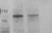
Western Blot: Eg5 Antibody [NB500-181]
Western Blot: Eg5 Antibody [NB500-181] - Cells were transfected with the pCMV6-ENTRY control or pCMV6-ENTRY KIF11 cDNA for 48 hrs and lysed. Equivalent amounts of cell lysates (5 ug per lane) were separated by SDS-PAGE and immunoblotted with anti-Eg5.![Immunocytochemistry/ Immunofluorescence: Eg5 Antibody [NB500-181]](https://resources.rndsystems.com/images/products/Eg5-Antibody-Immunocytochemistry-Immunofluorescence-NB500-181-img0003.jpg "Immunocytochemistry/ Immunofluorescence: Eg5 Antibody [NB500-181]")
Immunocytochemistry/ Immunofluorescence: Eg5 Antibody [NB500-181]
Immunocytochemistry/Immunofluorescence: Eg5 Antibody [NB500-181] - Staining of HeLa cells fixed in 3.5% paraformaldehyde. Cytoplasmic staining during interphase and spindle staining during mitosis.
Immunocytochemistry/ Immunofluorescence: Eg5 Antibody [NB500-181] -
Immunocytochemistry/ Immunofluorescence: Eg5 Antibody [NB500-181] - Spontaneous assembly of rosettes in iChM5 derivatives.Feeder free cultures iChM5Ap15 & iChM5Bp28 cells in chamber slides were immunostained as indicated. Left panel, emerging rosettes among self-renewing iPSCs; grayscale inset shows 1x magnification of immunostaining of Oct4 alone. Middle panel shows Nanog staining alone with inset at 2X magnification showing presumptive centrosomes (arrows). Bottom left panel shows forming rosettes immunopositive for Sox2 & Eg5. Bottom right panel shows low magnification image of immunostaining of Sox2 in this iChM5Ap15 culture, showing that virtually all cells were immunopositive. Grey scale inset shows representative forming rosette. Asterisks (*) in each panel indicates example of forming rosette. Scale bar, 50 microns. Image collected & cropped by CiteAb from the following publication (https://pubmed.ncbi.nlm.nih.gov/25426336), licensed under a CC-BY license. Not internally tested by Novus Biologicals.Applications for Eg5 Antibody - Azide and BSA Free
Application
Recommended Usage
Immunoblotting
reported in scientific literature (PMID 28392145)
Immunocytochemistry/ Immunofluorescence
1:1000
Immunoprecipitation
1:10-1:500
Western Blot
1:1000
Application Notes
A band at ~125 kDa can be detected by Western blot.
Reviewed Applications
Read 2 reviews rated 2.5 using NB500-181 in the following applications:
Formulation, Preparation, and Storage
Purification
Unpurified
Formulation
Whole antisera
Format
Azide and BSA Free
Preservative
No Preservative
Concentration
This product is unpurified. The exact concentration of antibody is not quantifiable.
Shipping
The product is shipped with polar packs. Upon receipt, store it immediately at the temperature recommended below.
Stability & Storage
Aliquot and store at -20C or -80C. Avoid freeze-thaw cycles.
Background: Eg5
Long Name
Kinesin-like protein KIF11
Alternate Names
KIF11, KNSL1, TRIP-5, TRIP5
Gene Symbol
KIF11
UniProt
Additional Eg5 Products
Product Documents for Eg5 Antibody - Azide and BSA Free
Certificate of Analysis
To download a Certificate of Analysis, please enter a lot or batch number in the search box below.
Product Specific Notices for Eg5 Antibody - Azide and BSA Free
This product is for research use only and is not approved for use in humans or in clinical diagnosis. Primary Antibodies are guaranteed for 1 year from date of receipt.
Citations for Eg5 Antibody - Azide and BSA Free
 Powered by Bioz
Customer Reviews for Eg5 Antibody - Azide and BSA Free (2)
2.5 out of 5
2 Customer Ratings
Have you used Eg5 Antibody - Azide and BSA Free?
Submit a review and receive an Amazon gift card!
$25/€18/£15/$25CAN/¥2500 Yen for a review with an image
$10/€7/£6/$10CAN/¥1110 Yen for a review without an image
Submit a review
Customer Images

Showing
1
-
2 of
2 reviews
Showing All
Filter By:
-
Application: Western BlotSample Tested: HeLa, Mouse brainSpecies: HumanVerified Customer | Posted 04/28/2014Control and a knockdown sample probed with Eg5

-
Application: Western BlotSample Tested: Mouse tissues, MiaPaCa2, BxPC3, T3M4Species: HumanVerified Customer | Posted 12/16/2013
There are no reviews that match your criteria.
Protocols
Find general support by application which include: protocols, troubleshooting, illustrated assays, videos and webinars.
- Appropriate Fixation of IHC/ICC Samples
- Cellular Response to Hypoxia Protocols
- ClariTSA™ Fluorophore Kits
- Detection & Visualization of Antibody Binding
- ICC Cell Smear Protocol for Suspension Cells
- ICC Immunocytochemistry Protocol Videos
- ICC for Adherent Cells
- Immunocytochemistry (ICC) Protocol
- Immunocytochemistry Troubleshooting
- Immunofluorescence of Organoids Embedded in Cultrex Basement Membrane Extract
- Immunohistochemistry (IHC) and Immunocytochemistry (ICC) Protocols
- Immunoprecipitation Protocol
- Preparing Samples for IHC/ICC Experiments
- Preventing Non-Specific Staining (Non-Specific Binding)
- Primary Antibody Selection & Optimization
- Protocol for VisUCyte™ HRP Polymer Detection Reagent
- Protocol for the Fluorescent ICC Staining of Cell Smears - Graphic
- Protocol for the Fluorescent ICC Staining of Cultured Cells on Coverslips - Graphic
- Protocol for the Preparation and Fluorescent ICC Staining of Cells on Coverslips
- Protocol for the Preparation and Fluorescent ICC Staining of Non-adherent Cells
- Protocol for the Preparation and Fluorescent ICC Staining of Stem Cells on Coverslips
- Protocol for the Preparation of a Cell Smear for Non-adherent Cell ICC - Graphic
- R&D Systems Quality Control Western Blot Protocol
- TUNEL and Active Caspase-3 Detection by IHC/ICC Protocol
- The Importance of IHC/ICC Controls
- Troubleshooting Guide: Western Blot Figures
- Western Blot Conditions
- Western Blot Protocol
- Western Blot Protocol for Cell Lysates
- Western Blot Troubleshooting
- Western Blot Troubleshooting Guide
- View all Protocols, Troubleshooting, Illustrated assays and Webinars
Loading...