EGLN3/PHD3 Antibody - BSA Free
Novus Biologicals | Catalog # NB100-139
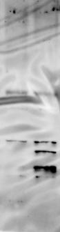
![Western Blot: EGLN3/PHD3 Antibody - BSA Free [NB100-139]](https://resources.rndsystems.com/images/products/EGLN3-PHD3-Antibody-Knockdown-Validated-NB100-139-img0017.jpg "Western Blot: EGLN3/PHD3 Antibody - BSA Free [NB100-139]")
Key Product Details
Validated by
Knockout/Knockdown
Species Reactivity
Validated:
Human, Mouse, Rat
Cited:
Human, Mouse, Rat
Applications
Validated:
Immunohistochemistry, Immunohistochemistry-Paraffin, Western Blot, Immunocytochemistry/ Immunofluorescence, Immunoprecipitation, Chromatin Immunoprecipitation, Mass Spectrometry, Electron Microscopy, Knockdown Validated
Cited:
Immunohistochemistry-Paraffin, Western Blot, Immunocytochemistry/ Immunofluorescence, Immunoprecipitation, Mass Spectrometry, Electron Microscopy
Label
Unconjugated
Antibody Source
Polyclonal Rabbit IgG
Format
BSA Free
Loading...
Product Specifications
Immunogen
Synthetic peptide corresponding to residues between 50-100 of human PHD3/HIF Prolyl Hydroxylase 3 using the numbering given in entry NP_071356.1 (GeneID 112399).
Reactivity Notes
Mouse reactivity reported in scientific literature (PMID: 24037093)
Clonality
Polyclonal
Host
Rabbit
Isotype
IgG
Theoretical MW
28 kDa.
Disclaimer note: The observed molecular weight of the protein may vary from the listed predicted molecular weight due to post translational modifications, post translation cleavages, relative charges, and other experimental factors.
Disclaimer note: The observed molecular weight of the protein may vary from the listed predicted molecular weight due to post translational modifications, post translation cleavages, relative charges, and other experimental factors.
Scientific Data Images for EGLN3/PHD3 Antibody - BSA Free
![Immunohistochemistry-Paraffin: EGLN3/PHD3 Antibody - BSA Free [NB100-139]](https://resources.rndsystems.com/images/products/EGLN3-PHD3-Antibody-Immunohistochemistry-Paraffin-NB100-139-img0010.jpg "Immunohistochemistry-Paraffin: EGLN3/PHD3 Antibody - BSA Free [NB100-139]")
Immunohistochemistry-Paraffin: EGLN3/PHD3 Antibody - BSA Free [NB100-139]
Immunohistochemistry-Paraffin: EGLN3/PHD3 Antibody [NB100-139] - Analysis of a formalin fixed tissue section of human renal cell carcinoma (clear cell type) using rabbit polyclonal EGLN3/PHD3 antibody with HRP-DAB detection and hematoxylin counterstaining. The antibody generated a strong nuclear staining of PHD3 primarily in the cancer cells while the stromal cells were largely negative for this protein.![Knockdown Validated: EGLN3/PHD3 Antibody - BSA Free [NB100-139]](https://resources.rndsystems.com/images/products/EGLN3-PHD3-Antibody-Knockdown-Validated-NB100-139-img0019.jpg "Western Blot: EGLN3/PHD3 Antibody - BSA Free [NB100-139]")
![Immunocytochemistry/ Immunofluorescence: EGLN3/PHD3 Antibody - BSA Free [NB100-139]](https://resources.rndsystems.com/images/products/EGLN3-PHD3-Antibody-Immunocytochemistry-Immunofluorescence-NB100-139-img0014.jpg "Immunocytochemistry/ Immunofluorescence: EGLN3/PHD3 Antibody - BSA Free [NB100-139]")
Immunocytochemistry/ Immunofluorescence: EGLN3/PHD3 Antibody - BSA Free [NB100-139]
Immunocytochemistry/Immunofluorescence: EGLN3/PHD3 Antibody [NB100-139] - EGLN3/PHD3 antibody was tested in HeLa cells with Dylight 488 (green). Nuclei and alpha-tubulin were counterstained with DAPI (blue) and Dylight 550 (red).![Knockdown Validated: EGLN3/PHD3 Antibody - BSA Free [NB100-139]](https://resources.rndsystems.com/images/products/EGLN3-PHD3-Antibody-Knockdown-Validated-NB100-139-img0016.jpg "Western Blot: EGLN3/PHD3 Antibody - BSA Free [NB100-139]")
![Western Blot: EGLN3/PHD3 AntibodyBSA Free [NB100-139]](https://resources.rndsystems.com/images/products/EGLN3-PHD3-Antibody-Western-Blot-NB100-139-img0018.jpg "Western Blot: EGLN3/PHD3 AntibodyBSA Free [NB100-139]")
Western Blot: EGLN3/PHD3 AntibodyBSA Free [NB100-139]
EGLN3-PHD3-Antibody-Western-Blot-NB100-139-img0018.jpg![Western Blot: EGLN3/PHD3 AntibodyBSA Free [NB100-139]](https://resources.rndsystems.com/images/products/EGLN3-PHD3-Antibody-Western-Blot-NB100-139-img0013.jpg "Western Blot: EGLN3/PHD3 AntibodyBSA Free [NB100-139]")
Western Blot: EGLN3/PHD3 AntibodyBSA Free [NB100-139]
Western Blot: EGLN3/PHD3 Antibody [NB100-139] - Analysis of HeLa lysates using NB100-139. Image courtesy of Dr Gregg Semenza (The Johns Hopkins University School of Medicine, Baltimore, MD USA)
Knockdown Validated: EGLN3/PHD3 Antibody - BSA Free [NB100-139] -
Cell cycle block under PHD3 depletion is accompanied by p27 induction. a PHD3 depletion induces a cell cycle block in G0/G1. HeLa & renal cell adenocarcinoma cells (786-O) were transfected with control (siScr) or PHD3 targeted (siPHD3) siRNA followed by synchronization at G0 & 24-h hypoxic exposure. Cell cycle progression was monitored by FACS analysis 8 h after cell cycle release. The combined means of three independent experiments are presented (±SEM) shown in the tables below. b PHD3 depletion induces p27 expression in HeLa cells & in 786-O cells under hypoxia (1 % O2) & normoxia (21 % O2) by siPHD3 & independent adenoviral shRNA against PHD3. p21 or p16 expression is not elevated by PHD3 knockdown. c Depletion of either PHD1 or PHD2 by siRNA does not increase p27 expression in 786-O cells Image collected & cropped by CiteAb from the following publication (https://pubmed.ncbi.nlm.nih.gov/26223520), licensed under a CC-BY license. Not internally tested by Novus Biologicals.
Western Blot: EGLN3/PHD3 Antibody - BSA Free [NB100-139] -
Representative picture of western blot in histopathologically unchanged tissue (N) & primary cancerous tissue (C) from patients with CRC. Immunodetection of bands was performed with Rp anti- PHD1, - PHD2, - PHD3 & - FIH Ab, followed by incubation with goat anti-rabbit HRP-conjugated Ab. The membrane was stripped & incubated with Rp anti-GAPDH Ab, followed by incubation with goat anti-rabbit HRP-conjugated Ab. Bands were revealed using SuperSignal West Femto Chemiluminescent Substrate, Thermo Fisher Scientific (Rockford, IL) & Biospectrum® Imaging System 500, UVP Ltd. (Upland, CA). Image collected & cropped by CiteAb from the following publication (https://pubmed.ncbi.nlm.nih.gov/24195777), licensed under a CC-BY license. Not internally tested by Novus Biologicals.
Knockdown Validated: EGLN3/PHD3 Antibody - BSA Free [NB100-139] -
PHD3 elevates p27 expression through a post-translational mechanism. a PHD3 depletion has no effect on p27 transcription under hypoxia. p27 mRNA levels were measured in HeLa cells using quantitative real-time PCR. Results shown as fold change vs normoxic control, four independent experiments (± SEM) (p = n.s.; n = 4). b Hypoxic p27 expression is HIF-1 alpha & EPAS1/HIF-2 alpha independent. QRT-PCR analysis of p27 & hypoxia-inducible glut-1 mRNA normalized to beta -actin using the indicated double knockdown after 24 h of hypoxia. Unlike glut-1 HIF knockdown has little effect on p27 transcription. Results from three independent experiments (±SEM) are shown (p = n.s.; n = 3). c PHD3 depletion induces p27 protein levels independently of HIF-1 alpha or EPAS1/HIF-2 alpha depletion in HeLa cells. d Quantification of p27 protein expression using indicated double knockdown. Results from three independent experiments (±SEM) are shown Image collected & cropped by CiteAb from the following publication (https://pubmed.ncbi.nlm.nih.gov/26223520), licensed under a CC-BY license. Not internally tested by Novus Biologicals.
Western Blot: EGLN3/PHD3 Antibody - BSA Free [NB100-139] -
PHD3 depletion stabilizes hypoxic p27 expression by increasing p27 half-life. a Cell cycle arrest at G0 & subsequent release shows an increase of p27 expression in siPHD3 exposed cells. b Quantification for p27 expression under PHD3 depletion at indicated time points after cell cycle release in HeLa & 786-O cells. Asterisk indicates significant difference (p < 0,05; n = 3). c Cell cycle arrest at G0 & inhibition of protein synthesis with cycloheximide indicate increased p27 stability in PHD3 depleted HeLa cells. d Quantification of p27 expression using siPHD3 or control at indicated time points. Four independent experiments (± SEM) are shown (p < 0,05; n = 4). e Analysis of p27 stability in 786–0 cells by cycloheximide chase during reoxygenation after 24 h hypoxia demonstrates markedly increased half-life of p27 upon PHD3 depletion Image collected & cropped by CiteAb from the following publication (https://pubmed.ncbi.nlm.nih.gov/26223520), licensed under a CC-BY license. Not internally tested by Novus Biologicals.
Western Blot: EGLN3/PHD3 Antibody - BSA Free [NB100-139] -
Western Blot: EGLN3/PHD3 Antibody - BSA Free [NB100-139] - Cell cycle block in PHD3 depleted cells is p27-dependent. a Reduced cell proliferation as judged by reduced cell amount of hypoxic siPHD3-depleted HeLa cells is rescued with concomitant p27 knockdown. b Quantification of cell survival from four independent experiments with 4–5 optical views per experiment. Asterisk indicates significant difference (p = 0,05; n = 4). Protein expression was monitored with western blotting from parallel samples. c Knockdown of p27 restores the effect of PHD3 depletion on cell cycle. HeLa cells exposed to the indicated double knockdown followed by 6 h hypoxia & FACS analysis. d Quantification of cell cycle phases from three independent experiments using indicated double knockdown Image collected & cropped by CiteAb from the following publication (https://pubmed.ncbi.nlm.nih.gov/26223520), licensed under a CC-BY license. Not internally tested by Novus Biologicals.
Western Blot: EGLN3/PHD3 Antibody - BSA Free [NB100-139] -
Western Blot: EGLN3/PHD3 Antibody - BSA Free [NB100-139] - 5-dAzaC effect on PHD3 transcript (A) & protein (B) levels in HCT116 & DLD-1 CRC cells. HCT116 & DLD-1 cells were cultured in DMEM for 6, 24 & 48 h either in the absence or in the presence of 5-dAzaC at a concentration of 1.00 or 5.00 μM under hypoxic or normoxic conditions. After incubation the cells were used for total RNA isolation & protein isolation. Total RNA was reverse-transcribed, & PHD3 cDNA levels were determined by RQ-PCR relative quantification analysis. RQ-PCR results were standardized by the geometric mean of PBGD & hMRPL19 cDNA levels. PHD3 cDNA levels are expressed as a multiplicity of the respective controls. Each sample was determined in triplicate & results are presented as the mean ± SE from three experiments *P < 0.05. The cell protein was separated by 10% SDS-PAGE, & transferred to a membrane that was then immunoblotted with Rp anti - PHD3 Ab & incubated with goat anti-rabbit HRP-conjugated Ab. The membrane was then stripped & reblotted with Rp anti-GAPDH Ab, followed by incubation with goat anti-rabbit HRP-conjugated Ab. The band densitometry readings were normalized to GAPDH loading control. The ratio PHD3 to GAPDH for control was assumed to be 1. Image collected & cropped by CiteAb from the following publication (https://pubmed.ncbi.nlm.nih.gov/24195777), licensed under a CC-BY license. Not internally tested by Novus Biologicals.
Western Blot: EGLN3/PHD3 Antibody - BSA Free [NB100-139] -
Western Blot: EGLN3/PHD3 Antibody - BSA Free [NB100-139] - DNA methylation & expression level of the PHD3 gene in HCT116 & DLD-1 CRC cells. A. HCT116 & DLD-1 cells were cultured under normoxic or hypoxic (1% O2) conditions for 48 hrs. Cells were then used for DNA isolation followed by bisulfite modification. Methylation percentage of three DNA fragments within the PHD3 CpG island (Additional file 1, Additional file 2) in HCT116 & DLD-1 cells under hypoxic & normoxic conditions was determined by Real Time PCR amplification of bisulfite treated standard & cell line DNA, followed by comparison of their HRM profiles. B. Cells were cultured in DMEM either in hypoxic (1%O2) or normoxic conditions for 48 hrs. After incubation, the cells were used for total RNA isolation & reverse transcription. The PHD3 cDNA levels were determined by RQ-PCR relative quantification analysis. RQ-PCR results were standardized by the geometric mean of PBGD & hMRPL19 cDNA levels. PHD3 cDNA levels are expressed as a multiplicity of these cDNA copies in the cell line’s calibrator. C. Cells were cultured in DMEM either in hypoxic (1%O2) (H) or normoxic (N) conditions for 48 hrs. Cells were then used for protein isolation. Proteins were separated by 10% SDS-PAGE, & transferred to a membrane that was then immunoblotted with Rp anti - PHD3 Ab & incubated with goat anti-rabbit HRP-conjugated Ab. The membrane was then stripped & reblotted with Rp anti-GAPDH Ab, followed by incubation with goat anti-rabbit HRP-conjugated Ab. The band densitometry readings were normalized to GAPDH loading control. The ratio of PHD3 to GAPDH for DLD-1 in normoxic conditions was assumed to be 1. Image collected & cropped by CiteAb from the following publication (https://pubmed.ncbi.nlm.nih.gov/24195777), licensed under a CC-BY license. Not internally tested by Novus Biologicals.
Western Blot: EGLN3/PHD3 Antibody - BSA Free [NB100-139] -
Western Blot: EGLN3/PHD3 Antibody - BSA Free [NB100-139] - PHD3 depletion increases S10 phosphorylation of p27 in hypoxia. a Western blot analysis of p27 expression in cells arrested at different cell cycle phases & exposed to PHD3 or control siRNA. PHD3 depletion has no effect on T187 phosphorylation of p27 at any cell cycle phase. b PHD3 depletion has little effect on p27 phosphorylated on T157 & T198. c S10 phosphorylated form of p27 is strongly induced in PHD3 depleted cells. d Quantification of total & indicated p27 phosphoproteins from four independent experiments normalized to beta -actin. Out of the phosphoproteins the effect on the expression of p27S10 is most prominent. Asterisk indicates significant difference (p < 0,005; n = 4) (e) Quantification of indicated p27 phosphoproteins from four independent experiments normalized to total p27 expression. The effect on the expression of p27S10 is most prominent & comparable to the total p27 expression. Asterisk indicates significant difference (p < 0,05; n = 4). Only minor effect on T157 & T198 are seen Image collected & cropped by CiteAb from the following publication (https://pubmed.ncbi.nlm.nih.gov/26223520), licensed under a CC-BY license. Not internally tested by Novus Biologicals.
Western Blot: EGLN3/PHD3 Antibody - BSA Free [NB100-139] -
Western Blot: EGLN3/PHD3 Antibody - BSA Free [NB100-139] - p27S10 phosphorylation is required for PHD3 depletion induced increase in p27 half-life. a Quantification of p27S10 phosphorylation from three independent experiments in 786-O cells. S10 phosphorylation demonstrated 2-fold increase at 6 h after cell cycle release in PHD3 depleted cells. b Serum starved 786-O cells were released from G0 block & S10 phosphorylation was monitored at the indicated timepoints under PHD3 depletion. PHD3 depletion had a prominent effect on S10 phosphorylation of p27. c Verification of subcellular localization of p27 plasmids. Immunofluorescence staining of Flag-p27wt / Flag-p27S10A transfected HeLa cells after 24 h hypoxia. As expected, p27wt is localized more into cytoplasmic than nuclear compartment. p27S10A is more localized into nucleus than into cytoplasm. Quantification of the subcellular localization from three optical fields (40x) (N; nuclear, C; cytoplasmic). d Flag-p27wt & S10A phosphorylation-deficient mutant (p27S10A) were transfected into siRNA-exposed HeLa cells. After 24 h of hypoxia CHX chase was performed. p27wt demonstrates strongly increased stability in siPHD3 exposed cells whereas p27S10A did not show any increased stability Image collected & cropped by CiteAb from the following publication (https://pubmed.ncbi.nlm.nih.gov/26223520), licensed under a CC-BY license. Not internally tested by Novus Biologicals.
Western Blot: EGLN3/PHD3 Antibody - BSA Free [NB100-139] -
Western Blot: EGLN3/PHD3 Antibody - BSA Free [NB100-139] - Experimental setup & the effect of PHD3 silencing on 786-O proteome. a siRNA-mediated silencing of PHD3 protein level in 786-O ccRCC cell line using two individual siRNA sequences in normoxic & in hypoxic (1% O2) condition. Quantification of three biological replicates, mean ± SEM, fold change to control (Scr). Asterisk indicates a statistically significant difference (*p < 0.05, **p < 0.01). b siRNA-mediated silencing of PHD3 mRNA expression, quantification of three individual experiments. Mean ± SEM, fold change to Scr (***p < 0.001). c Flow chart of the experimental procedure. 786-O cells were transfected with siPHD3#1 or with a non-targeting control siRNA (Scr) for 24 h followed by a hypoxic (1% O2) or normoxic (21% O2) exposure. Three independent experiments were performed; proteins were extracted, followed by in-gel digestion with trypsin. Purified peptides were ran through mass spectrometer. Protein identification was done with Mascot database search. Protein quantification was carried out with Progenesis QI, followed by testing the differential expression between sample groups using peptide-level expression change averaging (PECA). d Western blot validations of selected proteins in 786-O & RCC4 cell lines with two individual siRNA sequences targeting PHD3, representative analyses are shown Image collected & cropped by CiteAb from the following publication (http://cancerandmetabolism.biomedcentral.com/articles/10.1186/s40170-01…), licensed under a CC-BY license. Not internally tested by Novus Biologicals.
Western Blot: EGLN3/PHD3 Antibody - BSA Free [NB100-139] -
Western Blot: EGLN3/PHD3 Antibody - BSA Free [NB100-139] - PHD3 depletion stabilizes hypoxic p27 expression by increasing p27 half-life. a Cell cycle arrest at G0 & subsequent release shows an increase of p27 expression in siPHD3 exposed cells. b Quantification for p27 expression under PHD3 depletion at indicated time points after cell cycle release in HeLa & 786-O cells. Asterisk indicates significant difference (p < 0,05; n = 3). c Cell cycle arrest at G0 & inhibition of protein synthesis with cycloheximide indicate increased p27 stability in PHD3 depleted HeLa cells. d Quantification of p27 expression using siPHD3 or control at indicated time points. Four independent experiments (± SEM) are shown (p < 0,05; n = 4). e Analysis of p27 stability in 786–0 cells by cycloheximide chase during reoxygenation after 24 h hypoxia demonstrates markedly increased half-life of p27 upon PHD3 depletion Image collected & cropped by CiteAb from the following publication (https://pubmed.ncbi.nlm.nih.gov/26223520), licensed under a CC-BY license. Not internally tested by Novus Biologicals.
Western Blot: EGLN3/PHD3 Antibody - BSA Free [NB100-139] -
Western Blot: EGLN3/PHD3 Antibody - BSA Free [NB100-139] - PHD3 inhibition reduces the amount of hyperphosphorylated Rb & increases p27 in hypoxia.(A) SCC2 cells were transfected with the indicated siRNAs & exposed to normoxia or hypoxia for 24 to 48 hours followed by western blot analysis of PHD3, phosphorylated Rb (p-Rb) & cyclin B1. (B) HeLa cells were transfected with the indicated siRNAs, synchronized & exposed to normoxia or hypoxia for 6 to 24 hours after release. PHD3, phosphorylated Rb (p-Rb) & cyclin D1 were analyzed from samples by western blotting. (C) SCC2 cells were transfected with the indicated siRNAs & exposed to normoxia or hypoxia for 24 hours followed by western blot analysis of p21(Cip1). (D) Cells transfected with the indicated siRNAs & exposed to normoxia or hypoxia for 24 hours followed by western blot analysis of p16 & p27. Hypoxia was monitored by HIF-1a expression. (E) Cells transfected with the indicated siRNAs & exposed to normoxia or hypoxia for 24 & 48 hours followed by western blot analysis of p27. Image collected & cropped by CiteAb from the following publication (https://pubmed.ncbi.nlm.nih.gov/22087251), licensed under a CC-BY license. Not internally tested by Novus Biologicals.
Western Blot: EGLN3/PHD3 Antibody - BSA Free [NB100-139] -
Western Blot: EGLN3/PHD3 Antibody - BSA Free [NB100-139] - Cell cycle block under PHD3 depletion is accompanied by p27 induction. a PHD3 depletion induces a cell cycle block in G0/G1. HeLa & renal cell adenocarcinoma cells (786-O) were transfected with control (siScr) or PHD3 targeted (siPHD3) siRNA followed by synchronization at G0 & 24-h hypoxic exposure. Cell cycle progression was monitored by FACS analysis 8 h after cell cycle release. The combined means of three independent experiments are presented (±SEM) shown in the tables below. b PHD3 depletion induces p27 expression in HeLa cells & in 786-O cells under hypoxia (1 % O2) & normoxia (21 % O2) by siPHD3 & independent adenoviral shRNA against PHD3. p21 or p16 expression is not elevated by PHD3 knockdown. c Depletion of either PHD1 or PHD2 by siRNA does not increase p27 expression in 786-O cells Image collected & cropped by CiteAb from the following publication (https://pubmed.ncbi.nlm.nih.gov/26223520), licensed under a CC-BY license. Not internally tested by Novus Biologicals.
Western Blot: EGLN3/PHD3 Antibody - BSA Free [NB100-139] -
Western Blot: EGLN3/PHD3 Antibody - BSA Free [NB100-139] - p27S10 phosphorylation is required for PHD3 depletion induced increase in p27 half-life. a Quantification of p27S10 phosphorylation from three independent experiments in 786-O cells. S10 phosphorylation demonstrated 2-fold increase at 6 h after cell cycle release in PHD3 depleted cells. b Serum starved 786-O cells were released from G0 block & S10 phosphorylation was monitored at the indicated timepoints under PHD3 depletion. PHD3 depletion had a prominent effect on S10 phosphorylation of p27. c Verification of subcellular localization of p27 plasmids. Immunofluorescence staining of Flag-p27wt / Flag-p27S10A transfected HeLa cells after 24 h hypoxia. As expected, p27wt is localized more into cytoplasmic than nuclear compartment. p27S10A is more localized into nucleus than into cytoplasm. Quantification of the subcellular localization from three optical fields (40x) (N; nuclear, C; cytoplasmic). d Flag-p27wt & S10A phosphorylation-deficient mutant (p27S10A) were transfected into siRNA-exposed HeLa cells. After 24 h of hypoxia CHX chase was performed. p27wt demonstrates strongly increased stability in siPHD3 exposed cells whereas p27S10A did not show any increased stability Image collected & cropped by CiteAb from the following publication (https://pubmed.ncbi.nlm.nih.gov/26223520), licensed under a CC-BY license. Not internally tested by Novus Biologicals.
Western Blot: EGLN3/PHD3 Antibody - BSA Free [NB100-139] -
Western Blot: EGLN3/PHD3 Antibody - BSA Free [NB100-139] - PHD3 depletion stabilizes hypoxic p27 expression by increasing p27 half-life. a Cell cycle arrest at G0 & subsequent release shows an increase of p27 expression in siPHD3 exposed cells. b Quantification for p27 expression under PHD3 depletion at indicated time points after cell cycle release in HeLa & 786-O cells. Asterisk indicates significant difference (p < 0,05; n = 3). c Cell cycle arrest at G0 & inhibition of protein synthesis with cycloheximide indicate increased p27 stability in PHD3 depleted HeLa cells. d Quantification of p27 expression using siPHD3 or control at indicated time points. Four independent experiments (± SEM) are shown (p < 0,05; n = 4). e Analysis of p27 stability in 786–0 cells by cycloheximide chase during reoxygenation after 24 h hypoxia demonstrates markedly increased half-life of p27 upon PHD3 depletion Image collected & cropped by CiteAb from the following publication (https://pubmed.ncbi.nlm.nih.gov/26223520), licensed under a CC-BY license. Not internally tested by Novus Biologicals.
Western Blot: EGLN3/PHD3 Antibody - BSA Free [NB100-139] -
Western Blot: EGLN3/PHD3 Antibody - BSA Free [NB100-139] - Hypoxia induced dedifferentiation employs both HIF-dependent & independent mechanismsA. Mammosphere formation efficiency in MCF-7 cells transfected with siHIF1 alpha and/or siHIF2 alpha, & cultured in normoxia or hypoxia for 3 days. B. Percentage of ALDH+ cells in T47D cells transfected with siHIF1 alpha and/or siHIF2 alpha, & cultured in normoxia or hypoxia. C. Percentage of CD44+CD24−/low cells from MDA-MB-468 cells transfected with siHIF1 alpha and/or siHIF2 alpha, & cultured in normoxia or hypoxia. Data are presented as mean ±SEM of 8 independent experiments. (D, E) HIF1 alpha & HIF2 alpha mRNA D. & protein E. expression in CSCs & non-CSCs from MDA-MB-468 cells cultured in normoxia or hypoxia. F. Percentage of CD44+CD24−/low MDA-MB-468 cells grown in normoxia after silencing all three PHDs individually or collectively or in hypoxia. G. Protein expression of HIF1 alpha & HIF2 alpha in MDA-MB-468 cells transfected with a control siRNA or a siRNA directed to PHD3 & cultured in normoxia or hypoxia. H., I. PHD1, PHD2 & PHD3 mRNA H. & protein I. expression levels in CSCs & non-CSCs sorted from MDA-MB-468 cells. B, D, F & H show means ±SD of three independent experiments. Image collected & cropped by CiteAb from the following publication (https://www.oncotarget.com/lookup/doi/10.18632/oncotarget.5564), licensed under a CC-BY license. Not internally tested by Novus Biologicals.Applications for EGLN3/PHD3 Antibody - BSA Free
Application
Recommended Usage
Chromatin Immunoprecipitation
reported in scientific literature (PMID 24367580)
Electron Microscopy
reported in scientific literature (PMID 17003483)
Immunocytochemistry/ Immunofluorescence
1:500
Immunohistochemistry
1:250-1:1000
Immunohistochemistry-Paraffin
1:250-1:1000
Western Blot
1:1000-1:2000
Application Notes
In WB, PHD3 band can be seen at 27-30 kDa molecular weight range. Mass Spectrometry reported in scientific literature (PMID:34426491).
Reviewed Applications
Read 2 reviews rated 5 using NB100-139 in the following applications:
Formulation, Preparation, and Storage
Purification
Immunogen affinity purified
Formulation
PBS
Format
BSA Free
Preservative
0.02% Sodium Azide
Concentration
1 mg/ml
Shipping
The product is shipped with polar packs. Upon receipt, store it immediately at the temperature recommended below.
Stability & Storage
Store at 4C short term. Aliquot and store at -20C long term. Avoid freeze-thaw cycles.
Background: EGLN3/PHD3
Long Name
Egl Nine Homolog 3/Prolyl Hydroxylase Domain-containing Protein 3
Alternate Names
HIFPH3, PHD3
Gene Symbol
EGLN3
UniProt
Additional EGLN3/PHD3 Products
Product Documents for EGLN3/PHD3 Antibody - BSA Free
Certificate of Analysis
To download a Certificate of Analysis, please enter a lot or batch number in the search box below.
Product Specific Notices for EGLN3/PHD3 Antibody - BSA Free
This product is for research use only and is not approved for use in humans or in clinical diagnosis. Primary Antibodies are guaranteed for 1 year from date of receipt.
Related Research Areas
Citations for EGLN3/PHD3 Antibody - BSA Free
 Powered by Bioz
Customer Reviews for EGLN3/PHD3 Antibody - BSA Free (2)
5 out of 5
2 Customer Ratings
Have you used EGLN3/PHD3 Antibody - BSA Free?
Submit a review and receive an Amazon gift card!
$25/€18/£15/$25CAN/¥2500 Yen for a review with an image
$10/€7/£6/$10CAN/¥1110 Yen for a review without an image
Submit a review
Customer Images
Showing
1
-
2 of
2 reviews
Showing All
Filter By:
-
Application: Western BlotSample Tested:Species: MouseVerified Customer | Posted 05/08/2016

-
Application: Western BlotSample Tested: HeLa cell lysates, Sample Amount: 20 ugSpecies: HumanVerified Customer | Posted 06/24/2010
There are no reviews that match your criteria.
Protocols
View specific protocols for EGLN3/PHD3 Antibody - BSA Free (NB100-139):
Immunocytochemistry Protocol
Culture cells to appropriate density in 35 mm culture dishes or 6-well plates.
1. Remove culture medium and wash the cells briefly in PBS. Add 10% formalin to the dish and fix at room temperature for 10 minutes.
2. Remove the formalin and wash the cells in PBS.
3. Permeablize the cells with 0.1% Triton X100 or other suitable detergent for 10 min.
4. Remove the permeablization buffer and wash three times for 10 minutes each in PBS. Be sure to not let the specimen dry out.
5. To block nonspecific antibody binding, incubate in 10% normal goat serum from 1 hour to overnight at room temperature.
6. Add primary antibody at appropriate dilution and incubate overnight at 4C.
7. Remove primary antibody and replace with PBS. Wash three times for 10 minutes each.
8. Add secondary antibody at appropriate dilution. Incubate for 1 hour at room temperature.
9. Remove secondary antibody and replace with PBS. Wash three times for 10 minutes each.
10. Counter stain DNA with DAPi if required.
Immunohistochemistry-Paraffin Embedded Sections
Antigen Unmasking:
Bring slides to a boil in 10 mM sodium citrate buffer (pH 6.0) then maintain at a sub-boiling temperature for 10 minutes. Cool slides on bench-top for 30 minutes (keep slides in the sodium citrate buffer at all times).
Staining:
1. Wash sections in deionized water three times for 5 minutes each.
2. Wash sections in PBS for 5 minutes.
3. Block each section with 100-400 ul blocking solution (1% BSA in PBS) for 1 hour at room temperature.
4. Remove blocking solution and add 100-400 ul diluted primary antibody. Incubate overnight at 4 C.
5. Remove antibody solution and wash sections in wash buffer three times for 5 minutes each.
6. Add 100-400 ul HRP polymer conjugated secondary antibody. Incubate 30 minutes at room temperature.
7. Wash sections three times in wash buffer for 5 minutes each.
8. Add 100-400 ul DAB substrate to each section and monitor staining closely.
9. As soon as the sections develop, immerse slides in deionized water.
10. Counterstain sections in hematoxylin.
11. Wash sections in deionized water two times for 5 minutes each.
12. Dehydrate sections.
13. Mount coverslips.
Western Blot Protocol
1. Perform SDS-PAGE on samples to be analyzed, loading 10-25 ug of total protein per lane.
2. Transfer proteins to PVDF membrane according to the instructions provided by the manufacturer of the membrane and transfer apparatus.
3. Stain the membrane with Ponceau S (or similar product) to assess transfer success, and mark molecular weight standards where appropriate.
4. Rinse the blot TBS -0.05% Tween 20 (TBST).
5. Block the membrane in 5% Non-fat milk in TBST (blocking buffer) for at least 1 hour.
6. Wash the membrane in TBST three times for 10 minutes each.
7. Dilute primary antibody in blocking buffer and incubate overnight at 4C with gentle rocking.
8. Wash the membrane in TBST three times for 10 minutes each.
9. Incubate the membrane in diluted HRP conjugated secondary antibody in blocking buffer (as per manufacturer's instructions) for 1 hour at room temperature.
10. Wash the blot in TBST three times for 10 minutes each (this step can be repeated as required to reduce background).
11. Apply the detection reagent of choice in accordance with the manufacturer's instructions.
Find general support by application which include: protocols, troubleshooting, illustrated assays, videos and webinars.
- Antigen Retrieval Protocol (PIER)
- Antigen Retrieval for Frozen Sections Protocol
- Appropriate Fixation of IHC/ICC Samples
- Cellular Response to Hypoxia Protocols
- Chromogenic IHC Staining of Formalin-Fixed Paraffin-Embedded (FFPE) Tissue Protocol
- Chromogenic Immunohistochemistry Staining of Frozen Tissue
- ClariTSA™ Fluorophore Kits
- Detection & Visualization of Antibody Binding
- Fluorescent IHC Staining of Frozen Tissue Protocol
- Graphic Protocol for Heat-induced Epitope Retrieval
- Graphic Protocol for the Preparation and Fluorescent IHC Staining of Frozen Tissue Sections
- Graphic Protocol for the Preparation and Fluorescent IHC Staining of Paraffin-embedded Tissue Sections
- Graphic Protocol for the Preparation of Gelatin-coated Slides for Histological Tissue Sections
- ICC Cell Smear Protocol for Suspension Cells
- ICC Immunocytochemistry Protocol Videos
- ICC for Adherent Cells
- IHC Sample Preparation (Frozen sections vs Paraffin)
- Immunocytochemistry (ICC) Protocol
- Immunocytochemistry Troubleshooting
- Immunofluorescence of Organoids Embedded in Cultrex Basement Membrane Extract
- Immunofluorescent IHC Staining of Formalin-Fixed Paraffin-Embedded (FFPE) Tissue Protocol
- Immunohistochemistry (IHC) and Immunocytochemistry (ICC) Protocols
- Immunohistochemistry Frozen Troubleshooting
- Immunohistochemistry Paraffin Troubleshooting
- Immunoprecipitation Protocol
- Preparing Samples for IHC/ICC Experiments
- Preventing Non-Specific Staining (Non-Specific Binding)
- Primary Antibody Selection & Optimization
- Protocol for Heat-Induced Epitope Retrieval (HIER)
- Protocol for Making a 4% Formaldehyde Solution in PBS
- Protocol for VisUCyte™ HRP Polymer Detection Reagent
- Protocol for the Fluorescent ICC Staining of Cell Smears - Graphic
- Protocol for the Fluorescent ICC Staining of Cultured Cells on Coverslips - Graphic
- Protocol for the Preparation & Fixation of Cells on Coverslips
- Protocol for the Preparation and Chromogenic IHC Staining of Frozen Tissue Sections
- Protocol for the Preparation and Chromogenic IHC Staining of Frozen Tissue Sections - Graphic
- Protocol for the Preparation and Chromogenic IHC Staining of Paraffin-embedded Tissue Sections
- Protocol for the Preparation and Chromogenic IHC Staining of Paraffin-embedded Tissue Sections - Graphic
- Protocol for the Preparation and Fluorescent ICC Staining of Cells on Coverslips
- Protocol for the Preparation and Fluorescent ICC Staining of Non-adherent Cells
- Protocol for the Preparation and Fluorescent ICC Staining of Stem Cells on Coverslips
- Protocol for the Preparation and Fluorescent IHC Staining of Frozen Tissue Sections
- Protocol for the Preparation and Fluorescent IHC Staining of Paraffin-embedded Tissue Sections
- Protocol for the Preparation of Gelatin-coated Slides for Histological Tissue Sections
- Protocol for the Preparation of a Cell Smear for Non-adherent Cell ICC - Graphic
- R&D Systems Quality Control Western Blot Protocol
- TUNEL and Active Caspase-3 Detection by IHC/ICC Protocol
- The Importance of IHC/ICC Controls
- Troubleshooting Guide: Immunohistochemistry
- Troubleshooting Guide: Western Blot Figures
- Western Blot Conditions
- Western Blot Protocol
- Western Blot Protocol for Cell Lysates
- Western Blot Troubleshooting
- Western Blot Troubleshooting Guide
- View all Protocols, Troubleshooting, Illustrated assays and Webinars
Loading...
Associated Pathways
