EGR2 Antibody - BSA Free
Novus Biologicals | Catalog # NB100-92327
![Western Blot: EGR2 AntibodyBSA Free [NB100-92327]](https://resources.rndsystems.com/images/products/EGR2-Antibody-Western-Blot-NB100-92327-img0004.jpg "Western Blot: EGR2 AntibodyBSA Free [NB100-92327]")
Key Product Details
Species Reactivity
Validated:
Cited:
Applications
Validated:
Cited:
Label
Antibody Source
Format
Product Specifications
Immunogen
Clonality
Host
Isotype
Scientific Data Images for EGR2 Antibody - BSA Free
Western Blot: EGR2 AntibodyBSA Free [NB100-92327]
Western Blot: EGR2 Antibody [NB100-92327] - Analysis of pAb at 1:500 dilution Lane1:HEK293T whole cell lysate(40ug) Lane2:PC12 whole cell lysate(40ug) Lane3:NIH-3T3 whole cell lysate(40ug) Lane3:Jurkat whole cell lysate(40ug)![Immunocytochemistry/ Immunofluorescence: EGR2 Antibody - BSA Free [NB100-92327]](https://resources.rndsystems.com/images/products/EGR2-Antibody-Immunocytochemistry-Immunofluorescence-NB100-92327-img0005.jpg "Immunocytochemistry/ Immunofluorescence: EGR2 Antibody - BSA Free [NB100-92327]")
Immunocytochemistry/ Immunofluorescence: EGR2 Antibody - BSA Free [NB100-92327]
Immunocytochemistry/Immunofluorescence: EGR2 Antibody [NB100-92327] - HepG2 cells were fixed for 10 minutes using 10% formalin and then permeabilized for 5 minutes using 1X TBS + 0.5% Triton-X100. The cells were incubated with anti-EGR2 at 5ug/ml overnight at 4C and detected with an anti-rabbit Dylight 488 (Green) at a 1:500 dilution. Alpha tubulin (DM1A) NB100-690 was used as a co-stain at a 1:1000 dilution and detected with an anti-mouse Dylight 550 (Red) at a 1:500 dilution. Nuclei were counterstained with DAPI (Blue). Cells were imaged using a 40X objective.![Immunohistochemistry-Paraffin: EGR2 Antibody - BSA Free [NB100-92327]](https://resources.rndsystems.com/images/products/EGR2-Antibody-Immunohistochemistry-Paraffin-NB100-92327-img0006.jpg "Immunohistochemistry-Paraffin: EGR2 Antibody - BSA Free [NB100-92327]")
Immunohistochemistry-Paraffin: EGR2 Antibody - BSA Free [NB100-92327]
Immunohistochemistry-Paraffin: EGR2 Antibody [NB100-92327] - IHC analysis of a formalin fixed and paraffin embedded tissue section of mouse brain using EGR2 antibody at 1:200 dilution. The signal was detected using HRP-conjugated anti-rabbit secondary antibody and DAB based detection reagent, and the sections were further counterstained with hematoxylin. This EGR2 antibody generated primarily a nuclear staining signal in the neurons and the glial cells, however, some cytoplasmic staining was also observed. The cytoplasmic signal potentially represents the translational or 14-3-3sigma interacting cytoplasmic pool of EGR2.![Western Blot: EGR2 AntibodyBSA Free [NB100-92327]](https://resources.rndsystems.com/images/products/EGR2-Antibody-Western-Blot-NB100-92327-img0001.jpg "Western Blot: EGR2 AntibodyBSA Free [NB100-92327]")
Western Blot: EGR2 AntibodyBSA Free [NB100-92327]
Western Blot: EGR2 Antibody [NB100-92327] - Egr-2 Antibody in extracts from HUVEC cells.
ERG2 in Hep G2 Human Cell Line.
ERG2 was detected in immersion fixed Hep G2 human hepatocellular carcinoma cell line using Rabbit anti-ERG2 Affinity Purified Polyclonal Antibody conjugated to Alexa Fluor® 647 (Catalog # NB100-92327AF647) (light blue) at 10 µg/mL overnight at 4C. Cells were stained counterstained with DAPI (blue). Cells were imaged using a 40X objective.Applications for EGR2 Antibody - BSA Free
Immunocytochemistry/ Immunofluorescence
Immunohistochemistry
Immunohistochemistry-Paraffin
Western Blot
Reviewed Applications
Read 1 review rated 1 using NB100-92327 in the following applications:
Formulation, Preparation, and Storage
Purification
Formulation
Format
Preservative
Concentration
Shipping
Stability & Storage
Background: EGR2
Alternate Names
Gene Symbol
UniProt
Additional EGR2 Products
Product Documents for EGR2 Antibody - BSA Free
Certificate of Analysis
To download a Certificate of Analysis, please enter a lot or batch number in the search box below.
Product Specific Notices for EGR2 Antibody - BSA Free
This product is for research use only and is not approved for use in humans or in clinical diagnosis. Primary Antibodies are guaranteed for 1 year from date of receipt.
Citations for EGR2 Antibody - BSA Free
 Powered by Bioz
Customer Reviews for EGR2 Antibody - BSA Free (1)
Have you used EGR2 Antibody - BSA Free?
Submit a review and receive an Amazon gift card!
$25/€18/£15/$25CAN/¥2500 Yen for a review with an image
$10/€7/£6/$10CAN/¥1110 Yen for a review without an image
Submit a review
Customer Images
-
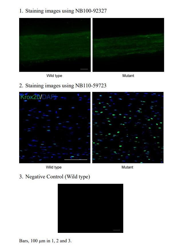
Application: Immunohistochemistry-FrozenSample Tested: Frozen mouse sciatic nerves and Frozen mouse sciatic nerveSpecies: MouseVerified Customer | Posted 12/05/2019Image 1. The staining image using NB100-92327. Image 2. The staining image using NB110-59723 (Previously worked well). Image 3. The staining image of Negative control for NB100-92327.
 Bio-Techne ResponseThis review was submitted through the legacy Novus Innovators Program, reflecting a new species or application tested on a primary antibody.
There are no reviews that match your criteria.
Protocols
Find general support by application which include: protocols, troubleshooting, illustrated assays, videos and webinars.
- Antigen Retrieval Protocol (PIER)
- Antigen Retrieval for Frozen Sections Protocol
- Appropriate Fixation of IHC/ICC Samples
- Cellular Response to Hypoxia Protocols
- Chromogenic IHC Staining of Formalin-Fixed Paraffin-Embedded (FFPE) Tissue Protocol
- Chromogenic Immunohistochemistry Staining of Frozen Tissue
- ClariTSA™ Fluorophore Kits
- Detection & Visualization of Antibody Binding
- Fluorescent IHC Staining of Frozen Tissue Protocol
- Graphic Protocol for Heat-induced Epitope Retrieval
- Graphic Protocol for the Preparation and Fluorescent IHC Staining of Frozen Tissue Sections
- Graphic Protocol for the Preparation and Fluorescent IHC Staining of Paraffin-embedded Tissue Sections
- Graphic Protocol for the Preparation of Gelatin-coated Slides for Histological Tissue Sections
- ICC Cell Smear Protocol for Suspension Cells
- ICC Immunocytochemistry Protocol Videos
- ICC for Adherent Cells
- IHC Sample Preparation (Frozen sections vs Paraffin)
- Immunocytochemistry (ICC) Protocol
- Immunocytochemistry Troubleshooting
- Immunofluorescence of Organoids Embedded in Cultrex Basement Membrane Extract
- Immunofluorescent IHC Staining of Formalin-Fixed Paraffin-Embedded (FFPE) Tissue Protocol
- Immunohistochemistry (IHC) and Immunocytochemistry (ICC) Protocols
- Immunohistochemistry Frozen Troubleshooting
- Immunohistochemistry Paraffin Troubleshooting
- Preparing Samples for IHC/ICC Experiments
- Preventing Non-Specific Staining (Non-Specific Binding)
- Primary Antibody Selection & Optimization
- Protocol for Heat-Induced Epitope Retrieval (HIER)
- Protocol for Making a 4% Formaldehyde Solution in PBS
- Protocol for VisUCyte™ HRP Polymer Detection Reagent
- Protocol for the Fluorescent ICC Staining of Cell Smears - Graphic
- Protocol for the Fluorescent ICC Staining of Cultured Cells on Coverslips - Graphic
- Protocol for the Preparation & Fixation of Cells on Coverslips
- Protocol for the Preparation and Chromogenic IHC Staining of Frozen Tissue Sections
- Protocol for the Preparation and Chromogenic IHC Staining of Frozen Tissue Sections - Graphic
- Protocol for the Preparation and Chromogenic IHC Staining of Paraffin-embedded Tissue Sections
- Protocol for the Preparation and Chromogenic IHC Staining of Paraffin-embedded Tissue Sections - Graphic
- Protocol for the Preparation and Fluorescent ICC Staining of Cells on Coverslips
- Protocol for the Preparation and Fluorescent ICC Staining of Non-adherent Cells
- Protocol for the Preparation and Fluorescent ICC Staining of Stem Cells on Coverslips
- Protocol for the Preparation and Fluorescent IHC Staining of Frozen Tissue Sections
- Protocol for the Preparation and Fluorescent IHC Staining of Paraffin-embedded Tissue Sections
- Protocol for the Preparation of Gelatin-coated Slides for Histological Tissue Sections
- Protocol for the Preparation of a Cell Smear for Non-adherent Cell ICC - Graphic
- R&D Systems Quality Control Western Blot Protocol
- TUNEL and Active Caspase-3 Detection by IHC/ICC Protocol
- The Importance of IHC/ICC Controls
- Troubleshooting Guide: Immunohistochemistry
- Troubleshooting Guide: Western Blot Figures
- Western Blot Conditions
- Western Blot Protocol
- Western Blot Protocol for Cell Lysates
- Western Blot Troubleshooting
- Western Blot Troubleshooting Guide
- View all Protocols, Troubleshooting, Illustrated assays and Webinars
FAQs for EGR2 Antibody - BSA Free
-
Q: We would like to try this antibody for staining in Xenopus. The mentioned immunogenic region shows high sequence identity with Xenopus EGR2. Would it be possible to obtain the exact epitope to predict cross reactivity?
A: I have now been informed that the immunogen sequence for this antibody corresponds to the amino acid range 391-440 of P11161. This shows high homology to the Xenopus sequence, so it is likely that the antibody would bind to Xenopus protein.