ERR alpha/NR3B1 Antibody - BSA Free
Novus Biologicals | Catalog # NBP1-47254
![Immunocytochemistry/ Immunofluorescence: ERR alpha/NR3B1 Antibody - BSA Free [NBP1-47254]](https://resources.rndsystems.com/images/products/ERR-alpha-NR3B1-Antibody-Immunocytochemistry-Immunofluorescence-NBP1-47254-img0012.jpg "Immunocytochemistry/ Immunofluorescence: ERR alpha/NR3B1 Antibody - BSA Free [NBP1-47254]")
Key Product Details
Species Reactivity
Validated:
Human, Mouse
Cited:
Human, Mouse
Predicted:
Canine (100%), Porcine (100%), Rat (100%). Backed by our 100% Guarantee.
Applications
Validated:
Immunohistochemistry, Immunohistochemistry-Paraffin, Western Blot, Immunocytochemistry/ Immunofluorescence
Cited:
Western Blot
Label
Unconjugated
Antibody Source
Polyclonal Rabbit IgG
Format
BSA Free
Loading...
Product Specifications
Immunogen
Synthetic peptide made to an N-terminal portion of human Estrogen Related Receptor alpha (within residues 1-50). [Swiss-Prot# P11474]
Reactivity Notes
Predicted to react with rat, pig and dog based on 100% sequence homology.
Localization
Nucleus, Cytoplasm
Clonality
Polyclonal
Host
Rabbit
Isotype
IgG
Theoretical MW
50 kDa.
Disclaimer note: The observed molecular weight of the protein may vary from the listed predicted molecular weight due to post translational modifications, post translation cleavages, relative charges, and other experimental factors.
Disclaimer note: The observed molecular weight of the protein may vary from the listed predicted molecular weight due to post translational modifications, post translation cleavages, relative charges, and other experimental factors.
Scientific Data Images for ERR alpha/NR3B1 Antibody - BSA Free
Immunocytochemistry/ Immunofluorescence: ERR alpha/NR3B1 Antibody - BSA Free [NBP1-47254]
Immunocytochemistry/Immunofluorescence: ERR alpha/NR3B1 Antibody [NBP1-47254] - HeLa cells were fixed in 4% paraformaldehyde for 10 minutes and permeabilized in 0.5% Triton X-100 in PBS for 5 minutes. The cells were incubated with conjugated to DyLight 488 (NBP1-47254G) at 5 ug/ml for 1 hour at room temperature. Nuclei were counterstained with DAPI (Blue). Cells were imaged using a 100X objective and digitally deconvolved.![Western Blot: ERR alpha/NR3B1 AntibodyBSA Free [NBP1-47254]](https://resources.rndsystems.com/images/products/ERR-alpha-NR3B1-Antibody-Western-Blot-NBP1-47254-img0011.jpg "Western Blot: ERR alpha/NR3B1 AntibodyBSA Free [NBP1-47254]")
Western Blot: ERR alpha/NR3B1 AntibodyBSA Free [NBP1-47254]
ERR-alpha-NR3B1-Antibody-Western-Blot-NBP1-47254-img0011.jpg![Immunohistochemistry-Paraffin: ERR alpha/NR3B1 Antibody - BSA Free [NBP1-47254]](https://resources.rndsystems.com/images/products/ERR-alpha-NR3B1-Antibody-Immunohistochemistry-Paraffin-NBP1-47254-img0010.jpg "Immunohistochemistry-Paraffin: ERR alpha/NR3B1 Antibody - BSA Free [NBP1-47254]")
Immunohistochemistry-Paraffin: ERR alpha/NR3B1 Antibody - BSA Free [NBP1-47254]
Immunohistochemistry-Paraffin: ERR alpha/NR3B1 Antibody [NBP1-47254] - IHC-P detection of ERR alpha/ESRRA protein in a section of normal human kidney using 5 ug/ml concentration of Estrogen Related Receptor alpha antibody. Some cells of tubules/ducts in the tested renal tissue depicted intense nuclear staining with an overall weak/diffused cytoplasmic staining. (10X Magnification)![Western Blot: ERR alpha/NR3B1 AntibodyBSA Free [NBP1-47254]](https://resources.rndsystems.com/images/products/ERR-alpha-NR3B1-Antibody-Western-Blot-NBP1-47254-img0008.jpg "Western Blot: ERR alpha/NR3B1 AntibodyBSA Free [NBP1-47254]")
Western Blot: ERR alpha/NR3B1 AntibodyBSA Free [NBP1-47254]
Western Blot: ERR alpha/NR3B1 Antibody [NBP1-47254] - Analysis of ERR alpha in A431 cell lysates![Immunocytochemistry/ Immunofluorescence: ERR alpha/NR3B1 Antibody - BSA Free [NBP1-47254]](https://resources.rndsystems.com/images/products/ERR-alpha-NR3B1-Antibody-Immunocytochemistry-Immunofluorescence-NBP1-47254-img0007.jpg "Immunocytochemistry/ Immunofluorescence: ERR alpha/NR3B1 Antibody - BSA Free [NBP1-47254]")
Immunocytochemistry/ Immunofluorescence: ERR alpha/NR3B1 Antibody - BSA Free [NBP1-47254]
Immunocytochemistry/Immunofluorescence: ERR alpha/NR3B1 Antibody [NBP1-47254] - ERR alpha antibody was tested in HeLa cells at a 1:100 dilution against Dylight 488 (Green). Alpha tubulin and nuclei were counterstained against Dylight 568 (Red) and DAPI (Blue), respectively.![Immunohistochemistry-Paraffin: ERR alpha/NR3B1 Antibody - BSA Free [NBP1-47254]](https://resources.rndsystems.com/images/products/ERR-alpha-NR3B1-Antibody-Immunohistochemistry-Paraffin-NBP1-47254-img0009.jpg "Immunohistochemistry-Paraffin: ERR alpha/NR3B1 Antibody - BSA Free [NBP1-47254]")
Immunohistochemistry-Paraffin: ERR alpha/NR3B1 Antibody - BSA Free [NBP1-47254]
Immunohistochemistry-Paraffin: ERR alpha/NR3B1 Antibody [NBP1-47254] - IHC-P detection of ERR alpha / ESRRA protein in a section of normal skin of human using 5 ug/ml concentration of Estrogen Related Receptor alpha antibody. The different cell types in the cellular epidermis as well as the blood vessels in the dermal layer showed positivity for ERR alpha, and the staining was found to be more intense in the nuclei compared to cytoplasm. (10X Magnification)Applications for ERR alpha/NR3B1 Antibody - BSA Free
Application
Recommended Usage
Immunocytochemistry/ Immunofluorescence
1:100
Immunohistochemistry
2 ug/ml
Immunohistochemistry-Paraffin
2 ug/ml
Western Blot
2 ug/ml
Application Notes
This Estrogen Related Receptor alpha antibody is useful for Western Blot, Immunohistochemistry-Paraffin and Immunocytochemistry/Immunofluorescence applications. ERR alpha/ ESRRA localize mainly to the nucleus, but there are reports on its cytoplasmic presence also, especially in cells with high nuclear expression. In a feedback loop mechanism, it has been shown that ERK8 /MAPK15 interacts with ERR alpha to induce its CRM1-dependent translocation to cytoplasm for inhibiting its transcriptional activity via regulation of its cellular localization (PMIDs 21190936, 18070081). The observed molecular weight of the protein may vary from the listed predicted molecular weight due to post translational modifications, post translation cleavages, relative charges, and other experimental factors.
Reviewed Applications
Read 1 review rated 3 using NBP1-47254 in the following applications:
Formulation, Preparation, and Storage
Purification
Immunogen affinity purified
Formulation
PBS and 30% Glycerol
Format
BSA Free
Preservative
0.1% Sodium Azide
Concentration
1 mg/ml
Shipping
The product is shipped with polar packs. Upon receipt, store it immediately at the temperature recommended below.
Stability & Storage
Store at 4C short term. Aliquot and store at -20C long term. Avoid freeze-thaw cycles.
Background: ERR alpha/NR3B1
Long Name
Estrogen-related Receptor alpha
Alternate Names
ERR1, ESRL1, ESRRA, NR3B1
Gene Symbol
ESRRA
UniProt
Additional ERR alpha/NR3B1 Products
Product Documents for ERR alpha/NR3B1 Antibody - BSA Free
Certificate of Analysis
To download a Certificate of Analysis, please enter a lot or batch number in the search box below.
Product Specific Notices for ERR alpha/NR3B1 Antibody - BSA Free
This product is for research use only and is not approved for use in humans or in clinical diagnosis. Primary Antibodies are guaranteed for 1 year from date of receipt.
Related Research Areas
Citations for ERR alpha/NR3B1 Antibody - BSA Free
 Powered by Bioz
Customer Reviews for ERR alpha/NR3B1 Antibody - BSA Free (1)
3 out of 5
1 Customer Rating
Have you used ERR alpha/NR3B1 Antibody - BSA Free?
Submit a review and receive an Amazon gift card!
$25/€18/£15/$25CAN/¥2500 Yen for a review with an image
$10/€7/£6/$10CAN/¥1110 Yen for a review without an image
Submit a review
Customer Images
Showing
1
-
1 of
1 review
Showing All
Filter By:
-
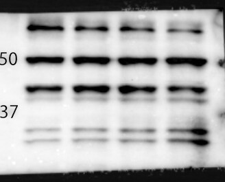
Application: Western BlotSample Tested: glioblastoma cell lysatesSpecies: HumanVerified Customer | Posted 08/02/2023human glioblastoma cell lysate. 10 ug loaded. condition: control, drug1, 2, and 3 treatment left numbers: size marker. bands of 50kDa is true band. There are multi bands.

There are no reviews that match your criteria.
Protocols
View specific protocols for ERR alpha/NR3B1 Antibody - BSA Free (NBP1-47254):
ERR alpha/NR3B1 Antibody:
Western Blot Protocol
1. Perform SDS-PAGE (4-12% MOPS) on samples to be analyzed, loading 35 ug of total protein per lane.
2. Transfer proteins to Nitrocellulose according to the instructions provided by the manufacturer of the transfer apparatus.
3. Rinse membrane with dH2O and then stain the blot using Ponceau S for 1-2 minutes to access the transfer of proteins onto the nitrocellulose membrane. Rinse the blot in water to remove excess stain and mark the lane locations and locations of molecular weight markers using a pencil.
4. Rinse the blot in TBS for approximately 5 minutes.
5. Block the membrane using 5% NFDM + 1% BSA in TBS + Tween, 1 hour at RT.
6. Rinse the membrane in dH2O and then wash the membrane in wash buffer [TBS + 0.1% Tween] 3 times for 10 minutes each.
7. Dilute the rabbit anti-ERR alpha primary antibody (NBP1-45282) in blocking buffer and incubate 1 hour at room temperature.
8. Rinse the membrane in dH2O and then wash the membrane in wash buffer [TBS + 0.1% Tween] 3 times for 10 minutes each.
9. Apply the diluted rabbit-IgG HRP-conjugated secondary antibody in blocking buffer (as per manufacturers instructions) and incubate 1 hour at room temperature.
10. Wash the blot in wash buffer [TBS + 0.1% Tween] 3 times for 10 minutes each (this step can be repeated as required to reduce background).
11. Apply the detection reagent of choice in accordance with the manufacturers instructions (Pierce ECL).
Note: Tween-20 can be added to the blocking or antibody dilution buffer at a final concentration of 0.05-0.2%, provided it does not interfere with antibody-antigen binding.
Find general support by application which include: protocols, troubleshooting, illustrated assays, videos and webinars.
- Antigen Retrieval Protocol (PIER)
- Antigen Retrieval for Frozen Sections Protocol
- Appropriate Fixation of IHC/ICC Samples
- Cellular Response to Hypoxia Protocols
- Chromogenic IHC Staining of Formalin-Fixed Paraffin-Embedded (FFPE) Tissue Protocol
- Chromogenic Immunohistochemistry Staining of Frozen Tissue
- ClariTSA™ Fluorophore Kits
- Detection & Visualization of Antibody Binding
- Fluorescent IHC Staining of Frozen Tissue Protocol
- Graphic Protocol for Heat-induced Epitope Retrieval
- Graphic Protocol for the Preparation and Fluorescent IHC Staining of Frozen Tissue Sections
- Graphic Protocol for the Preparation and Fluorescent IHC Staining of Paraffin-embedded Tissue Sections
- Graphic Protocol for the Preparation of Gelatin-coated Slides for Histological Tissue Sections
- ICC Cell Smear Protocol for Suspension Cells
- ICC Immunocytochemistry Protocol Videos
- ICC for Adherent Cells
- IHC Sample Preparation (Frozen sections vs Paraffin)
- Immunocytochemistry (ICC) Protocol
- Immunocytochemistry Troubleshooting
- Immunofluorescence of Organoids Embedded in Cultrex Basement Membrane Extract
- Immunofluorescent IHC Staining of Formalin-Fixed Paraffin-Embedded (FFPE) Tissue Protocol
- Immunohistochemistry (IHC) and Immunocytochemistry (ICC) Protocols
- Immunohistochemistry Frozen Troubleshooting
- Immunohistochemistry Paraffin Troubleshooting
- Preparing Samples for IHC/ICC Experiments
- Preventing Non-Specific Staining (Non-Specific Binding)
- Primary Antibody Selection & Optimization
- Protocol for Heat-Induced Epitope Retrieval (HIER)
- Protocol for Making a 4% Formaldehyde Solution in PBS
- Protocol for VisUCyte™ HRP Polymer Detection Reagent
- Protocol for the Fluorescent ICC Staining of Cell Smears - Graphic
- Protocol for the Fluorescent ICC Staining of Cultured Cells on Coverslips - Graphic
- Protocol for the Preparation & Fixation of Cells on Coverslips
- Protocol for the Preparation and Chromogenic IHC Staining of Frozen Tissue Sections
- Protocol for the Preparation and Chromogenic IHC Staining of Frozen Tissue Sections - Graphic
- Protocol for the Preparation and Chromogenic IHC Staining of Paraffin-embedded Tissue Sections
- Protocol for the Preparation and Chromogenic IHC Staining of Paraffin-embedded Tissue Sections - Graphic
- Protocol for the Preparation and Fluorescent ICC Staining of Cells on Coverslips
- Protocol for the Preparation and Fluorescent ICC Staining of Non-adherent Cells
- Protocol for the Preparation and Fluorescent ICC Staining of Stem Cells on Coverslips
- Protocol for the Preparation and Fluorescent IHC Staining of Frozen Tissue Sections
- Protocol for the Preparation and Fluorescent IHC Staining of Paraffin-embedded Tissue Sections
- Protocol for the Preparation of Gelatin-coated Slides for Histological Tissue Sections
- Protocol for the Preparation of a Cell Smear for Non-adherent Cell ICC - Graphic
- R&D Systems Quality Control Western Blot Protocol
- TUNEL and Active Caspase-3 Detection by IHC/ICC Protocol
- The Importance of IHC/ICC Controls
- Troubleshooting Guide: Immunohistochemistry
- Troubleshooting Guide: Western Blot Figures
- Western Blot Conditions
- Western Blot Protocol
- Western Blot Protocol for Cell Lysates
- Western Blot Troubleshooting
- Western Blot Troubleshooting Guide
- View all Protocols, Troubleshooting, Illustrated assays and Webinars
Loading...