Erythropoietin R Antibody - BSA Free
Novus Biologicals | Catalog # NBP1-19388
![Western Blot: Erythropoietin R AntibodyBSA Free [NBP1-19388]](https://resources.rndsystems.com/images/products/Erythropoietin-R-Antibody-Western-Blot-NBP1-19388-img0004.jpg "Western Blot: Erythropoietin R AntibodyBSA Free [NBP1-19388]")
Key Product Details
Species Reactivity
Validated:
Cited:
Applications
Validated:
Cited:
Label
Antibody Source
Format
Product Specifications
Immunogen
Reactivity Notes
Localization
Specificity
Clonality
Host
Isotype
Scientific Data Images for Erythropoietin R Antibody - BSA Free
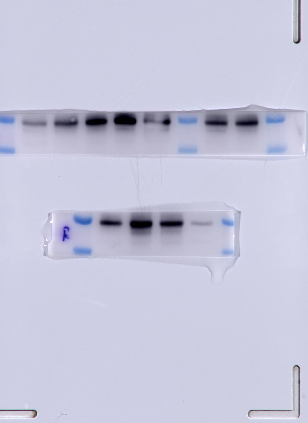
Western Blot: Erythropoietin R AntibodyBSA Free [NBP1-19388]
Western Blot: Erythropoietin R Antibody [NBP1-19388] - Western blot analysis of K562 (left) and SH-SY5Y (right) cell lysate using EPO receptor antibody at a concentration of 2 ug/ml.![Immunocytochemistry/ Immunofluorescence: Erythropoietin R Antibody - BSA Free [NBP1-19388]](https://resources.rndsystems.com/images/products/Erythropoietin-R-Antibody-Immunocytochemistry-Immunofluorescence-NBP1-19388-img0003.jpg "Immunocytochemistry/ Immunofluorescence: Erythropoietin R Antibody - BSA Free [NBP1-19388]")
Immunocytochemistry/ Immunofluorescence: Erythropoietin R Antibody - BSA Free [NBP1-19388]
Immunocytochemistry/Immunofluorescence: Erythropoietin R Antibody [NBP1-19388] - EPO receptor antibody was tested in HeLa cells with DyLight 488 (green). Nuclei and alpha-tubulin were counterstained with DAPI (blue) and Dylight 550 (red).![Immunohistochemistry-Paraffin: Erythropoietin R Antibody - BSA Free [NBP1-19388]](https://resources.rndsystems.com/images/products/Erythropoietin-R-Antibody-Immunohistochemistry-Paraffin-NBP1-19388-img0002.jpg "Immunohistochemistry-Paraffin: Erythropoietin R Antibody - BSA Free [NBP1-19388]")
Immunohistochemistry-Paraffin: Erythropoietin R Antibody - BSA Free [NBP1-19388]
Immunohistochemistry-Paraffin: Erythropoietin R Antibody [NBP1-19388] - IHC analysis of the EPO receptor in human placenta.![Simple Western: Erythropoietin R AntibodyBSA Free [NBP1-19388]](https://resources.rndsystems.com/images/products/Erythropoietin-R-Antibody-Simple-Western-NBP1-19388-img0005.jpg "Simple Western: Erythropoietin R AntibodyBSA Free [NBP1-19388]")
Simple Western: Erythropoietin R AntibodyBSA Free [NBP1-19388]
Simple Western: Erythropoietin R Antibody [NBP1-19388] - Simple Western lane view shows a specific band for EPO Receptor in 0.5 mg/ml of Human Brain lysate. This experiment was performed under reducing conditions using the 12-230 kDa separation system.Applications for Erythropoietin R Antibody - BSA Free
Immunocytochemistry/ Immunofluorescence
Immunohistochemistry
Immunohistochemistry-Paraffin
Simple Western
Western Blot
See Simple Western Antibody Database for Simple Western validation: Tested in Human Brain lysate 0.5 mg/mL, separated by Size, antibody dilution of 1:10, apparent MW was 49 kDa.
Reviewed Applications
Read 2 reviews rated 5 using NBP1-19388 in the following applications:
Formulation, Preparation, and Storage
Purification
Formulation
Format
Preservative
Concentration
Shipping
Stability & Storage
Background: Erythropoietin R
Long Name
Alternate Names
Gene Symbol
UniProt
Additional Erythropoietin R Products
Product Documents for Erythropoietin R Antibody - BSA Free
Certificate of Analysis
To download a Certificate of Analysis, please enter a lot or batch number in the search box below.
Product Specific Notices for Erythropoietin R Antibody - BSA Free
This product is for research use only and is not approved for use in humans or in clinical diagnosis. Primary Antibodies are guaranteed for 1 year from date of receipt.
Related Research Areas
Citations for Erythropoietin R Antibody - BSA Free
 Powered by Bioz
Customer Reviews for Erythropoietin R Antibody - BSA Free (2)
Have you used Erythropoietin R Antibody - BSA Free?
Submit a review and receive an Amazon gift card!
$25/€18/£15/$25CAN/¥2500 Yen for a review with an image
$10/€7/£6/$10CAN/¥1110 Yen for a review without an image
Submit a review
Customer Images

-
Application: Western BlotSample Tested: k562 cellsSpecies: HumanVerified Customer | Posted 10/05/2022EPOR expression in K562 cells

-
Application: Western BlotSample Tested: blood vessel tissueSpecies: MouseVerified Customer | Posted 11/17/2017
There are no reviews that match your criteria.
Protocols
View specific protocols for Erythropoietin R Antibody - BSA Free (NBP1-19388):
Immunocytochemistry Protocol
Culture cells to appropriate density in 35 mm culture dishes or 6-well plates.
1. Remove culture medium and add 10% formalin to the dish. Fix at room temperature for 30 minutes.
2. Remove the formalin and add ice cold methanol. Incubate for 5-10 minutes.
3. Remove methanol and add washing solution (i.e. PBS). Be sure to not let the specimen dry out. Wash three times for 10 minutes.
4. To block nonspecific antibody binding incubate in 10% normal goat serum from 1 hour to overnight at room temperature.
5. Add primary antibody at appropriate dilution and incubate at room temperature from 2 hours to overnight at room temperature.
6. Remove primary antibody and replace with washing solution. Wash three times for 10 minutes.
7. Add secondary antibody at appropriate dilution. Incubate for 1 hour at room temperature.
8. Remove antibody and replace with wash solution, then wash for 10 minutes. Add Hoechst 33258 to wash solution at 1:25,0000 and incubate for 10 minutes. Wash a third time for 10 minutes.
9. Cells can be viewed directly after washing. The plates can also be stored in PBS containing Azide covered in Parafilm (TM). Cells can also be cover-slipped using Fluoromount, with appropriate sealing.
*The above information is only intended as a guide. The researcher should determine what protocol best meets their needs. Please follow safe laboratory procedures.
Immunohistochemistry-Paraffin Embedded Sections
Antigen Unmasking:
Bring slides to a boil in 10 mM sodium citrate buffer (pH 6.0) then maintain at a sub-boiling temperature for 10 minutes. Cool slides on bench-top for 30 minutes.
Staining:
1. Wash sections in deionized water three times for 5 minutes each.
2. Wash sections in wash buffer for 5 minutes.
3. Block each section with 100-400 ul blocking solution for 1 hour at room temperature.
4. Remove blocking solution and add 100-400 ul diluted primary antibody. Incubate overnight at 4 C.
5. Remove antibody solution and wash sections in wash buffer three times for 5 minutes each.
6. Add 100-400 ul biotinylated diluted secondary antibody. Incubate 30 minutes at room temperature.
7. Remove secondary antibody solution and wash sections three times with wash buffer for 5 minutes each.
8. Add 100-400 ul Streptavidin-HRP reagent to each section and incubate for 30 minutes at room temperature.
9. Wash sections three times in wash buffer for 5 minutes each.
10. Add 100-400 ul DAB substrate to each section and monitor staining closely.
11. As soon as the sections develop, immerse slides in deionized water.
12. Counterstain sections in hematoxylin.
13. Wash sections in deionized water two times for 5 minutes each.
14. Dehydrate sections.
15. Mount coverslips.
Western Blot Protocol
1. Perform SDS-PAGE on samples to be analyzed, loading 25 ug of total protein per lane.
2. Transfer proteins to membrane according to the instructions provided by the manufacturer of the membrane and transfer apparatus.
3. Stain according to standard Ponceau S procedure (or similar product) to assess transfer success, and mark molecular weight standards where appropriate.
4. Rinse the blot.
5. Block the membrane using standard blocking buffer for at least 1 hour.
6. Wash the membrane in wash buffer three times for 10 minutes each.
7. Dilute anti-HDAC5 primary antibody in blocking buffer and incubate 1 hour at room temperature.
8. Wash the membrane in wash buffer three times for 10 minutes each.
9. Apply the diluted HRP conjugated secondary antibody in blocking buffer (as per manufacturers instructions) and incubate 1 hour at room temperature.
10. Wash the blot in wash buffer three times for 10 minutes each (this step can be repeated as required to reduce background).
11. Apply the detection reagent of choice in accordance with the manufacturers instructions.
Note: Tween-20 can be added to the blocking or antibody dilution buffer at a final concentration of 0.05-0.2%.
Find general support by application which include: protocols, troubleshooting, illustrated assays, videos and webinars.
- Antigen Retrieval Protocol (PIER)
- Antigen Retrieval for Frozen Sections Protocol
- Appropriate Fixation of IHC/ICC Samples
- Cellular Response to Hypoxia Protocols
- Chromogenic IHC Staining of Formalin-Fixed Paraffin-Embedded (FFPE) Tissue Protocol
- Chromogenic Immunohistochemistry Staining of Frozen Tissue
- ClariTSA™ Fluorophore Kits
- Detection & Visualization of Antibody Binding
- Fluorescent IHC Staining of Frozen Tissue Protocol
- Graphic Protocol for Heat-induced Epitope Retrieval
- Graphic Protocol for the Preparation and Fluorescent IHC Staining of Frozen Tissue Sections
- Graphic Protocol for the Preparation and Fluorescent IHC Staining of Paraffin-embedded Tissue Sections
- Graphic Protocol for the Preparation of Gelatin-coated Slides for Histological Tissue Sections
- ICC Cell Smear Protocol for Suspension Cells
- ICC Immunocytochemistry Protocol Videos
- ICC for Adherent Cells
- IHC Sample Preparation (Frozen sections vs Paraffin)
- Immunocytochemistry (ICC) Protocol
- Immunocytochemistry Troubleshooting
- Immunofluorescence of Organoids Embedded in Cultrex Basement Membrane Extract
- Immunofluorescent IHC Staining of Formalin-Fixed Paraffin-Embedded (FFPE) Tissue Protocol
- Immunohistochemistry (IHC) and Immunocytochemistry (ICC) Protocols
- Immunohistochemistry Frozen Troubleshooting
- Immunohistochemistry Paraffin Troubleshooting
- Preparing Samples for IHC/ICC Experiments
- Preventing Non-Specific Staining (Non-Specific Binding)
- Primary Antibody Selection & Optimization
- Protocol for Heat-Induced Epitope Retrieval (HIER)
- Protocol for Making a 4% Formaldehyde Solution in PBS
- Protocol for VisUCyte™ HRP Polymer Detection Reagent
- Protocol for the Fluorescent ICC Staining of Cell Smears - Graphic
- Protocol for the Fluorescent ICC Staining of Cultured Cells on Coverslips - Graphic
- Protocol for the Preparation & Fixation of Cells on Coverslips
- Protocol for the Preparation and Chromogenic IHC Staining of Frozen Tissue Sections
- Protocol for the Preparation and Chromogenic IHC Staining of Frozen Tissue Sections - Graphic
- Protocol for the Preparation and Chromogenic IHC Staining of Paraffin-embedded Tissue Sections
- Protocol for the Preparation and Chromogenic IHC Staining of Paraffin-embedded Tissue Sections - Graphic
- Protocol for the Preparation and Fluorescent ICC Staining of Cells on Coverslips
- Protocol for the Preparation and Fluorescent ICC Staining of Non-adherent Cells
- Protocol for the Preparation and Fluorescent ICC Staining of Stem Cells on Coverslips
- Protocol for the Preparation and Fluorescent IHC Staining of Frozen Tissue Sections
- Protocol for the Preparation and Fluorescent IHC Staining of Paraffin-embedded Tissue Sections
- Protocol for the Preparation of Gelatin-coated Slides for Histological Tissue Sections
- Protocol for the Preparation of a Cell Smear for Non-adherent Cell ICC - Graphic
- R&D Systems Quality Control Western Blot Protocol
- TUNEL and Active Caspase-3 Detection by IHC/ICC Protocol
- The Importance of IHC/ICC Controls
- Troubleshooting Guide: Immunohistochemistry
- Troubleshooting Guide: Western Blot Figures
- Western Blot Conditions
- Western Blot Protocol
- Western Blot Protocol for Cell Lysates
- Western Blot Troubleshooting
- Western Blot Troubleshooting Guide
- View all Protocols, Troubleshooting, Illustrated assays and Webinars
FAQs for Erythropoietin R Antibody - BSA Free
-
Q: EPO Receptor Antibody (NBP1-19388) is suitable for immunohistochemistry (paraffin) and you also say that it can be used for IF. I just want to confirm that this particular antibody can be visualized by other systems also?
A: This antibody has only been validated for use in WB and IHC-Paraffin, not Immunofluorescence. This was incorrect information on the datasheet and I have now corrected this.
Associated Pathways
