Exosome Detection (Western Blot) Antibody Pack
Novus Biologicals | Catalog # NBP3-11740
![Western Blot: Exosome Detection (Western Blot) Antibody Pack [NBP3-11740]](https://resources.rndsystems.com/images/products/Exosome-Detection-Western-Blot-Antibody-Pack-Western-Blot-NBP3-11740-img0001.jpg "Western Blot: Exosome Detection (Western Blot) Antibody Pack [NBP3-11740]")
Loading...
Key Product Details
Species
Human
Applications
Immunocytochemistry/ Immunofluorescence, Immunohistochemistry, Western Blot
Product Summary for Exosome Detection (Western Blot) Antibody Pack
This pack contains 1 vial each of: NBP2-77452SS (0.025 mg); MAB1663-SP (25 ug); MAB25292-SP (25 ug); NB100-65805SS (0.025 ml); NBP2-42225SS (0.025 ml); NBP1-47546SS (0.025 ml); MAB1455-SP (25 ug), HAF007 and HAF008.
Loading...
Product Specifications
Specificity
Please see each vial's data sheet for specificity information specific to each product.
Application Notes
See individual datasheets of components for their validated applications
Reactivity Notes
See individual datasheets of components for their validated species
Scientific Data Images for Exosome Detection (Western Blot) Antibody Pack
Western Blot: Exosome Detection (Western Blot) Antibody Pack [NBP3-11740]
Exosome-Detection-Western-Blot-Antibody-Pack-Western-Blot-NBP3-11740-img0001.jpg![Western Blot: Exosome Detection (Western Blot) Antibody Pack [NBP3-11740]](https://resources.rndsystems.com/images/products/Exosome-Detection-Western-Blot-Antibody-Pack-Western-Blot-NBP3-11740-img0003.jpg "Western Blot: Exosome Detection (Western Blot) Antibody Pack [NBP3-11740]")
Western Blot: Exosome Detection (Western Blot) Antibody Pack [NBP3-11740]
Western Blot: Exosome Detection (Western Blot) Antibody Pack [NBP3-11740] - Western blot shows lysates of TIME human endothelial cell line and human platelets. PVDF membrane was probed with 2 ug/mL of Mouse Anti-Human CD9 Monoclonal Antibody (Catalog # MAB25292) followed by HRP-conjugated Anti-Mouse IgG Secondary Antibody (Catalog # HAF018). A specific band was detected for CD9 at approximately 24 kDa (as indicated).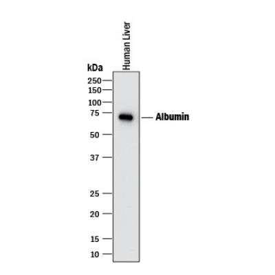
Exosome Detection (Western Blot) Antibody Pack [NBP3-11740] - Western blot shows lysate of human liver tissue. PVDF membrane was probed with 1 ug/mL of Mouse Anti-Human Serum Albumin Monoclonal Antibody (Catalog # MAB1455) followed by HRP-conjugated Anti-Mouse IgG Secondary Antibody (Catalog # HAF018). A specific band was detected for Albumin at approximately 65-70 kDa (as indicated).
Kit Contents for Exosome Detection (Western Blot) Antibody Pack
- MAB1455-SP
- MAB1663-SP
- MAB25292-SP
- HAF007: Mouse IgG Horseradish Peroxidase-conjugated Antibody
- NB100-65805SS
- NBP1-47546SS
- NBP2-42225SS
- NBP2-77452SS
- HAF008: Rabbit IgG Horseradish Peroxidase-conjugated Antibody
Formulation, Preparation, and Storage
Formulation
See individual datasheets
Concentration
Concentration of individual antibodies may be found on the vial label. If unlisted please contact technical services.
Shipping
The product is shipped with polar packs. Upon receipt, store it immediately at the temperature recommended below.
Storage
Storage is content dependent.
Background: Exosome Detection (Western Blot)
Alternate Names
EV detection, Exosome Markers, extracellular vesicle detection
Additional Exosome Detection (Western Blot) Products
Product Documents for Exosome Detection (Western Blot) Antibody Pack
Certificate of Analysis
To download a Certificate of Analysis, please enter a lot or batch number in the search box below.
Product Specific Notices for Exosome Detection (Western Blot) Antibody Pack
This product is for research use only and is not approved for use in humans or in clinical diagnosis. Antibody Packs are guaranteed for 1 year from date of receipt.
Customer Reviews for Exosome Detection (Western Blot) Antibody Pack
There are currently no reviews for this product. Be the first to review Exosome Detection (Western Blot) Antibody Pack and earn rewards!
Have you used Exosome Detection (Western Blot) Antibody Pack?
Submit a review and receive an Amazon gift card!
$25/€18/£15/$25CAN/¥2500 Yen for a review with an image
$10/€7/£6/$10CAN/¥1110 Yen for a review without an image
Submit a review
Protocols
Find general support by application which include: protocols, troubleshooting, illustrated assays, videos and webinars.
- Antigen Retrieval Protocol (PIER)
- Antigen Retrieval for Frozen Sections Protocol
- Appropriate Fixation of IHC/ICC Samples
- Cellular Response to Hypoxia Protocols
- Chromogenic IHC Staining of Formalin-Fixed Paraffin-Embedded (FFPE) Tissue Protocol
- Chromogenic Immunohistochemistry Staining of Frozen Tissue
- ClariTSA™ Fluorophore Kits
- Detection & Visualization of Antibody Binding
- Fluorescent IHC Staining of Frozen Tissue Protocol
- Graphic Protocol for Heat-induced Epitope Retrieval
- Graphic Protocol for the Preparation and Fluorescent IHC Staining of Frozen Tissue Sections
- Graphic Protocol for the Preparation and Fluorescent IHC Staining of Paraffin-embedded Tissue Sections
- Graphic Protocol for the Preparation of Gelatin-coated Slides for Histological Tissue Sections
- ICC Cell Smear Protocol for Suspension Cells
- ICC Immunocytochemistry Protocol Videos
- ICC for Adherent Cells
- IHC Sample Preparation (Frozen sections vs Paraffin)
- Immunocytochemistry (ICC) Protocol
- Immunocytochemistry Troubleshooting
- Immunofluorescence of Organoids Embedded in Cultrex Basement Membrane Extract
- Immunofluorescent IHC Staining of Formalin-Fixed Paraffin-Embedded (FFPE) Tissue Protocol
- Immunohistochemistry (IHC) and Immunocytochemistry (ICC) Protocols
- Immunohistochemistry Frozen Troubleshooting
- Immunohistochemistry Paraffin Troubleshooting
- Preparing Samples for IHC/ICC Experiments
- Preventing Non-Specific Staining (Non-Specific Binding)
- Primary Antibody Selection & Optimization
- Protocol for Heat-Induced Epitope Retrieval (HIER)
- Protocol for Making a 4% Formaldehyde Solution in PBS
- Protocol for VisUCyte™ HRP Polymer Detection Reagent
- Protocol for the Fluorescent ICC Staining of Cell Smears - Graphic
- Protocol for the Fluorescent ICC Staining of Cultured Cells on Coverslips - Graphic
- Protocol for the Preparation & Fixation of Cells on Coverslips
- Protocol for the Preparation and Chromogenic IHC Staining of Frozen Tissue Sections
- Protocol for the Preparation and Chromogenic IHC Staining of Frozen Tissue Sections - Graphic
- Protocol for the Preparation and Chromogenic IHC Staining of Paraffin-embedded Tissue Sections
- Protocol for the Preparation and Chromogenic IHC Staining of Paraffin-embedded Tissue Sections - Graphic
- Protocol for the Preparation and Fluorescent ICC Staining of Cells on Coverslips
- Protocol for the Preparation and Fluorescent ICC Staining of Non-adherent Cells
- Protocol for the Preparation and Fluorescent ICC Staining of Stem Cells on Coverslips
- Protocol for the Preparation and Fluorescent IHC Staining of Frozen Tissue Sections
- Protocol for the Preparation and Fluorescent IHC Staining of Paraffin-embedded Tissue Sections
- Protocol for the Preparation of Gelatin-coated Slides for Histological Tissue Sections
- Protocol for the Preparation of a Cell Smear for Non-adherent Cell ICC - Graphic
- R&D Systems Quality Control Western Blot Protocol
- TUNEL and Active Caspase-3 Detection by IHC/ICC Protocol
- The Importance of IHC/ICC Controls
- Troubleshooting Guide: Immunohistochemistry
- Troubleshooting Guide: Western Blot Figures
- Western Blot Conditions
- Western Blot Protocol
- Western Blot Protocol for Cell Lysates
- Western Blot Troubleshooting
- Western Blot Troubleshooting Guide
- View all Protocols, Troubleshooting, Illustrated assays and Webinars
Loading...